. 2023 Feb 22;8(4):e165713.
doi: 10.1172/jci.insight.165713.

Insulin-like growth factor 1 reduces coronary atherosclerosis in pigs with familial hypercholesterolemia

Affiliations

Insulin-like growth factor 1 reduces coronary atherosclerosis in pigs with familial hypercholesterolemia

Sergiy Sukhanov et al. JCI Insight. .

Abstract

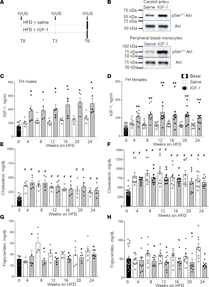
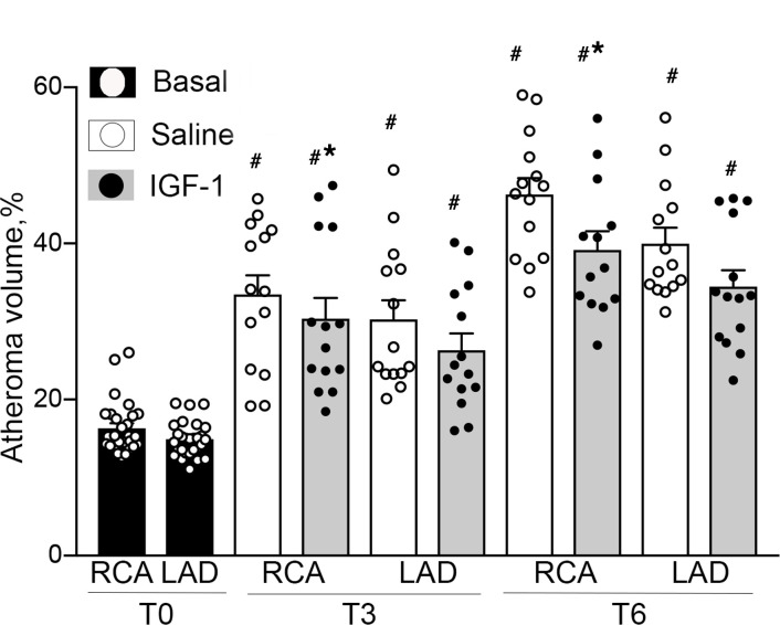
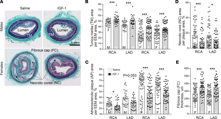
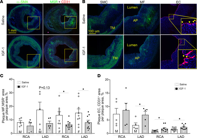
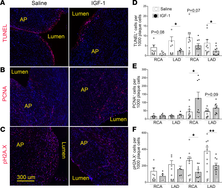
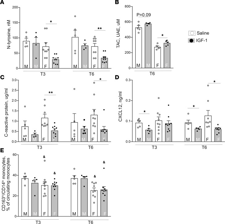
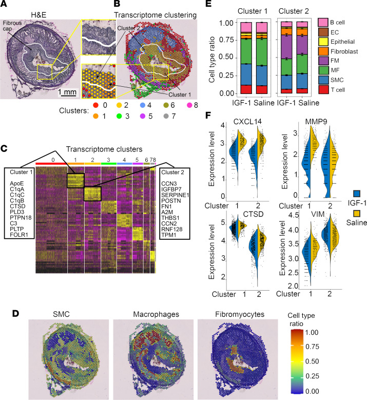
Although murine models of coronary atherosclerotic disease have been used extensively to determine mechanisms, limited new therapeutic options have emerged. Pigs with familial hypercholesterolemia (FH pigs) develop complex coronary atheromas that are almost identical to human lesions. We reported previously that insulin-like growth factor 1 (IGF-1) reduced aortic atherosclerosis and promoted features of stable plaque in a murine model. We administered human recombinant IGF-1 or saline (control) in atherosclerotic FH pigs for 6 months. IGF-1 decreased relative coronary atheroma in vivo (intravascular ultrasound) and reduced lesion cross-sectional area (postmortem histology). IGF-1 increased plaque's fibrous cap thickness, and reduced necrotic core, macrophage content, and cell apoptosis, consistent with promotion of a stable plaque phenotype. IGF-1 reduced circulating triglycerides, markers of systemic oxidative stress, and CXCL12 chemokine levels. We used spatial transcriptomics (ST) to identify global transcriptome changes in advanced plaque compartments and to obtain mechanistic insights into IGF-1 effects. ST analysis showed that IGF-1 suppressed FOS/FOSB factors and gene expression of MMP9 and CXCL14 in plaque macrophages, suggesting possible involvement of these molecules in IGF-1's effect on atherosclerosis. Thus, IGF-1 reduced coronary plaque burden and promoted features of stable plaque in a pig model, providing support for consideration of clinical trials.

Keywords: Atherosclerosis; Cardiology; Growth factors; Plaque formation; Vascular Biology.


Conflict of interest statement

Conflict of interest: The authors have declared that no conflict of interest exists.

Figures

Figure 1
Figure 1. Phenotype of FH pigs.
(A) Experimental design. FH pigs were injected daily with 50 μg/kg human recombinant IGF-1 or saline (control) (males, n = 5/group; females, n = 9/group) and fed with high-fat diet (HFD) for 6 months. Coronary atherosclerosis was quantified by intravascular ultrasound (IVUS) before injections (T0), after 3 months (T3), and after 6 months (T6, at sacrificing). (B) IGF-1 stimulated specific downstream signaling in porcine carotid artery and in peripheral blood mononuclear cells (PBMC). IGF-1 (or saline) was injected into pig, carotids and blood were collected 4 hours following injections, and Akt phosphorylation was quantified by immunoblotting. (CH) Blood was collected at basal level (T0) and each month during injections (total 7 time points). Total plasma IGF-1 level was quantified by ELISA in FH males (C) and females (D). Cholesterol (E and F) and triglyceride (G and H) levels in IGF-1– and saline-injected male (left) and female (right) FH pigs. Males: n = 10 for basal, and n = 5/group for each time point. Females: n = 18 for basal, and n = 9/group for each time point. *P < 0.05, **P < 0.01, vs. saline based on t test, #P < 0.05 vs. basal level based on 3-way ANOVA.
Figure 2
Figure 2. IGF-1 reduces coronary atheroma volume.
FH pigs (both sexes) were injected with IGF-1 or saline (control) (males, 5/group; females, 9/group). The coronary atheroma volume was quantified in RCAs and LADs by serial IVUS before injections (T0), after 3 months (T3), and after 6 months (T6). Relative atheroma volume (%) was defined as plaque + media volume divided per the vessel (external elastic membrane, EEM) volume × 100%. Since sex does not influence IGF-1’s effect on relative atheroma volume, atheroma measurements of both sexes were combined and shown. n = 28/group for T0, and n = 14 per RCA or LAD per group for T3 and T6. *P < 0.05 vs. saline, and #P < 0.05 vs. T0 based on 2-way ANOVA.
Figure 3
Figure 3. IGF-1 reduces coronary atherosclerosis and promotes features of stable atherosclerotic plaque.
IGF-1 increased vascular media (A and B), reduced atherosclerotic plaque cross-sectional area (CSA) (C), decreased necrotic core (D), and elevated thickness of fibrous cap (E). RCA and LAD were isolated from IGF-1– and saline-injected FH pigs and further cut onto 6 sequential fragments for embedding in paraffin. Trichrome-stained cross sections were obtained from each fragment and used for morphological analysis. n = 30 per RCA or LAD per group for males and n = 54 for females. (A) Representative images of RCA sections obtained from FH males and females. Tunica media (TM), atherosclerotic plaque (AP), fibrous cap (FC), and necrotic cores (NCs) were manually outlined to quantify TM and AP CSA, and results were normalized per EEM area. The thickness of FC was calculated as the mean length of 5 arbitrary lines distributed across the cap area. *P < 0.05, ***P < 0.005 vs. saline based on unpaired 2-tailed t test.
Figure 4
Figure 4. IGF-1 suppresses MF-like cells and upregulates EC-like cells in coronary plaques.
Serial RCA and LAD sections were immunostained with α–smooth muscle actin (α-SMA), macrophage scavenger receptor A (MSR), and CD31 antibody to identify SMC-like, MF-like, and EC-like cells, respectively. The primary antibody signal was amplified by biotin/streptavidin or tyramide systems conjugated to Alexa Fluor 488 (for α-SMA and MSR) or Alexa Fluor 594 (CD31). (A) Representative images of RCA sections obtained from IGF-1– or saline-injected FH females. Yellow square outlines plaque area magnified in B. (B) SMC, MF, and EC marker–immunopositive cells. Yellow arrows in insert indicate breaks in endothelial layer. (C and D) Quantitative data. n = 5 per RCA or LAD per group for males and n = 9 for females. *P < 0.05 vs. saline based on unpaired 2-tailed t test.
Figure 5
Figure 5. IGF-1 suppresses plaque cell apoptosis, promotes cell proliferation, and decreases DNA damage.
Cell apoptosis was quantified by TUNEL assay, and cell proliferation and DNA damage were quantified by immunostaining with PCNA antibody and pH2A.X antibody, respectively. (AC) Representative images of RCA sections obtained from IGF-1– or saline-injected FH females. (DF) Quantitative data. n = 5 per RCA or LAD per group for males and n = 9 for females. *P < 0.05, **P < 0.01 vs. saline based on unpaired 2-tailed t test.
Figure 6
Figure 6. IGF-1 downregulates markers of systemic oxidative stress and decreases C-reactive protein and chemokine CXCL12.
Markers of systemic oxidative stress (A and B), C-reactive protein (CRP) (C), and chemokine CXCL12 (D). IGF-1 did not alter circulating monocyte subsets (E). Circulating N-tyrosine, CXCL12, and CRP levels were quantified by ELISA and TAC, by using colorimetric assay. TAC assay results shown in urinary acid (standard) equivalents (UAE). (E) Whole blood was mixed with a cocktail of antibodies against CD163-PE, CD14–Alexa Fluor 488, and porcine CD172a and subsequently with streptavidin-APC/Cy7. CD172a-positive leukocytes were size-gated and further differentiated into subsets based on CD163 and CD14 expression levels using FACS. n = 5 per time point per group for males and n = 9 for females for N-tyrosine, CRP, CXCL12, and monocyte assay. n = 5/males and females for TAC assay. *P < 0.05 and **P < 0.01 vs. saline, &P < 0.05 vs. males based on 3-way ANOVA.
Figure 7
Figure 7. IGF-1 alters global transcriptomic profile of coronary plaque.
The RCA cryosections were obtained from IGF-1–injected (n = 2) and saline-injected (n = 2) FH females, and spatially resolved global transcriptome was assessed by ST. ST spots in IGF-1 and saline specimens were grouped into 9 clusters (numbered 0–8) based on their transcriptome, and the heatmap with top 10 upregulated genes/cluster was generated. (A) H&E-stained image with FC outline (white curve). (B) Representative transcriptome clustering. Yellow squares in A and B outline FC fragment shown magnified between panels. Cluster 1 and 2 were identified within a histologically homogeneous FC area. (C) Heatmap. (D) Cell type ratio was calculated for each ST spot to identify spots enriched by SMCs, MFs, or fibromyocytes (FMs). (E) Cell type ratio for IGF-1 versus saline specimens for cluster 1 and 2. (F) Violin plots show expression levels of CXCL14, MMP9, cathepsin D (CTSD), and vimentin (VIM) comparing IGF-1 versus saline specimens in cluster 1 and 2. IGF-1 downregulated CXCL14, MMP9, VIM, and CTSD within cluster 1 and decreased expression of CXCL14, MMP9, and CTSD within cluster 2 compared with saline.

References

    1. Mozaffarian D, et al. Executive summary: heart disease and stroke statistics--2016 update: a report from the American Heart Association. Circulation. 2016;133(4):447–454. doi: 10.1161/CIR.0000000000000366. - DOI - PubMed
    1. Tsao CW, et al. Heart disease and stroke statistics-2022 update: a report from the American Heart Association. Circulation. 2022;145(8):e153–e639. doi: 10.1161/CIR.0000000000001052. - DOI - PubMed
    1. Arbab-Zadeh A. Coronary atheroma burden is the main determinant of patient outcome: but how much detail is needed? Circ Cardiovasc Imaging. 2018;11(7):e007992. doi: 10.1161/CIRCIMAGING.118.007992. - DOI - PubMed
    1. Sui W, et al. Bladder drug mirabegron exacerbates atherosclerosis through activation of brown fat-mediated lipolysis. Proc Natl Acad Sci U S A. 2019;116(22):10937–10942. doi: 10.1073/pnas.1901655116. - DOI - PMC - PubMed
    1. Zhang C, et al. Angiotensin-converting enzyme 2 attenuates atherosclerotic lesions by targeting vascular cells. Proc Natl Acad Sci U S A. 2010;107(36):15886–15891. doi: 10.1073/pnas.1001253107. - DOI - PMC - PubMed

Publication types

Substances