. 2023 Jan 6;379(6627):71-78.
doi: 10.1126/science.abq7317. Epub 2023 Jan 5.

Cilia function as calcium-mediated mechanosensors that instruct left-right asymmetry

Affiliations

Cilia function as calcium-mediated mechanosensors that instruct left-right asymmetry

Lydia Djenoune et al. Science. .

Abstract

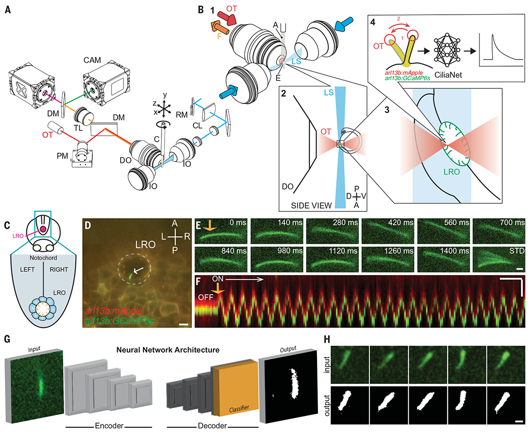
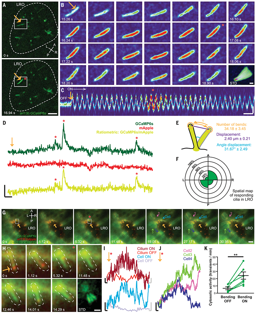
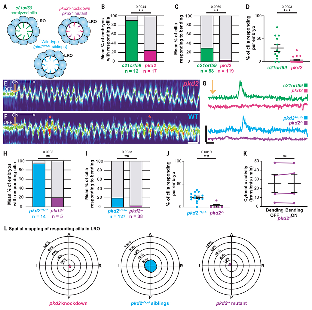
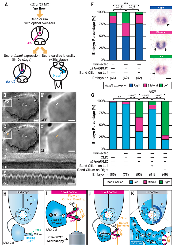
The breaking of bilateral symmetry in most vertebrates is critically dependent upon the motile cilia of the embryonic left-right organizer (LRO), which generate a directional fluid flow; however, it remains unclear how this flow is sensed. Here, we demonstrated that immotile LRO cilia are mechanosensors for shear force using a methodological pipeline that combines optical tweezers, light sheet microscopy, and deep learning to permit in vivo analyses in zebrafish. Mechanical manipulation of immotile LRO cilia activated intraciliary calcium transients that required the cation channel Polycystin-2. Furthermore, mechanical force applied to LRO cilia was sufficient to rescue and reverse cardiac situs in zebrafish that lack motile cilia. Thus, LRO cilia are mechanosensitive cellular levers that convert biomechanical forces into calcium signals to instruct left-right asymmetry.


Conflict of interest statement

Competing interests: Authors declare that they have no competing interests.

Figures

Fig. 1.
Fig. 1.. CiliaSPOT is a precise and tunable platform for cilia mechanosensing studies.
(A) Mechanical drawing of the CiliaSPOT microscope highlighting key components. For detailed description of the setup, see Materials and Methods and fig. S3. (B) Trapping and analyzing ciliary responses in the LRO of zebrafish embryos. (1) Close-up view showing the zebrafish embryo mounted in an agarose column extruded from a glass capillary. The cilia are fluorescently excited by the light sheet. (2) Side view of the optical trap and the LRO of a zebrafish embryo mounted in agarose. (3) Trapping of a single cilium in the LRO (green). (4) The trapped cilium is bent in an oscillatory fashion while being imaged. Images are then processed and analyzed by the CiliaNet machine learning algorithm to track and measure cilia responses to bending. A, agarose column; C, capillary; CL, cylindrical lens; CAM, camera; DM, dichroic mirror; DO, detection objective; E, embryo; F, fluorescence signal; IO, illumination objective; LS, light sheet; OT, optical trapping laser; PM, piezo mirror; RM, resonant mirror; TL, tube lens. (C) Illustration representing the LRO in the zebrafish embryo. (D) Representative image of an embryo expressing the ratiometric ciliary calcium reporter (arl13b:mApple;arl13b:GCaMP6s) with an intraciliary calcium oscillation (ICO, white arrow) within the LRO (dashed line). Scale: 10 μm. A, anterior; P, posterior; L, left; R, right; LRO, left-right organizer. (E) Representative montage of a fluorescent LRO cilium being bent by the optical trap (orange arrow) in vivo. Numbers indicate time after start of the bend. STD represents the standard deviation Z-projection of the montage. Scale: 2 μm. (F) Representative kymograph of an LRO cilium being bent by the optical trap (orange arrow). Note here that the GCaMP6s and mApple signals are kept slightly shifted for illustration purposes (see Materials and Methods and fig. S4). Scales: vertical: 2 μm; horizontal: 2 s. (G) Illustration of CiliaNet segmentation workflow. (H) Representative montage of sequential images of an LRO cilium dynamically trapped and moved by the optical tweezers (input) annotated by CiliaNet (output). Scale: 2 μm.
Fig. 2.
Fig. 2.. Oscillatory mechanical stimuli on LRO cilia activate intraciliary calcium transients.
(A) Representative images of the LRO of a c21orf59 morphant zebrafish. Dashed line: LRO. Scale: 10 μm. (B) Representative montage of the GCaMP6s-positive LRO cilium highlighted in (A). Scale: 2 μm. (C) Kymograph of the cilium shown in (B) before (“OFF”) and during optical bending (“ON”, white arrow, starting at orange arrow). Scales: vertical: 2 μm; horizontal: 2 s. (D) Intraciliary intensity over time plots of a single LRO cilium exhibiting intraciliary calcium oscillations of different amplitudes in response to optical bending. Scales: vertical: 50% ΔF/F; horizontal: 5 s. (E) Optical bending characteristics associated with responding LRO cilia. Mean ± S.E.M, n = 23 responses analyzed. (F) Spatial mapping of ciliary responses in the c21orf59 embryos. Mean percentage of ciliary responses to optical bending in each region of the LRO (n = 88 cilia from 12 embryos). No statistical differences were observed between LRO regions (Fisher’s exact tests with Bonferroni correction, all P > 0.05). (G) Representative montage of cytosolic calcium responses (colored arrowheads) following the intraciliary calcium response of a LRO cilium (white box) to oscillatory optical bending (orange arrow). Scale: 10 μm. (H) Representative montage of the cilium (dashed line) highlighted in (G). Scale: 2 μm. (I) GCaMP6s intensity over time plots of the cilium bent in (G) and (H) and in the connected cell (G), before (OFF) and after (ON) the start of the optical mechanical stimulation. Black scales for ON traces: vertical: 20% ΔF/F; horizontal: 2 s. Gray scales for OFF traces: vertical: 20% ΔF/F; horizontal: 0.3 s. (J) GCaMP6s intensity over time plots of the responding cells highlighted in (G). (K) Mean frequency of cytosolic activity (number of calcium transients per minute) at rest before bending (bending OFF) and during bending by the optical tweezers (bending ON) in c21orf59 embryos (n = 6 morphants). **P < 0.01 (P-value = 0.0044), paired two-tailed t-test. A, anterior; P, posterior; L, left; R, right; LRO, left-right organizer. Orange arrows: optical mechanical stimulation; red asterisks: start of the intraciliary response; numbers: time after start of the bend; STD: standard deviation Z-projection of montage.
Fig. 3.
Fig. 3.. Ciliary mechanosensation requires Polycystin-2.
(A) Illustrations of the different models used in this study. (B to D) Percentage of embryos (B), cilia (C), and cilia per embryo (D) responding to optical bending in c21orf59 (green, 88 cilia from 12 embryos) and pkd2 morphants (magenta, 119 cilia from 17 embryos). Data shown are pooled from four independent experiments. Statistical comparison was analyzed by unpaired two-tailed t-tests; **P < 0.01 and ***P < 0.001. (E and F) Representative kymographs of a LRO cilium from a pkd2 morphant (E) and a WT (F) embryo showing their oscillatory motions and calcium activity in response to the optical bending (ON, white arrow, starting at orange arrow). Scales: vertical: 2 μm; horizontal: 2 s. (G) Representative GCaMP6s intraciliary intensity over time plots of a single LRO cilium in response to optical bending in c21orf59 (green) and pkd2 (magenta) morphants, and in a pkd2 mutant (purple) and WT sibling (blue). Scales: vertical: 100% ΔF/F; horizontal: 5 s. (H to J) Percentage of embryos (H), cilia (I), and cilia per embryo (J) responding to optical bending in WT siblings (total of 127 cilia from 14 embryos) and pkd2 homozygous mutants (total of 38 cilia from 5 embryos). Data shown are pooled from five independent experiments. Statistical comparison was analyzed by unpaired two-tailed t-tests; **P < 0.01. (K) Mean frequency of cytosolic activity (number of calcium transients per minute) at rest before bending (bending OFF) compared with when LRO cilia are being bent by the optical tweezers (bending ON) in pkd2 mutants. n = 4 mutants. ns, not significant, paired two-tailed t-test. (L) Spatial mapping of ciliary responses to optical bending in the pkd2 knockdown (magenta), pkd2 homozygous mutant (purple) and WT sibling (pkd2+/+;+/−, blue) zebrafish LROs. The rose diagrams represent the mean percentage of ciliary responses to optical bending in each region of the LRO. (pkd2 knockdown = 119 cilia from 17 embryos; pkd2 homozygous mutants = 38 cilia from 5 embryos; WT siblings pkd2+/+;+/− = 127 cilia from 14 embryos). A, anterior; P, posterior; L, left; R, right.
Fig. 4.
Fig. 4.. Ciliary mechanosensation is determinative for LR asymmetry.
(A) Schematic depicting the approach followed to assess dand5 expression and cardiac laterality in c21orf59 morphants (MO) after oscillatory optical bending of one LRO cilium. (B to C’) Representative images of LRO cilia (arrows) from c21orf59 embryos in the absence [(B) white arrow, OFF] or presence of optical tweezers [(C) orange arrow, ON]. [(B’) and (C’)] Cilium highlighted in white box in [(B) and (C)]. Scales: [(B) and (C)]: 5 μm; [(B’) and (C’)]: 2 μm. (D and E) Representative kymographs of LRO cilia from c21orf59 embryos in the absence [(D) OFF] or presence of oscillatory optical tweezers [(E) ON]. Scales: vertical: 1 μm; horizontal: 1 s. (F) Graph with illustrative pictures, representing percentage of uninjected and c21orf59 morphants displaying normal right-sided (dark blue) and abnormal left-sided (green) or bilateral (magenta) dand5 expression. n = total number of embryos analyzed. Statistical comparison was analyzed by a Pearson’s chi-square test (Bonferroni corrected); *P < 0.05 and ns: P ≥ 0.05. Scale: 50 μm. (G) Graph represents percentage of uninjected, control morpholino-injected (CMO) and c21orf59 morphants displaying normal left-sided (light blue) and abnormal right-sided (green) or middle (magenta) positioned hearts. n = total number of embryos analyzed. Data shown are pooled from three independent experiments. Statistical comparison was analyzed by one-way ANOVA with Tukey’s multiple comparison test; ***P < 0.001, ****P < 0.0001 and ns: P ≥ 0.05. (H to K) Model for calcium-mediated ciliary mechanosensation in the LRO during LR development. At early stages of LR patterning, counterclockwise left-biased flow (curved orange arrow) or ciliary optical bending triggers Pkd2-dependent intraciliary calcium signaling (in cilia, magenta ICOs; in cells, dark blue) on the side of the LRO subjected to ciliary mechanical stimulation (1). Cilia-to-cytosolic LRO calcium (2) is then transmitted to neighboring cells of the mesendoderm in a side-biased manner (3), which in turn ultimately direct asymmetric gene expression (4) leading to LR patterning A, anterior; P, posterior; L, left; R, right.

Comment in

References

    1. McGrath J, Somlo S, Makova S, Tian X, Brueckner M, Cell 114, 61–73 (2003). - PubMed
    1. Okada Y et al., Mol. Cell 4, 459–468 (1999). - PubMed
    1. Nonaka S, Shiratori H, Saijoh Y, Hamada H, Nature 418,96–99 (2002). - PubMed
    1. Nonaka S et al., Cell 95, 829–837 (1998). - PubMed
    1. Yoshiba S et al., Science 338, 226–231 (2012). - PMC - PubMed