Review
. 2023 Jul 1;103(3):1693-1787.
doi: 10.1152/physrev.00017.2022. Epub 2023 Jan 5.

The molecular athlete: exercise physiology from mechanisms to medals

Affiliations
Review

The molecular athlete: exercise physiology from mechanisms to medals

Regula Furrer et al. Physiol Rev. .

Abstract

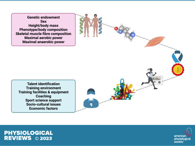
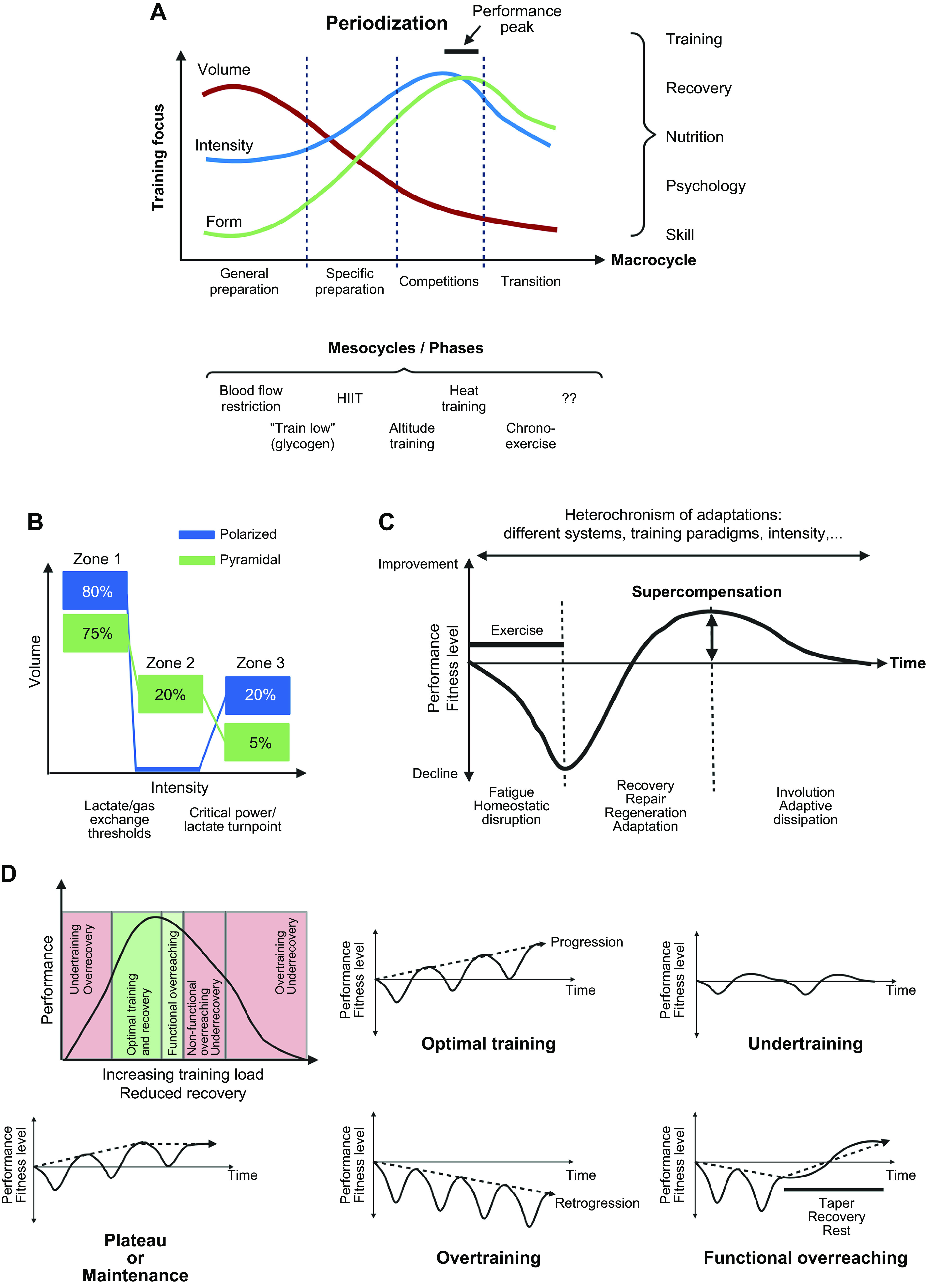
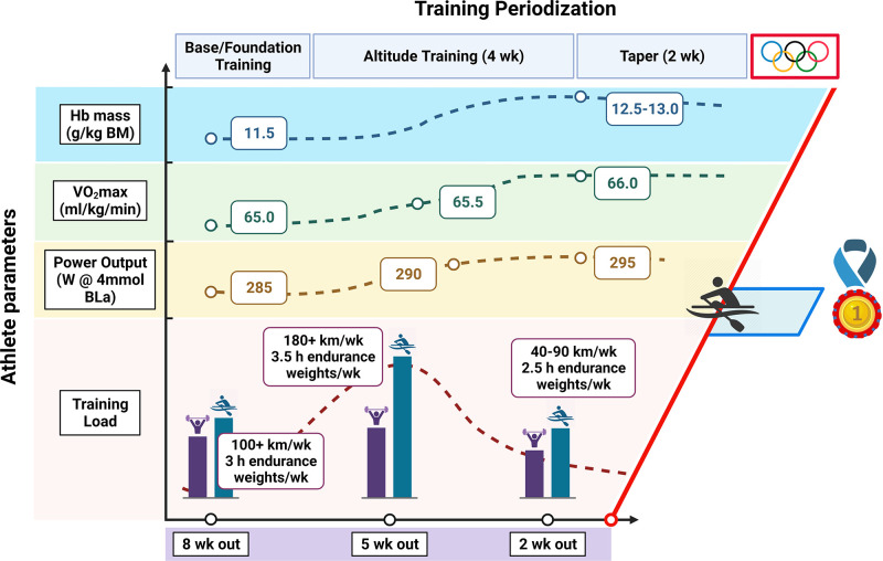
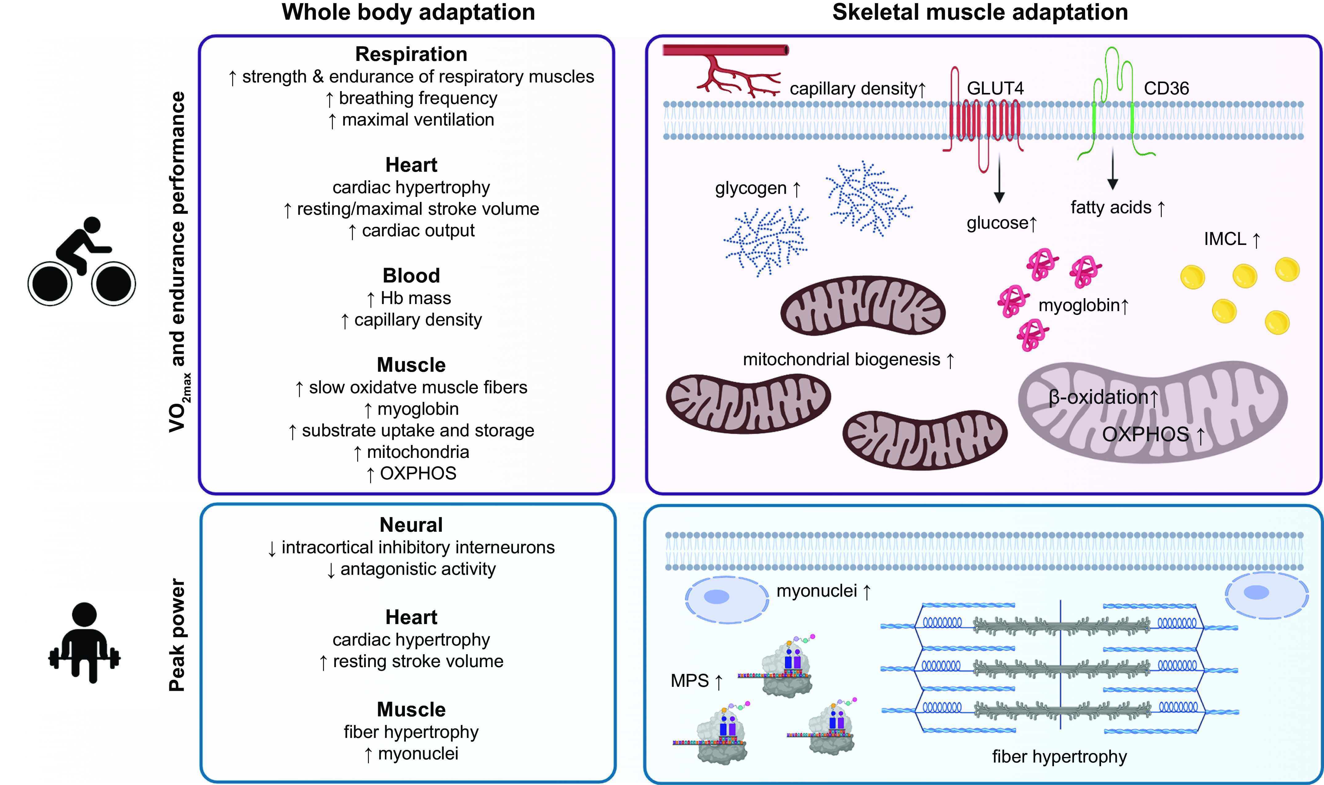
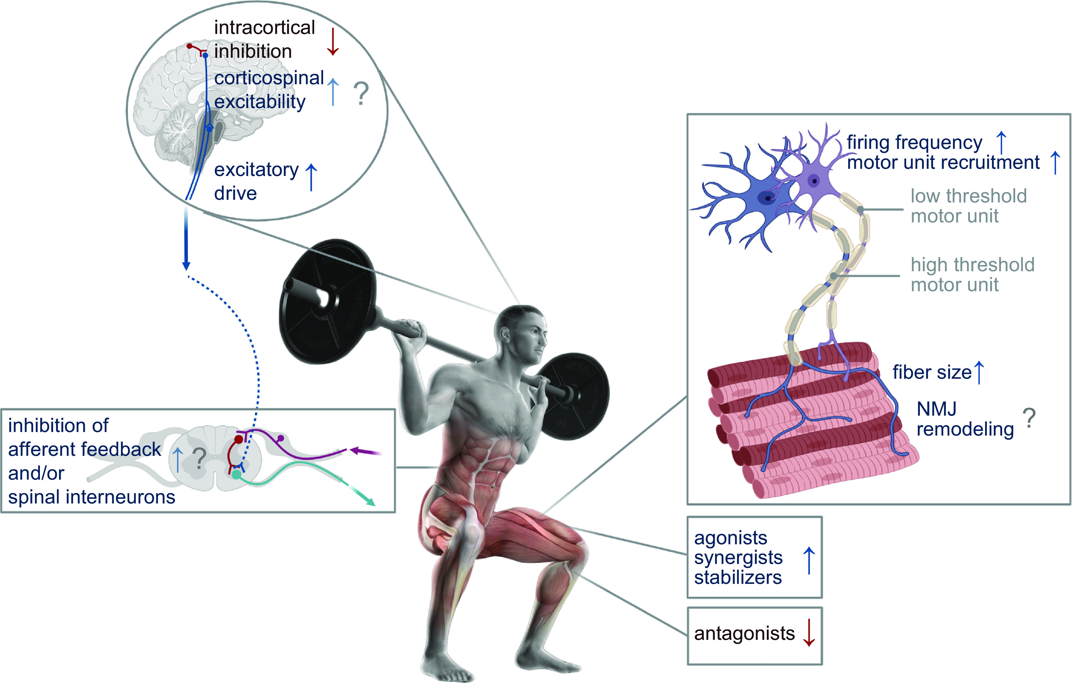
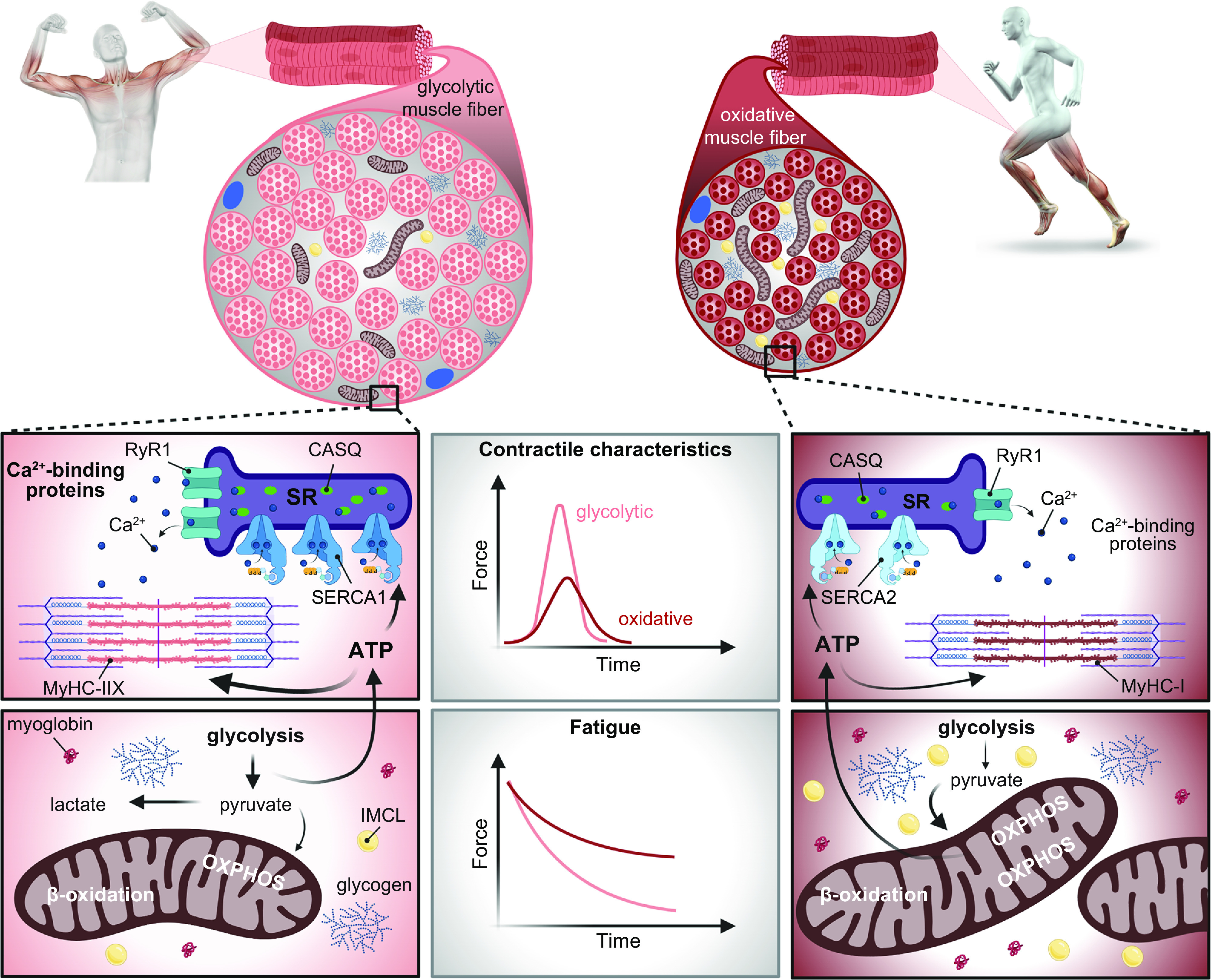
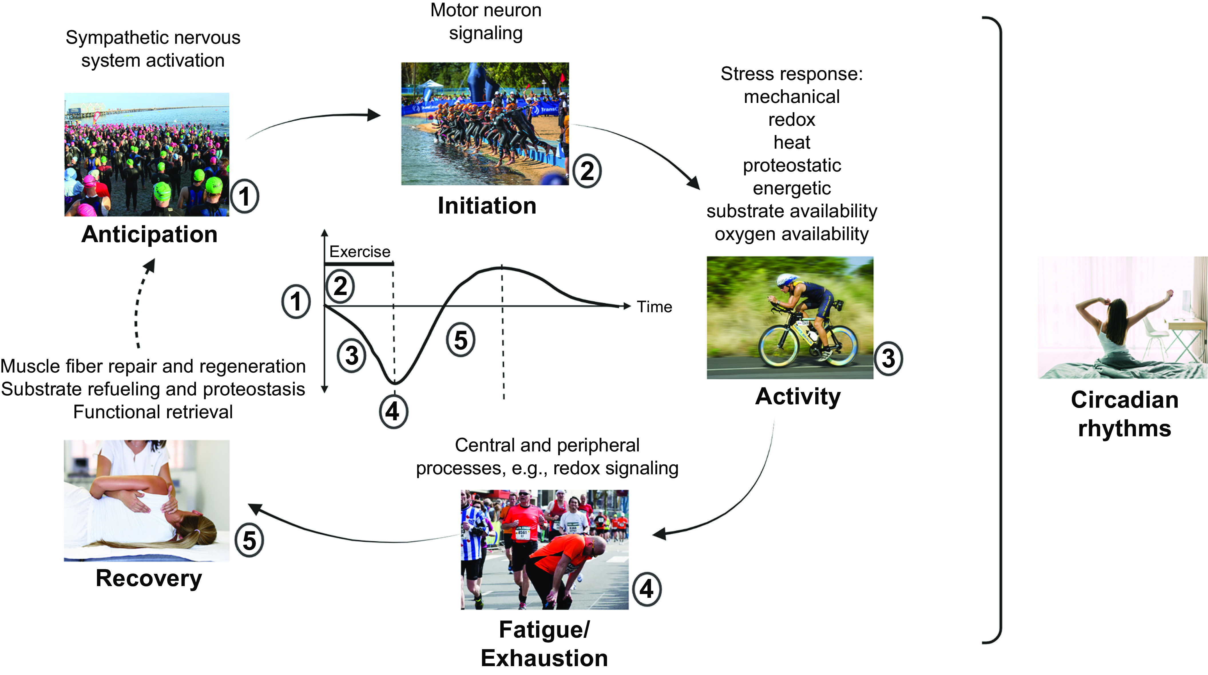
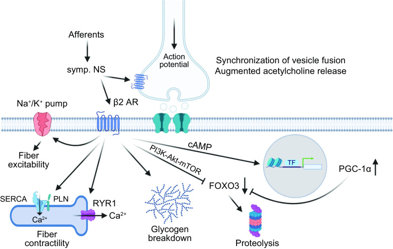
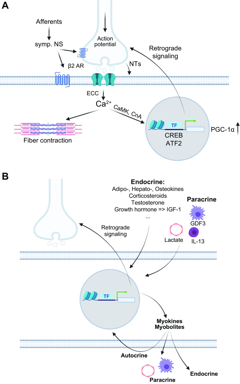
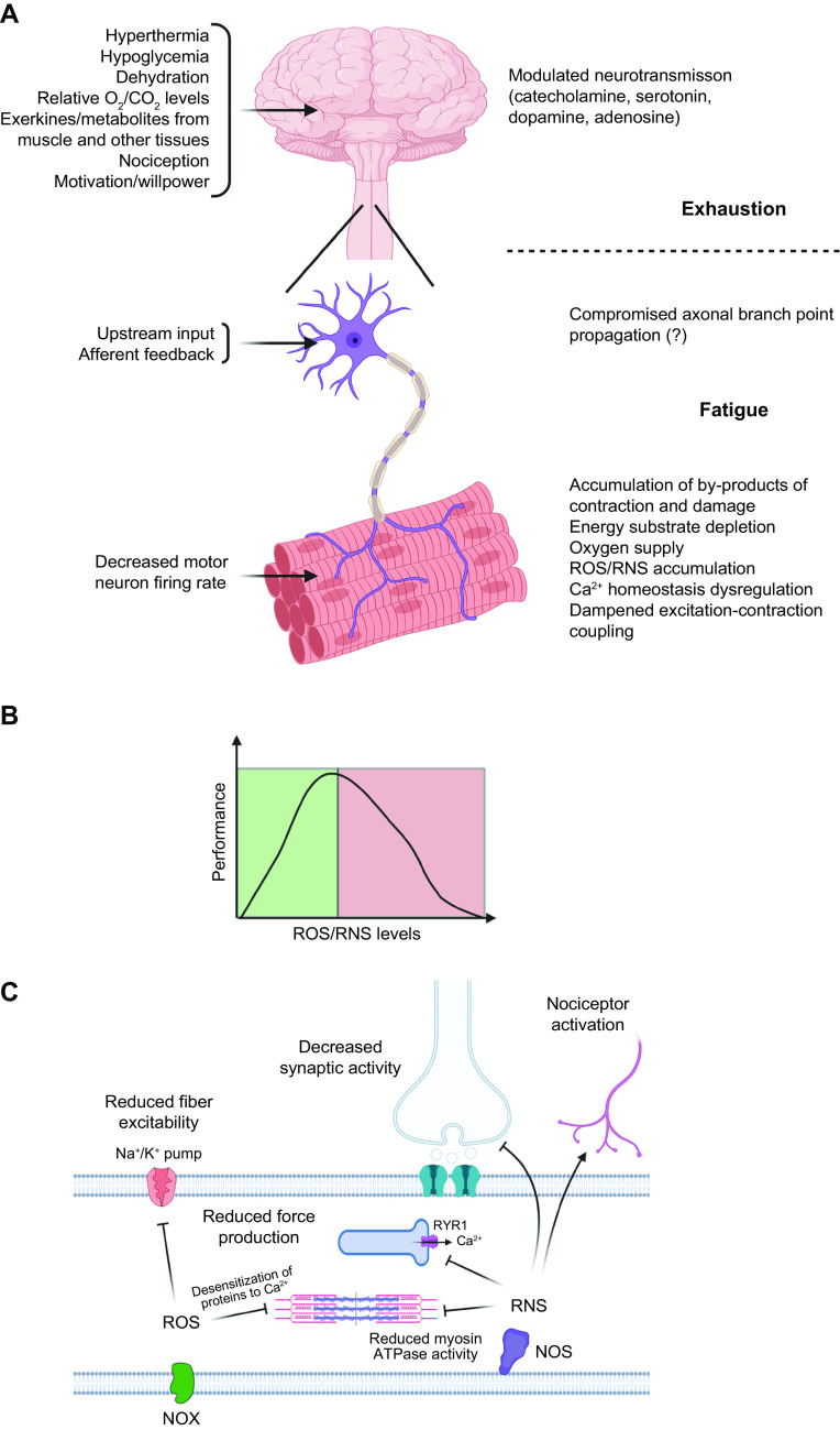
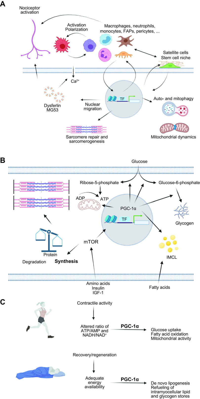
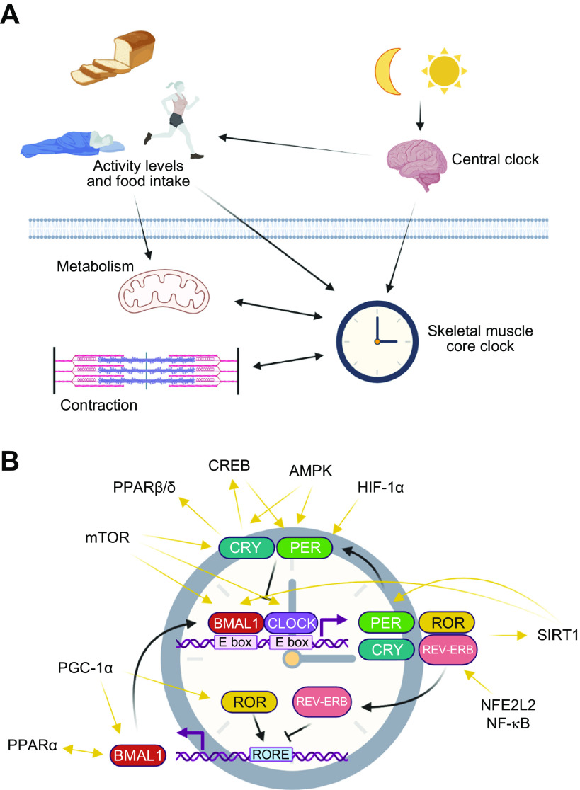
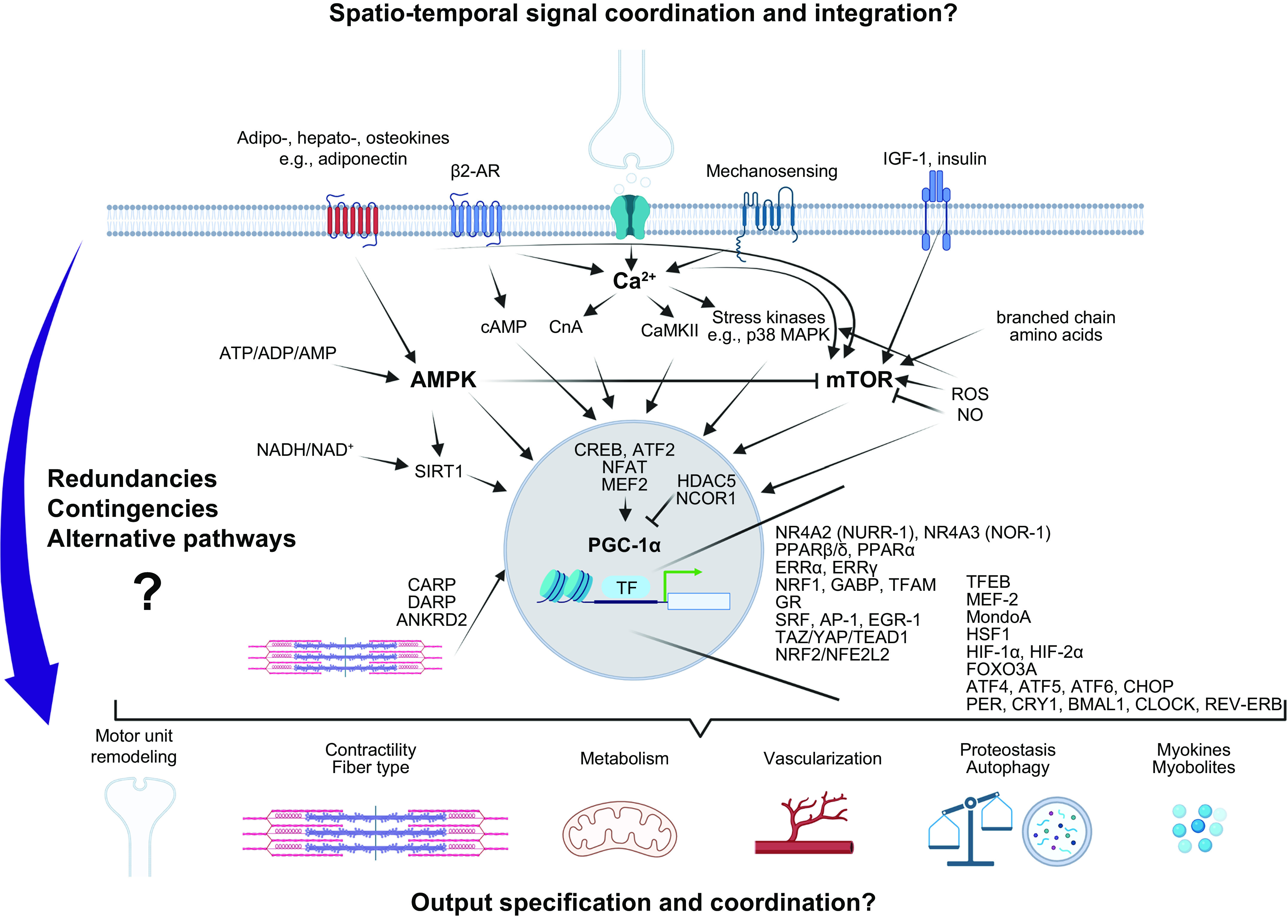
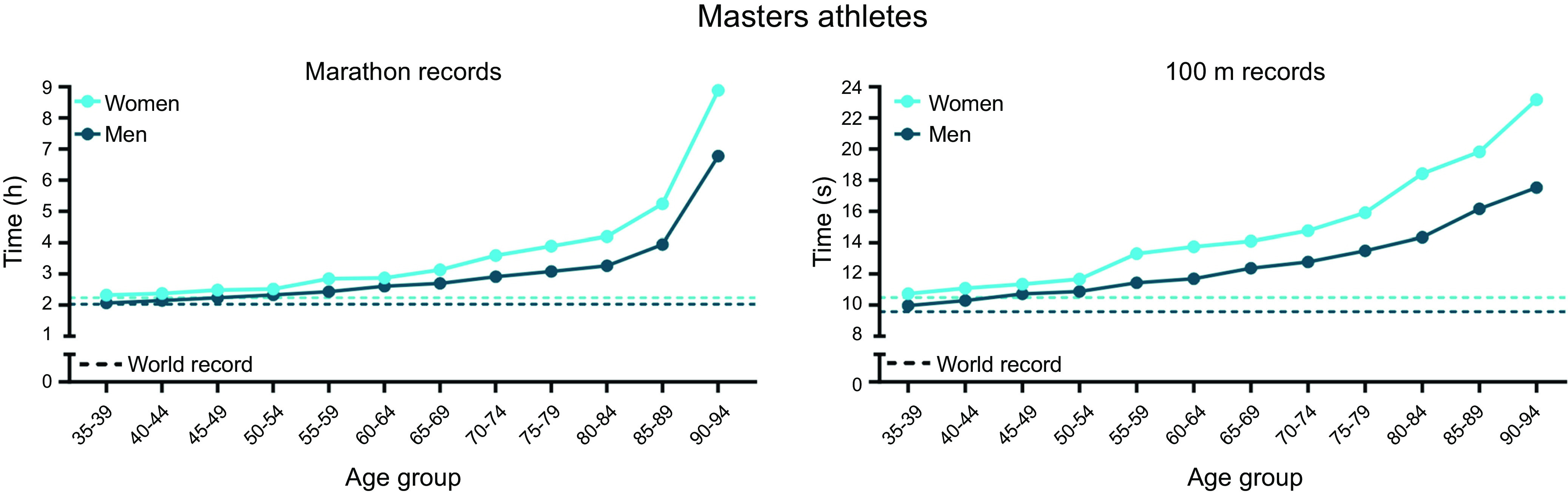
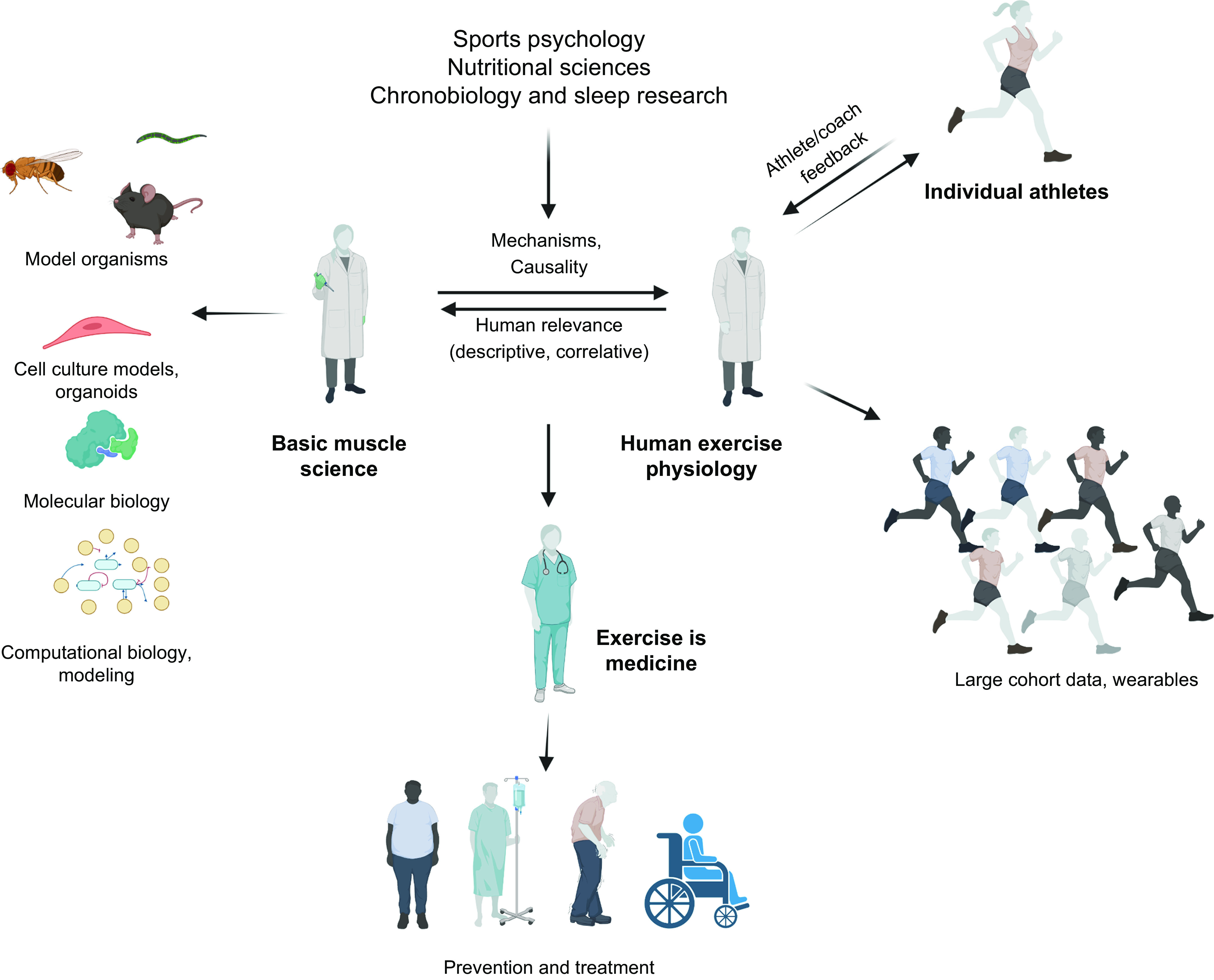
Human skeletal muscle demonstrates remarkable plasticity, adapting to numerous external stimuli including the habitual level of contractile loading. Accordingly, muscle function and exercise capacity encompass a broad spectrum, from inactive individuals with low levels of endurance and strength to elite athletes who produce prodigious performances underpinned by pleiotropic training-induced muscular adaptations. Our current understanding of the signal integration, interpretation, and output coordination of the cellular and molecular mechanisms that govern muscle plasticity across this continuum is incomplete. As such, training methods and their application to elite athletes largely rely on a "trial-and-error" approach, with the experience and practices of successful coaches and athletes often providing the bases for "post hoc" scientific enquiry and research. This review provides a synopsis of the morphological and functional changes along with the molecular mechanisms underlying exercise adaptation to endurance- and resistance-based training. These traits are placed in the context of innate genetic and interindividual differences in exercise capacity and performance, with special consideration given to aging athletes. Collectively, we provide a comprehensive overview of skeletal muscle plasticity in response to different modes of exercise and how such adaptations translate from "molecules to medals."

Keywords: athlete; endurance training; exercise; resistance training; skeletal muscle.


Conflict of interest statement

C.H. is an associate editor of Physiological Reviews and was not involved in and did not have access to information regarding the peer-review process or final disposition of this article. An alternate editor oversaw the peer-review and decision-making process for this article. None of the other authors has any conflicts of interest, financial or otherwise, to disclose.

Figures

None
Graphical abstract
FIGURE 1.
FIGURE 1.
From evolution to modern-day athleticism. Evolutionary selection of 5 main traits has facilitated the prolonged upright, bounding, bipedal locomotion in humans. Energetic barriers are lowered by long, spring-like tendons (in particular the Achilles tendon), the longitudinal plantar foot arch, ankle ligaments, long legs, in particular femur length, and short toes (to increase stride length and reduce vertical trajectories for better locomotor economy), thinner heart ventricles and larger cavity, increased hind limb muscle mass, and other adaptations. Skeletal strength is conferred, e.g., by larger joint areas in lower but not upper limbs to dissipate impact forces. Stabilization for bipedal movement is mediated by large erector spinae and gluteus muscles opposed to reduced forearm mass and an elongated, narrow waist and broad shoulders to facilitate counterrotation of thorax and arms, while decreased facial length helps head stabilization or an integrated system of bio-tensegrity for embedded perturbation repelling. Eccrine sweat glands, with a particular high density in the head for brain cooling, reduced body hair, dense skin vascularization, mouth breathing, and a large nasal epithelial area all contribute to thermoregulation. Finally, coevolution of locomotion and the brain resulted in expanded cerebello-cerebralcortical circuitry (for anticipation, pre-preparation, sensory integration, pre-planned multilevel compensation to deal with perturbations and destabilizations), cognitive capabilities (to recognize landmarks, long-range orientation, recognizing prey and predators, tracking and speculative tracking/anticipation), perception, fine motor control, and balance. See Refs. – for more information. Modern-day athletic peak performances most likely exceed these general evolutionary traits because of efficient training strategies and paradigms, nutrition and supplements, technological innovations, i.e., pertaining to equipment and facilities, and genetic and epigenetic predispositions. Figure created with BioRender.com, with permission.
FIGURE 2.
FIGURE 2.
Innovations that contributed to the progress in the development of world records over time (light blue for women, dark blue for men). Speed skating is one of many cases in which the progression of world records is driven by innovations (63). For example, the invention to improve ice quality (natural vs artificial ice) by refrigerated ovals (first 1958), spraying tiny droplets of water to smoothen the surface (first 1960), followed by the ice resurfacer “Zamboni” (Olympics 1960) and eventually indoor rinks all contributed to new records. Additionally, the development of gear such as tight-fit suits to improve aerodynamics and the invention of the clap skate that enabled a longer contact with the ice as well as further enhanced aerodynamics due to the crouched posture pushed the progress in world record development (http://www.speedskatingstats.com/index.php?file=records). Image on left was taken at the 1932 Winter Olympics and is from Henriksen & Steen (public domain, via Wikimedia Commons); image on right was originally posted to Flickr by adrian8_8.
FIGURE 3.
FIGURE 3.
Elite athletic performance is determined by the complex interaction of intrinsic and extrinsic factors. Undisputedly, genetic predisposition, even though poorly defined and understood, contributes to athletic prowess and trainability. In fact, the “right” genes might even be a prerequisite for elite, world-class performance. The epigenetic landscape is at least in part inherited but, in contrast to the genome, can also be influenced by behavior, including prior athletic experience, nutrition, and other lifestyle factors. A higher-than-average motivation and drive, the willpower to overcome obstacles, adversities, and setbacks, perseverance, and the willingness to forgo activities common for non-athletic peers are essential. These factors as well as daily training are shaped by body perception and prior athletic experience, including a multidisciplinary/multisport practice in youths. Most likely, nutrition, ergogenic aids, and gut microbiomes mutually interact in an intimate manner, collectively affecting trainability and performance. Optimal training strategies not only comprise personalized planning but should also integrate adequate consideration of recovery and injury prevention and, if the situation arises, rehabilitation. State-of-the-art equipment and facilities are part of a permissive environment, which is also strongly shaped by socio-economic status and social interactions with coaches, medical and other staff, team members, parents, siblings, friends, and rivals. This network of supporting people helps to optimize knowledge and education for proper planning and implementation. Finally, peak performance also relies on proper and personalized sleep patterns, matched to the individual chronotype. The use of doping might confer performance enhancements in the short term but is linked to long-term health detriments and is counter to the ethos of a fair and clean sport. Figure created with BioRender.com, with permission.
FIGURE 4.
FIGURE 4.
Training principles and strategies. A: to achieve peak performance at the time of competition, training volume, intensity, and form/specificity have to be adapted in different cycles/phases. Specific paradigms, e.g., high-intensity interval training (HIIT), “train low,” and others, are likewise periodized and matched to the prevailing volume/intensity/form requirements. Importantly, the periodization of training has to be matched to that of nutrition (e.g., low glucose vs. carb loading), recovery, psychological aspects, and skill acquisition. B: within shorter cycles, e.g., weekly planning, polarized or pyramidal partitioning of training volume at different intensities (e.g., defined by lactate/ventilatory thresholds between zones 1 and 2 and the critical power/lactate turnpoint between zones 2 and 3) helps to improve performance and reduce overtraining. C: training adaptation is initiated by a homeostatic disruption induced by exercise. After exercise cessation, recovery and repair mechanisms not only result in a return to baseline but trigger adaptive mechanisms, optimally in a supercompensatory manner, which should help to protect muscle better from future perturbations. However, in the absence of continued stimuli, i.e., detraining, this supercompensatory response is abolished by an adaptive dissipation. The amplitude and temporal aspects of this curve are strongly influenced by the training paradigm and related parameters. Moreover, within the same system, biochemical processes, cell types, or tissues can react in a different manner (heterochronism of adaptation). D: performance gains are controlled by the balance between training load and recovery. A suboptimal planning can result in either undertraining with little or no gains or overtraining, in which performance decreases (retrogression) and the risk for injuries increases. In proper conditions, a functional overreach helps to maximize progression and overcome training plateaus. Figure created with BioRender.com, with permission.
FIGURE 5.
FIGURE 5.
Periodization of training for an elite athlete. Schematic representation of periodization of training along with physiological data collected during preparation for the 2016 Rio Olympic Games for a gold medal-winning female rower. BLa, blood lactate; BM, body mass. See glossary for other abbreviations. Figure created with BioRender.com, with permission.
FIGURE 6.
FIGURE 6.
Whole body adaptations that contribute specifically to higher peak power or endurance performance. Although mainly neural and muscular adaptations improve peak power, for endurance performance various organs and tissues show major changes. To maximize V˙O2max and thereby endurance performance, changes in respiratory and cardiovascular function as well as adaptations in skeletal muscle are required. In skeletal muscle, the high mitochondrial density, elevated substrate (i.e., fatty acids and glucose) uptake and storage, myoglobin content, and increased vascularization all contribute to the elevated performance of endurance athletes. Strength training-induced adaptations include increased muscle protein synthesis (MPS) resulting in fiber hypertrophy and optimally myonuclear accretion. CD36, platelet glycoprotein 4; GLUT4, glucose transporter type 4; IMCL, intramyocellular lipids. See glossary for other abbreviations. Sport icon vectors were created by ibrandify/Freepik. Image created with BioRender.com, with permission.
FIGURE 7.
FIGURE 7.
Neuromuscular adaptation to training. The number of activated motor units, their firing frequency, as well as size and contractile properties of the muscle fibers determine total force-generating capacity. In trained individuals, neural adaptations include an increased excitatory drive that can lead to an elevated firing frequency and higher number of activated motor units. In addition, the enhanced activation of agonists, synergists, and stabilizers together with the reduced coactivation of antagonists contribute to the increased force production after training. For many of these adaptations only data in rodent models exist, and/or controversial findings in humans have been reported. NMJ, neuromuscular junction. Illustration of person was created by kjpargeter/Freepik. Image created with BioRender.com, with permission.
FIGURE 8.
FIGURE 8.
Contractile and metabolic properties of a strength/power-trained and an endurance-trained athlete. Characteristics of fast and powerful muscles of strength/power athletes that are accompanied by a more fatigable muscle as compared with endurance-trained muscles with elevated oxidative capacity that are more fatigue resistant. ATP, adenosine triphosphate; CASQ, calsequestrin; IMCL, intramyocellular lipids; MyHC, myosin heavy chain; OXPHOS, oxidative phosphorylation; RyR1, ryanodine receptor 1; SERCA, sarcoplasmic/endoplasmic reticulum Ca2+-ATPase; SR, sarcoplasmic reticulum. Illustrations of people were created by kjpargeter/Freepik. Image created with BioRender.com, with permission.
FIGURE 9.
FIGURE 9.
Temporal engagement of different pathways and processes in skeletal muscle in exercise. Anticipation of activity is linked to elevated sympathetic nervous system tone. Motor neuron firing triggers muscle contractions at the start of and during an activity. While contracting, muscle will be affected by different stressors and the related reaction/mitigation, for example, mechanical stress, reactive oxygen and nitrogen species (redox) production, heat, altered proteostasis and protein unfolding, metabolic changes, substrate availability, and oxygen provisioning. The exact temporal sequence of engagement of and interactions between these pathways are unknown. Different mechanisms contribute to muscle fatigue, exhaustion, and exercise cessation. Subsequently, muscle repair, regeneration, and refueling are important for functional retrieval. Many of these processes are modulated by circadian input. Images: triathlon anticipation from Wikimeda Commons (CC-BY-SA 3.0, creator: Wiech), triathlon start from Wikimedia Commons (CC-BY-SA 2.0, creator: IQRemix), triathlon cycling from PxHere.com (CC0 Public Domain), exhaustion from Wikimedia Commons (CC-BY-SA 4.0, creator: Wallco26), massage from Freepick.com (author: javi_indy), waking up from Freepik.com (author: diana.grytsku).
FIGURE 10.
FIGURE 10.
Neuroendocrine signaling by the sympathetic nervous system in exercise anticipation and muscle contraction. Sympathetic activation of the motor neuron and skeletal muscle cells results in modulation of fiber excitability and contractility, metabolic and proteostatic remodeling, and the activation of a transcriptional program. These events prepare muscle cells for upcoming contractions and help to maintain contractile activity upon engagement. β2 AR, β2-adrenoreceptors; Akt, protein kinase B; cAMP, cyclic AMP; FOXO3, forkhead transcription factor O3; mTOR, mammalian target of rapamycin; PGC-1α, peroxisome proliferator-activated receptor γ coactivator 1α; PI3K, phophoinositide 3-kinase; PLN, phospholamban; RYR1, ryanodine receptor 1; SERCA, sarcoplasmic/endoplasmic reticulum Ca2+-ATPase; symp. NS, sympathetic nervous system; TF, transcription factor. Image created with BioRender.com, with permission.
FIGURE 11.
FIGURE 11.
Motor neuron signaling and (neuro)endocrine effectors of contraction. A: motor neuronal signaling triggers excitation-contraction coupling and thereby evokes a rise in intramyocellular calcium (Ca2+), which enables fiber contractions, activates various signaling pathways, and modulates a transcriptional response, including retrograde feedback to the motor neuron. Motor neuron activity is modulated by sympathetic tone and includes various neurotrophic factors besides the neurotransmitter acetylcholine. B: exerkines, originating from tissues including muscle (myokines), liver (hepatokines), adipose tissue (adipokines), and bone (osteokines) as well as other hormones coordinate a systemic response to contractile activity. Many of these factors exert auto-, para-, and endocrine effects. In addition, signals can be propagated by exercise-linked changes in different metabolites (myobolites or myometabokines). β2 AR, β2-adrenoreceptors; ATF2, activating factor 2; CaMK, calcium/calmodulin-dependent protein kinase; CnA, calcineurin A; CREB, cAMP-responsive element binding protein; ECC, excitation-contraction coupling; GDF3, growth differentiation factor 3; IGF-1, insulin-like growth factor 1; IL-13, interleukin 13; NTs, neurotrophic factors; PGC-1α, peroxisome proliferator-activated receptor γ coactivator 1α; symp. NS, sympathetic nervous system; TF, transcription factor. Image created with BioRender.com, with permission.
FIGURE 12.
FIGURE 12.
Mechanosensing and mechanostress mitigation in the contracting muscle fiber. Mechanical stress is sensed and translated by structures at the cell membrane and intramyocellular components in the cytosol, cytoskeleton, sarcomeres, or nucleus. As a consequence, resistance to shear stress is increased, stiffness and integrity of sarcomeric structures adapted, and a broad program of immediate-early and delayed primary genes initiated. AP-1, activating protein 1; EGR-1, early growth response gene 1; ERK, extracellular signal-regulated kinase; FAC, focal adhesion complex; JNK, c-Jun NH2-terminal protein kinase; mTORC1, mammalian target of rapamycin complex 1; p38 MAPK, p38 mitogen-activated protein kinase; PGC-1α, peroxisome proliferator-activated receptor γ coactivator 1α; SAPK, stress-activated protein kinase; SRF, serum response factor; TEAD, TEA domain transcription factor; TF, transcription factor; TRPC, mechano-gated Ca2+ transient receptor potential channels; YAP, yes-associated protein. Image created with BioRender.com, with permission.
FIGURE 13.
FIGURE 13.
Redox stress by reactive oxygen and nitrogen species in muscle contraction. Redox regulation of muscle contractility and stress response by reactive oxygen (ROS) and nitrogen (RNS) species during muscle contraction. ROS and RNS are primarily produced by enzymes at the muscle cell membrane. Subsequently, a number of downstream effects are promoted, including an increase in energy substrate and oxygen availability, enhancement of force generation, improvement of the resilience against oxidative stress, and modulation of a transcriptional program for muscle remodeling. cGMP, cyclic guanylate monophosphate; FAC, focal adhesion complex; mTORC1, mammalian target of rapamycin complex 1; NOS, nitric oxide synthase; NOX, NADPH oxidase; NRF2/NFE2L2, nuclear factor erythroid-derived 2-like 2; PGC-1α, peroxisome proliferator-activated receptor γ coactivator 1α; TF, transcription factor; TRPC, mechano-gated Ca2+ transient receptor potential channels. Image created with BioRender.com, with permission.
FIGURE 14.
FIGURE 14.
Energy homeostasis, substrate and oxygen signaling. A: metabolic stress signaling in muscle contraction to increase energy provisioning. A decrease in energy substrate availability leads to the activation of energy sensors that reduce anabolic processes consuming ATP and induce catabolic pathways to produce more ATP. This broad response comprises direct modulation of protein and enzymatic activities by posttranslational modification as well as the control of a broad transcriptional program. B: energy substrate signaling senses substrate levels and leads to metabolic partitioning. Thereby, muscle cell metabolism is coordinated with substrate availability and anabolism balanced with catabolism. C: reduced oxygen availability in skeletal muscle is sensed by hypoxia-inducible factor (HIF)-1α and HIF-2α in acute and chronic settings, respectively. HIF-1α rapidly reduces pathways that consume O2, while promoting anaerobic glycolysis to generate ATP. HIF-2α promotes muscle oxygen extraction and provisioning. AMPK, AMP-activated protein kinase; ERRα, estrogen-related receptor α; FAD: flavin adenine dinucleotide; IGF-1, insulin-like growth factor 1; mTOR, mammalian target of rapamycin; NAD, nicotinamide adenine dinucleotide; PGC-1α, peroxisome proliferator-activated receptor γ coactivator 1α; PPARα/-β/δ, peroxisome proliferator-activated receptor α/-β/δ; SIRT1, sirtuin 1; TF, transcription factor. Image created with BioRender.com, with permission.
FIGURE 15.
FIGURE 15.
Heat and proteostatic stress. A: thermosensing and the heat stress response mitigate misfolding of proteins. Heat is produced by various processes in the contracting muscle fiber, including shear stress, mitochondrial activity, Ca2+ release and reuptake, and ATP metabolism in contraction cycling. Heat is sensed in the cell and a broad transcriptional program engaged to increase mitigating measures, e.g., chaperones to reduce thermally induced protein misfolding. B: proteostatic stress and the ensuing response pathways reduce protein load, misfolding, and organelle health. Dedicated pathways in the endoplasmic reticulum and mitochondria are engaged by proteostatic dysbalances, e.g., excessive protein accumulation or misfolding. At least in part, these two pathways converge to initiate a transcriptional program aimed at reversing protein misfolding, alleviating proteostatic stress by reducing gene expression and protein synthesis while enhancing protein degradation, and by ensuring organelle functionality. ATF4/5/6α, activating transcription factor 4/5/6α; CHOP, C/EBP homologous protein; HSF1, heat shock factor 1; HSP, heat shock protein; IRE1α, inositol-requiring enzyme 1α; PERK, RNA-dependent protein kinase-like ER eukaryotic translation initiation factor 2α kinase; PGC-1α, peroxisome proliferator-activated receptor γ coactivator 1α; TF, transcription factor; UPRER, endoplasmatic reticulum unfolded protein response; UPRmt, mitochondrial unfolded protein response. Image created with BioRender.com, with permission.
FIGURE 16.
FIGURE 16.
Exhaustion and muscle fatigue. A: central and peripheral contributors to exhaustion (volitional) and fatigue (involuntary). Altered neurotransmission in the brain ensures protection of this and other organs from hyperthermia, hypoglycemia, dehydration, shifted O2/CO2 levels, and further potentially detrimental processes in exercise. To a limited extent, these effects can be overcome by willpower and motivation. Peripheral factors of fatigue involve the motor neuron and muscle fibers. Although impairments in action potential propagation and neuromuscular junction transmission seem minor in healthy individuals, input from upstream brain regions and afferent feedback modulate motor neuron firing rate. Muscle-intrinsic contractility is affected by energy substrate and oxygen availability, accumulation of by-products of contraction and damage, elevation of reactive oxygen (ROS) and nitrogen (RNS) species, a dampening of excitation-contraction coupling including the Na+-K+ pump, and intramyocellular Ca2+ homeostasis. B: concentration-dependent, biphasic bell-shaped effect of ROS and RNS on muscle performance. During contractions, ROS and RNS sustain and enhance contractile activity. However, once levels exceed a poorly defined threshold, a different set of processes is engaged that limits performance. C: exceeding a certain concentration, ROS and RNS contribute to muscle fatigue by affecting fiber excitability and contractility, synaptic activation, and nociception. A modulation of intramyocellular Ca2+ homeostasis thereby plays a central role. Putatively, this “rheostat” helps to avoid overexertion and to minimize muscle tissue damage. NOS, nitric oxide synthase; NOX, NADPH oxidase; RYR1, ryanodine receptor 1. Image created with BioRender.com, with permission.
FIGURE 17.
FIGURE 17.
Muscle repair, regeneration, and refueling. A: multicellular interactions and intramyocellular processes that control muscle repair. Resident and infiltrating cells of different types are engaged by various signals in a temporally highly orchestrated manner. Thereby, damaged material is removed, muscle fibers repaired or de novo formed, and functional retrieval achieved. Activation of nociceptor signaling should prevent further exertion and damage during repair and regeneration. Moreover, the multicellular processes are complemented by intramyocellular pathways to restore membrane and sarcomere integrity as well as organelle function. B: postexercise refueling of glycogen, intramyocellular lipids (IMCL), and protein structures. Depleted intramyocellular energy substrate stores are replenished after exercise depending on a systemic, anabolic context, i.e., the availability of the corresponding substrates and signaling of anabolic hormones. Because of the energetic demand for protein, glycogen, and IMCL synthesis, these processes are coordinated with mitochondrial activity and ATP production. C: temporal specification of catabolic and anabolic processes by peroxisome proliferator-activated receptor γ coactivator 1α (PGC-1α). To avoid a futile cycle, the catabolic activity of PGC-1α during contractions must be separated from the anabolic function in regeneration. The mechanistic underpinnings of the transcriptional specification of this coactivator (and other regulators with multipurpose roles) remain unknown. FAP, fibro-adipogenic precursor; MG53, mitsugumin 53; mTOR, mammalian target of rapamycin; TF, transcription factor. Image created with BioRender.com, with permission.
FIGURE 18.
FIGURE 18.
Circadian regulation of skeletal muscle function and plasticity. A: circadian regulation of activity and muscle function. Circadian regulation of sleep-wake cycles, feeding, physical activity, or other zeitgebers is sensed and translated by the master clock in the suprachiasmatic nucleus as well as peripheral clocks in almost every cell in the human body. As a consequence, various physiological processes, e.g., muscle cell metabolism or contractile performance, can be affected in a circadian manner. B: interactions between core clock genes/proteins with regulators of muscle function and plasticity. Such cross talk pertains to the regulation of gene expression, protein-protein interactions, and/or enzymatic activity. Thereby, physiological processes in muscle might be affected by the core clock. Inversely, muscle fiber metabolism, contractile activity, oxygen availability, redox balance, and other perturbations could modulate the skeletal muscle clock. AMPK, AMP-dependent protein kinase; BMAL1, brain and muscle ARNT-like 1; CLOCK, circadian locomotor output cycles kaput; CREB, cAMP response element binding protein; CRY, cryptochrome circadian regulator; HIF-1α, hypoxia-inducible factor 1α; mTOR, mammalian target of rapamycin; NFE2L2, nuclear factor erythroid-derived 2-like 2; NF-κB, nuclear factor κB; PER, period; PGC-1α, peroxisome proliferator-activated receptor γ coactivator 1α; PPARα/β/δ, peroxisome proliferator-activated receptor α/β/δ; REV-ERB, nuclear receptor subfamily 1 group D member 1/2; ROR, retinoic acid-related orphan receptor; RORE, ROR elements; SIRT1, sirtuin 1. Image created with BioRender.com, with permission.
FIGURE 19.
FIGURE 19.
Molecular mechanisms in contracting muscle fibers. A small selection of molecular sensors and mediators as well as a simplified summary of the presumed interactions are depicted. Nota bene: the spatio-temporal integration and coordination of these pathways, functional redundancies (e.g., overlapping or parallel pathways), contingencies (back-up processes with the same function), alternatives (back-up processes leading to similar adaptations using different functions), specification and coordination of downstream effects and adaptations, in particular in chronic settings, as well as many other aspects are still only understood at a very rudimentary level. β-AR, β-adrenoreceptor; CARP, cardiac ankyrin-repeat protein; CnA, calcineurin A; CREB, cAMP-dependent binding protein; DARP, diabetes-related ankyrin-repeat protein; ERRα/γ, estrogen-related receptor α/γ; GR, glucocorticoid receptor; HSF1, heat shock factor 1; NO, nitric oxide; PPARα/β/δ, peroxisome proliferator-activated receptor α/β/δ; ROS, reactive oxygen species; SIRT1, sirtuin 1. See text for other abbreviations. Image created with BioRender.com, with permission.
FIGURE 20.
FIGURE 20.
Age-related decline in records of sprint and endurance events. Records for marathons (https://world-masters-athletics.com/championships/non-stadia/) and 100 m sprints (747) of masters athletes represent the age-induced reduction in tissue and organ function that despite high training loads leads to a decrease in performance. However, they also highlight the potential of the human body to achieve incredible performances at advanced age with adequate training.
FIGURE 21.
FIGURE 21.
The future of exercise science for safe, evidence-based, and personalized approaches. To overcome existing hurdles and efficiently leverage the power of novel techniques and approaches, a close interaction between basic muscle research, applied human exercise physiology, as well as athletes and coaches should be aimed for. Model organisms, cell culture, and molecular and computational biology might provide insights into cause-effect relationships, epistasis, etiologies, and mechanisms complementing the descriptive and correlative studies in human volunteers. Inversely, data from large cohorts that will become available because of widespread use of wearables and trackers as well as those obtained in and based on feedback from athletes and coaches will reveal processes and pathways to be explored in mechanistic detail. The molecular athlete should furthermore be informed by sports psychology, e.g., in regard to motivation, perseverance, compliance and adherence, nutritional sciences, chronobiology, sleep research, and other fields of relevance to training. Finally, a mutual exchange between the observations in training and those in various pathologies associated with an inactive lifestyle or inadequate muscle functionality will help to push the boundaries of physical activity interventions in the prevention and treatment of numerous diseases. Image created with BioRender.com, with permission.

References

    1. Marino FE, Sibson BE, Lieberman DE. The evolution of human fatigue resistance. J Comp Physiol B 192: 411–422, 2022. doi:10.1007/s00360-022-01439-4. - DOI - PMC - PubMed
    1. Bramble DM, Lieberman DE. Endurance running and the evolution of Homo. Nature 432: 345–352, 2004. doi:10.1038/nature03052. - DOI - PubMed
    1. Hunter P. The evolution of human endurance: research on the biology of extreme endurance gives insights into its evolution in humans and animals. EMBO Rep 20: e49396, 2019. doi:10.15252/embr.201949396. - DOI - PMC - PubMed
    1. Kim DS, Wheeler MT, Ashley EA. The genetics of human performance. Nat Rev Genet 23: 40–54, 2022. doi:10.1038/s41576-021-00400-5. - DOI - PubMed
    1. Liebenberg L. The relevance of persistence hunting to human evolution. J Hum Evol 55: 1156–1159, 2008. doi:10.1016/j.jhevol.2008.07.004. - DOI - PubMed

Publication types