. 2023 Feb 8;43(6):1018-1026.
doi: 10.1523/JNEUROSCI.0962-22.2022. Epub 2023 Jan 5.

Ameliorating Hemianopia with Multisensory Training

Affiliations

Ameliorating Hemianopia with Multisensory Training

Benjamin A Rowland et al. J Neurosci. .

Abstract

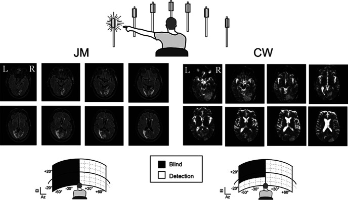
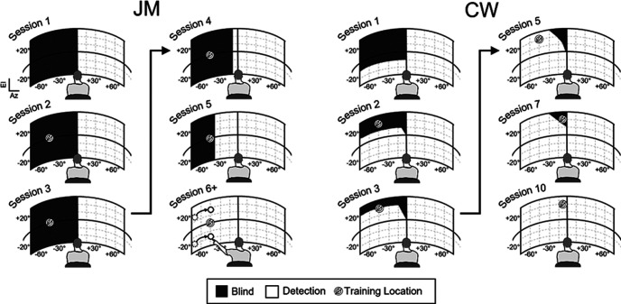
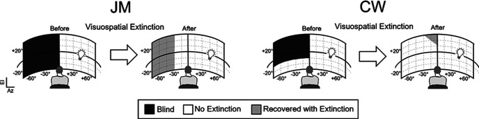
Hemianopia (unilateral blindness), a common consequence of stroke and trauma to visual cortex, is a debilitating disorder for which there are few treatments. Research in an animal model has suggested that visual-auditory stimulation therapy, which exploits the multisensory architecture of the brain, may be effective in restoring visual sensitivity in hemianopia. It was tested in two male human patients who were hemianopic for at least 8 months following a stroke. The patients were repeatedly exposed to congruent visual-auditory stimuli within their blinded hemifield during 2 h sessions over several weeks. The results were dramatic. Both recovered the ability to detect and describe visual stimuli throughout their formerly blind field within a few weeks. They could also localize these stimuli, identify some of their features, and perceive multiple visual stimuli simultaneously in both fields. These results indicate that the multisensory therapy is a rapid and effective method for restoring visual function in hemianopia.SIGNIFICANCE STATEMENT Hemianopia (blindness on one side of space) is widely considered to be a permanent disorder. Here, we show that a simple multisensory training paradigm can ameliorate this disorder in human patients.

Keywords: cortical blindness; cross-modal; hemianopsia; superior colliculus; training; visual-auditory.


Figures

Figure 1.
Figure 1.
Top: The apparatus used in multisensory training and flash detection/localization. LED and speaker assemblies were mounted on top of a set of movable and height-adjustable towers (7 depicted). During training, visual-auditory stimuli were repeatedly presented at a location within the blind field. During testing, patients were asked to verbally report and/or point to the location of a flashed stimulus. Middle and Bottom: MRI scans and initial testing for both patients. In both patients, a right PCA infarct created blindness on the left side of space. Unshaded areas in the plots (Az = azimuth, El = elevation) indicate areas responsive to light flashes, black shaded regions indicate where those flashes could not be detected. Note that CW had vision spared in left space below −10° of elevation.
Figure 2.
Figure 2.
Recovery of visual detection and localization. Depicted are each patients' ability to detect the flashed stimulus at the beginning of select sessions in the experiment. Unshaded regions indicate areas where responses were elicited by the flash stimulus and black shaded regions indicate where they were not elicited. The icon (a flash in a circle) indicates the location of the visual-auditory training stimulus in the previous session. Dashed and solid circles (connected by arrows) in the last of JM's figures (see session 6) represent his translocation of visual stimuli in the far periphery of the rehabilitated field, which were systematically biased to a compressed, more central range.
Figure 3.
Figure 3.
Visuospatial extinction: before and after. Visual extinction was evaluated by simultaneously presented two lights within the same hemifield or in different hemifields. Depicted above are the regions where a bright light could or could not be detected in the left hemifield when an equally-bright light was simultaneously presented in the right hemifield (depicted with a light bulb). Different plots illustrate the observations before (blind regions shown with black shading) and after rehabilitation. Areas of space in which both lights could be perceived are unshaded. Areas of space after rehabilitation in which individual lights could be detected, but where detection was suppressed by the simultaneous presence of a light in the right hemifield, are shaded in gray.

Similar articles

Cited by

References

    1. Ajina S, Jünemann K, Sahraie A, Bridge H (2021) Increased visual sensitivity and occipital activity in patients with hemianopia following vision rehabilitation. J Neurosci 41:5994–6005. 10.1523/JNEUROSCI.2790-20.2021 - DOI - PMC - PubMed
    1. Baleydier C (1977) A bilateral cortical projection to the superior colliculus in the cat. Neurosci Lett 4:9–14. 10.1016/0304-3940(77)90116-1 - DOI - PubMed
    1. Benedek G, Perény J, Kovács G, Fischer-Szátmári L, Katoh YY (1997) Visual, somatosensory, auditory and nociceptive modality properties in the feline suprageniculate nucleus. Neuroscience 78:179–189. 10.1016/s0306-4522(96)00562-3 - DOI - PubMed
    1. Benedek G, Keri S, Nagy A, Braunitzer G, Norita M (2019) A multimodal pathway including the basal ganglia in the feline brain. Physiol Int 106:95–113. 10.1556/2060.106.2019.09 - DOI - PubMed
    1. Bi G, Poo M (2001) Synaptic modification by correlated activity: Hebb's postulate revisited. Annu Rev Neurosci 24:139–166. 10.1146/annurev.neuro.24.1.139 - DOI - PubMed

Publication types