. 2023 Feb 8;43(6):918-935.
doi: 10.1523/JNEUROSCI.0729-22.2022. Epub 2023 Jan 5.

A Unique Role for Protocadherin γC3 in Promoting Dendrite Arborization through an Axin1-Dependent Mechanism

Affiliations

A Unique Role for Protocadherin γC3 in Promoting Dendrite Arborization through an Axin1-Dependent Mechanism

David M Steffen et al. J Neurosci. .

Abstract

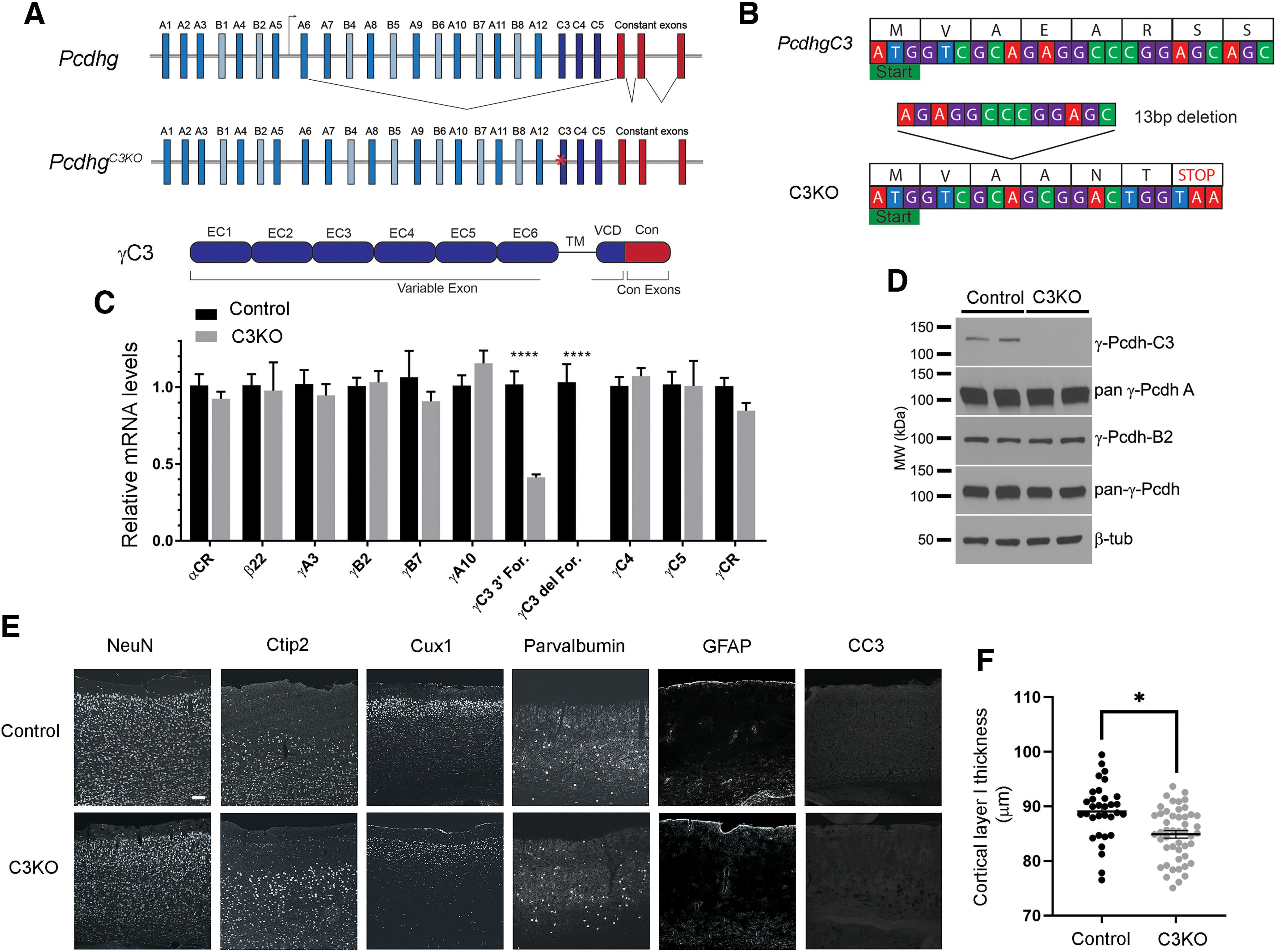
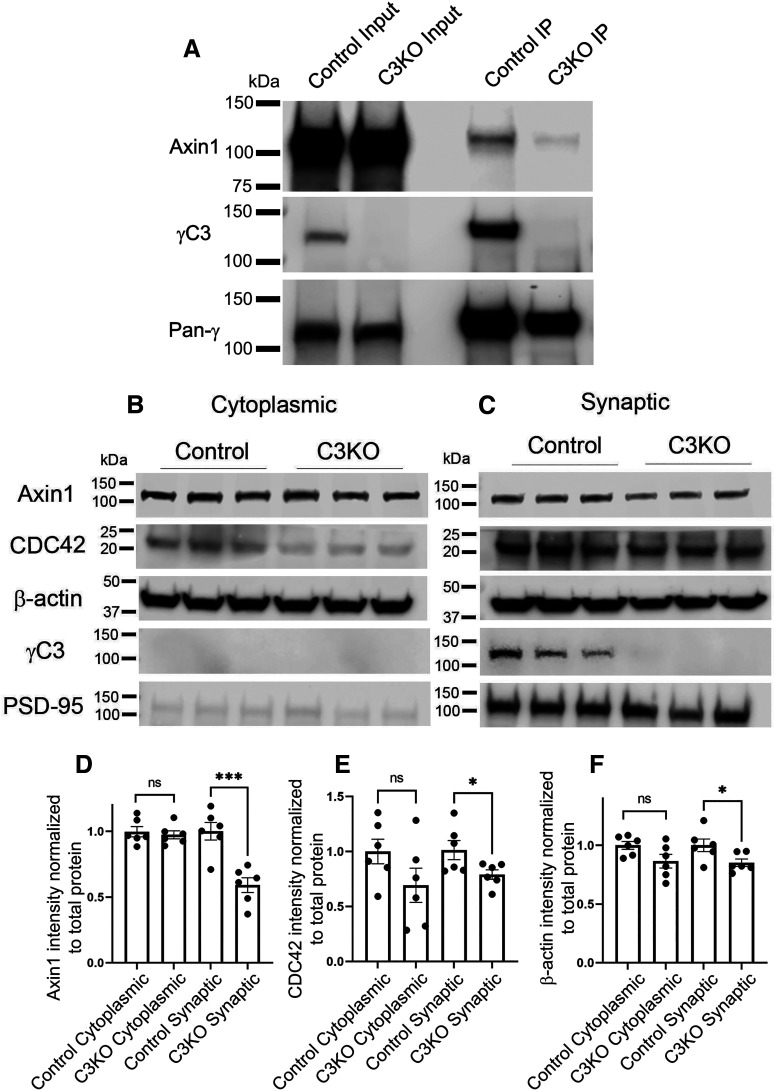
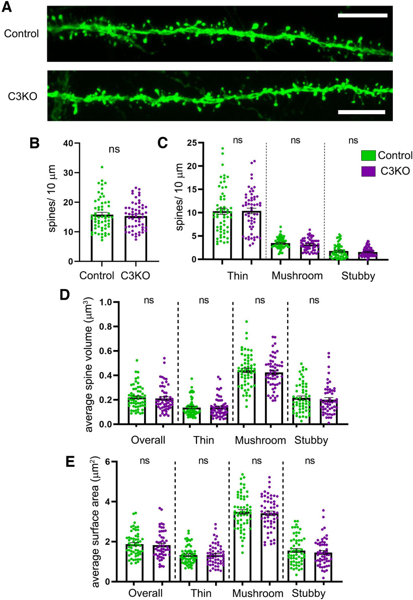
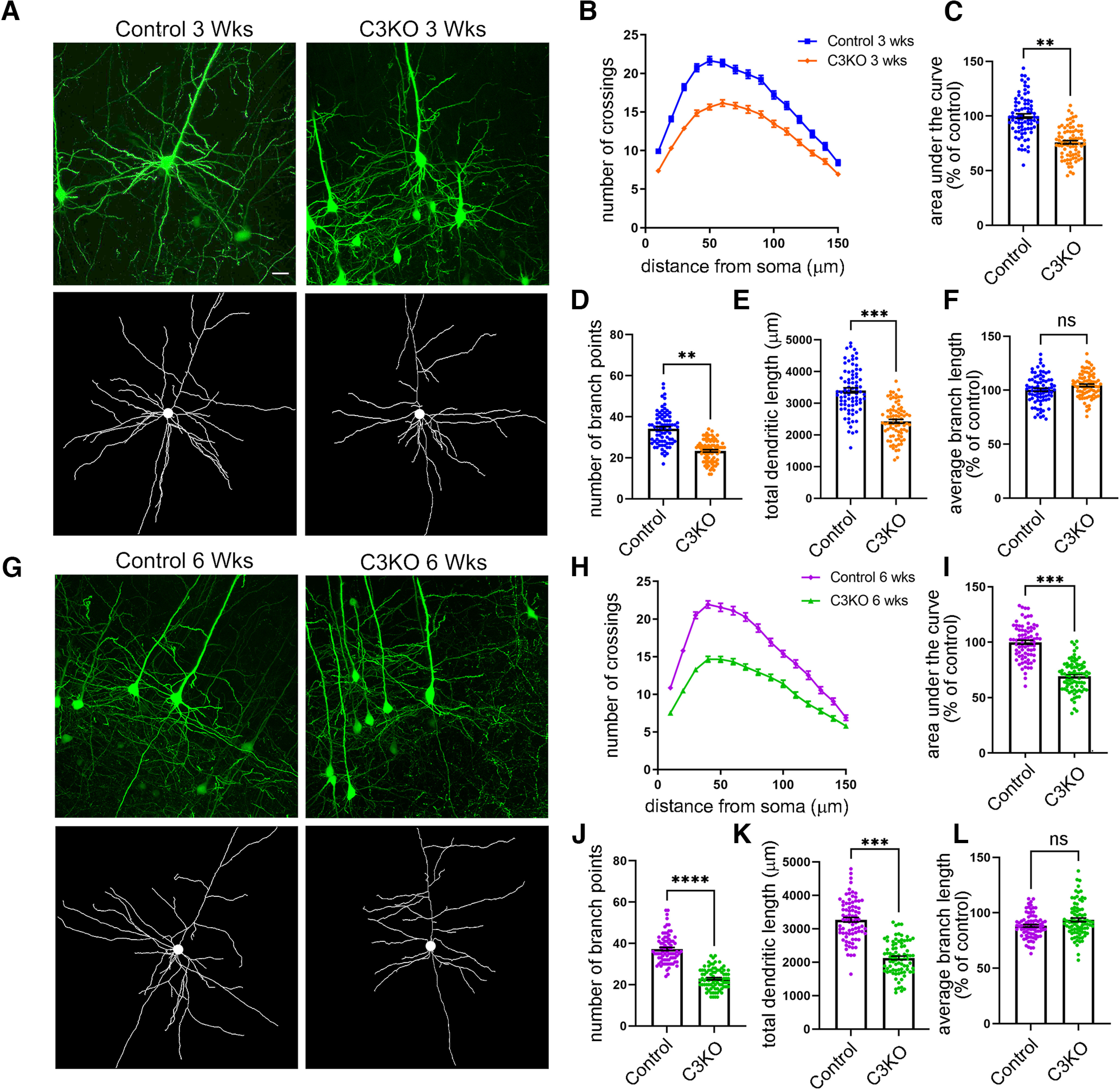
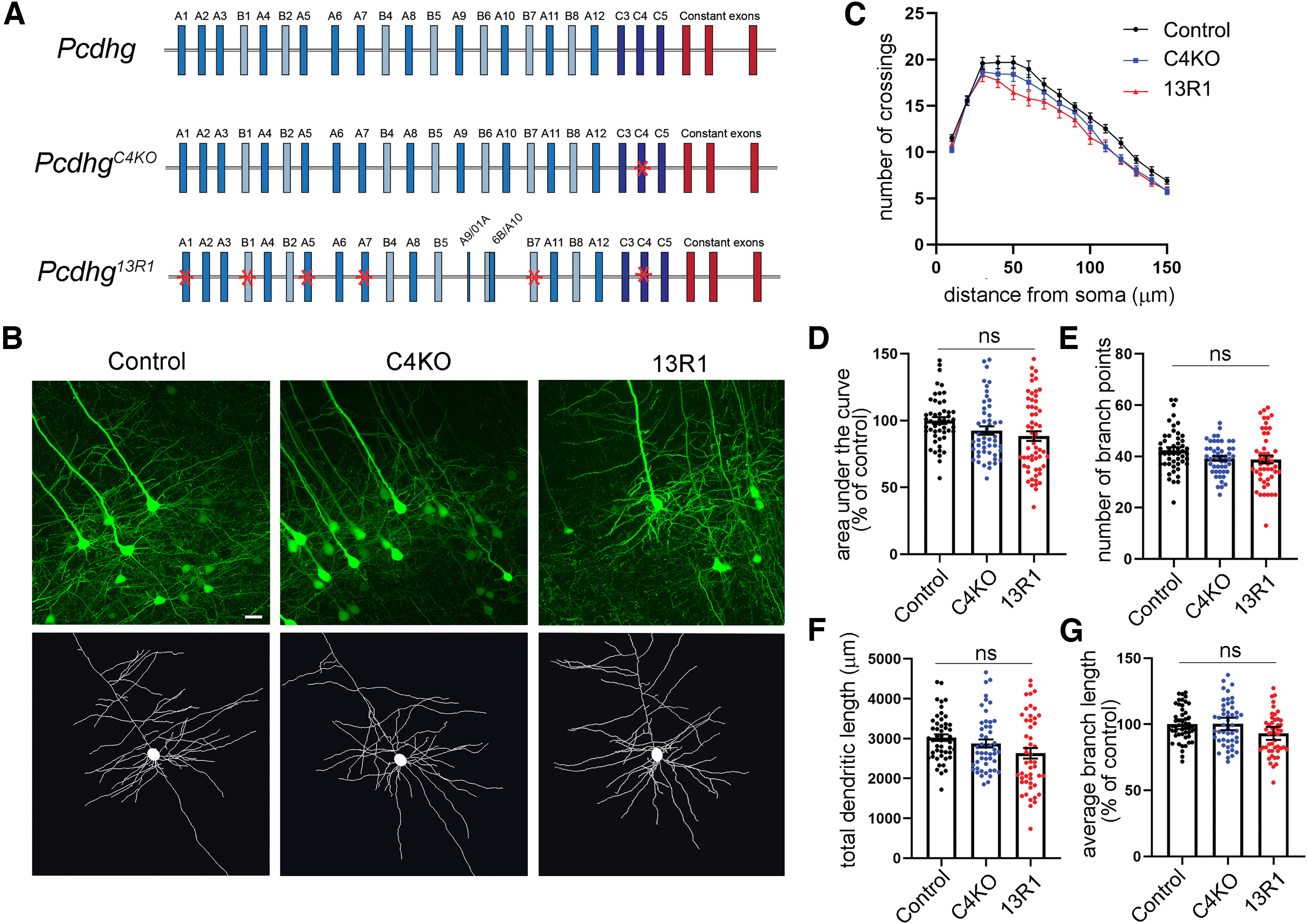
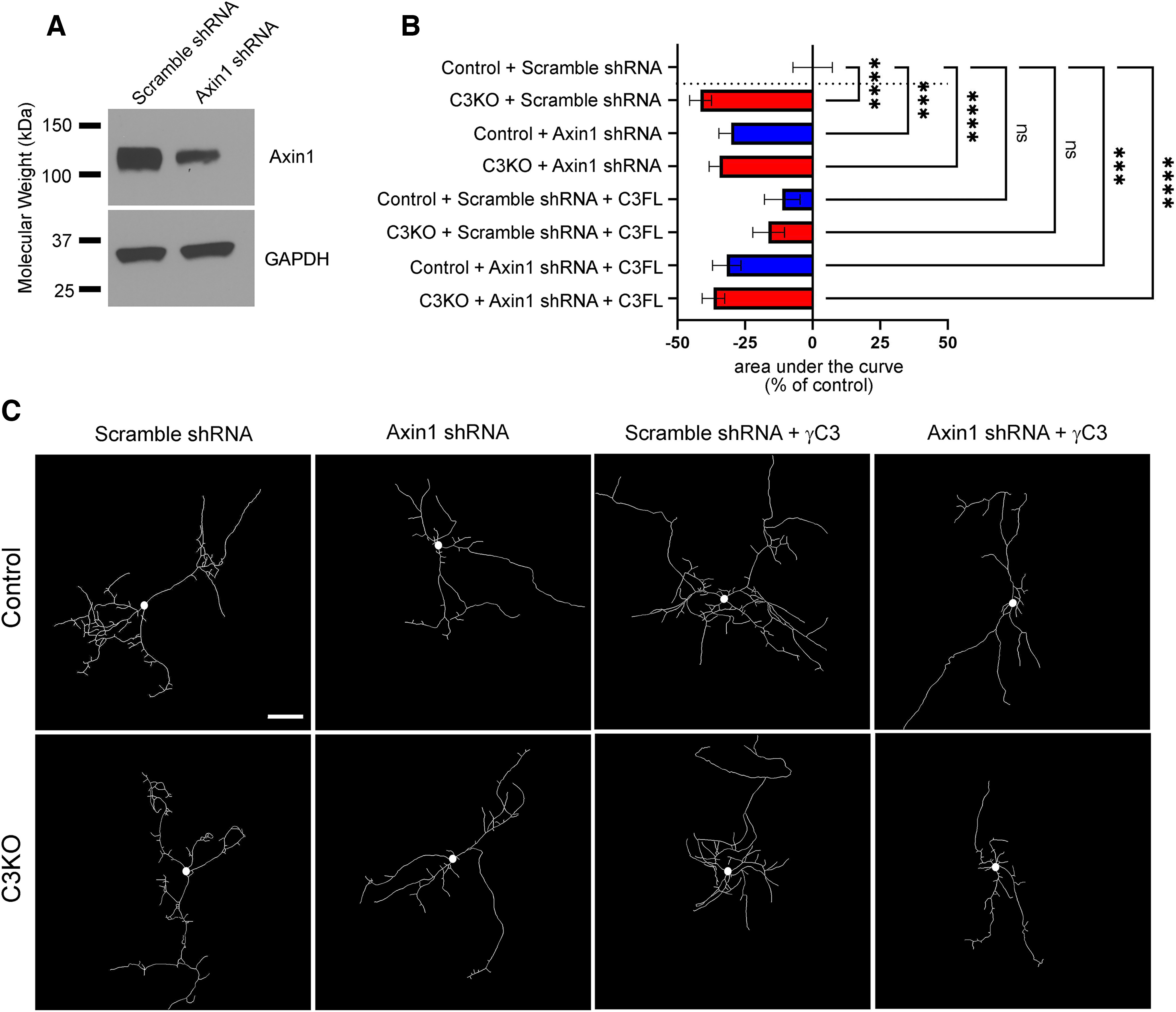
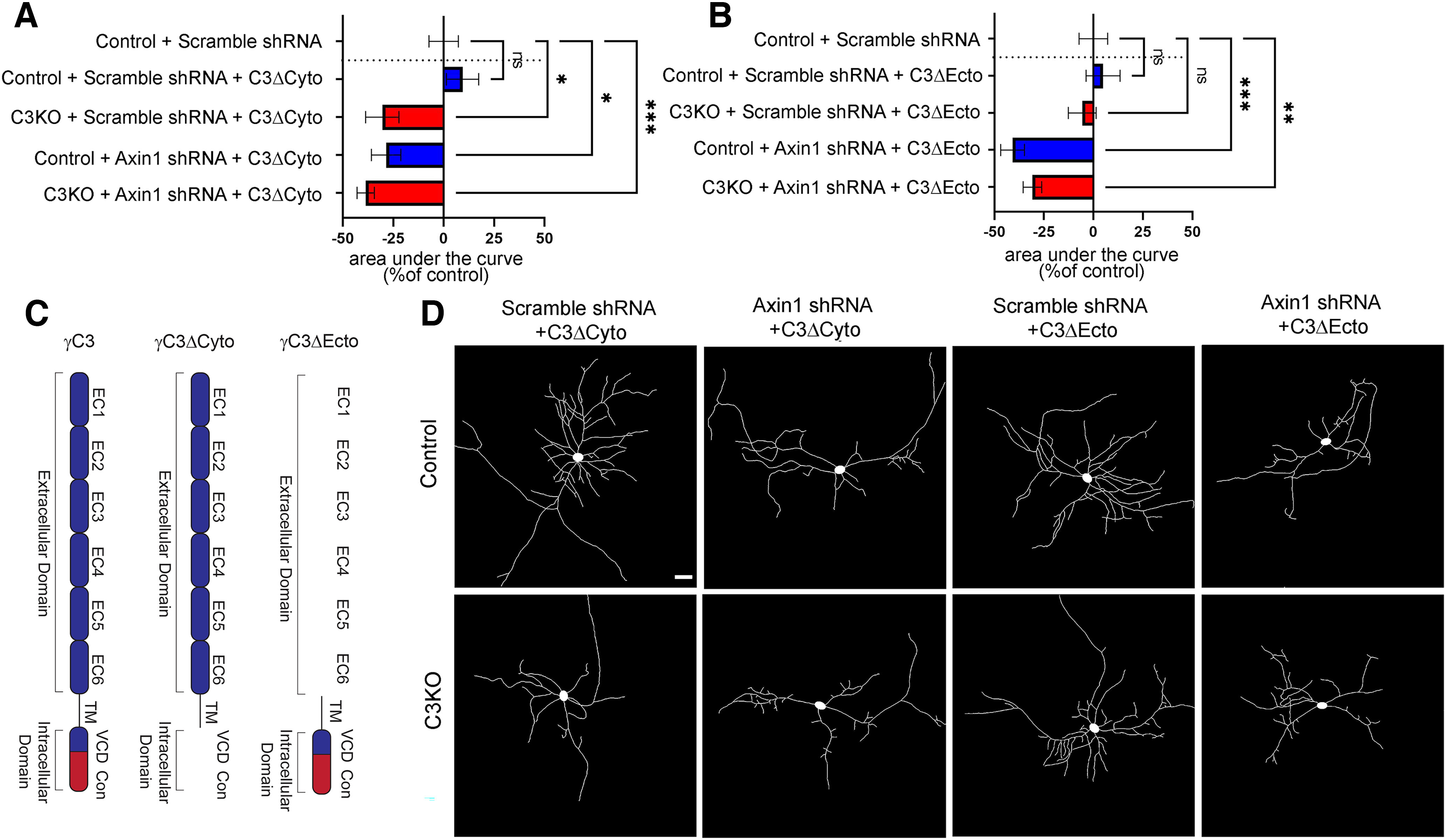
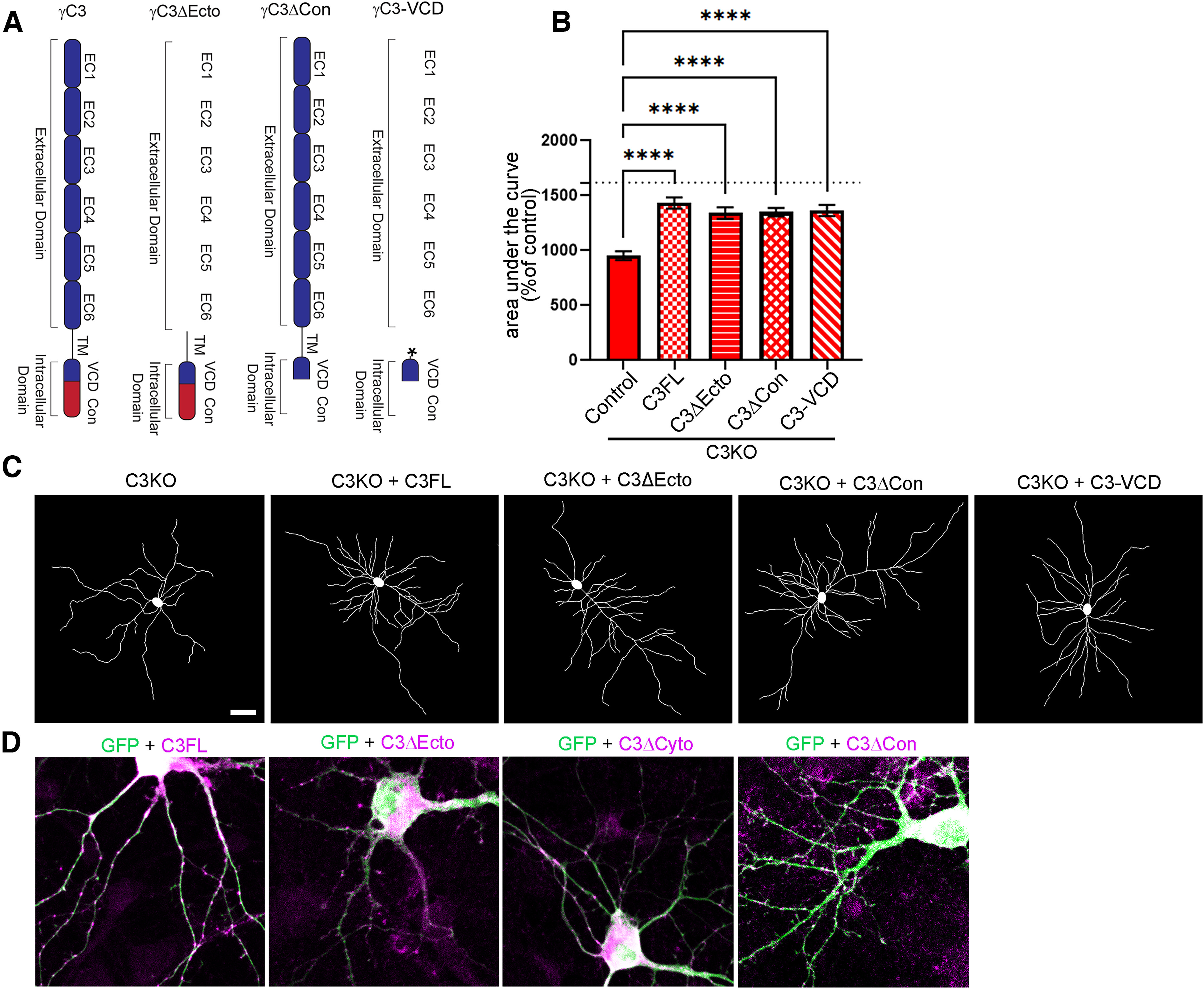
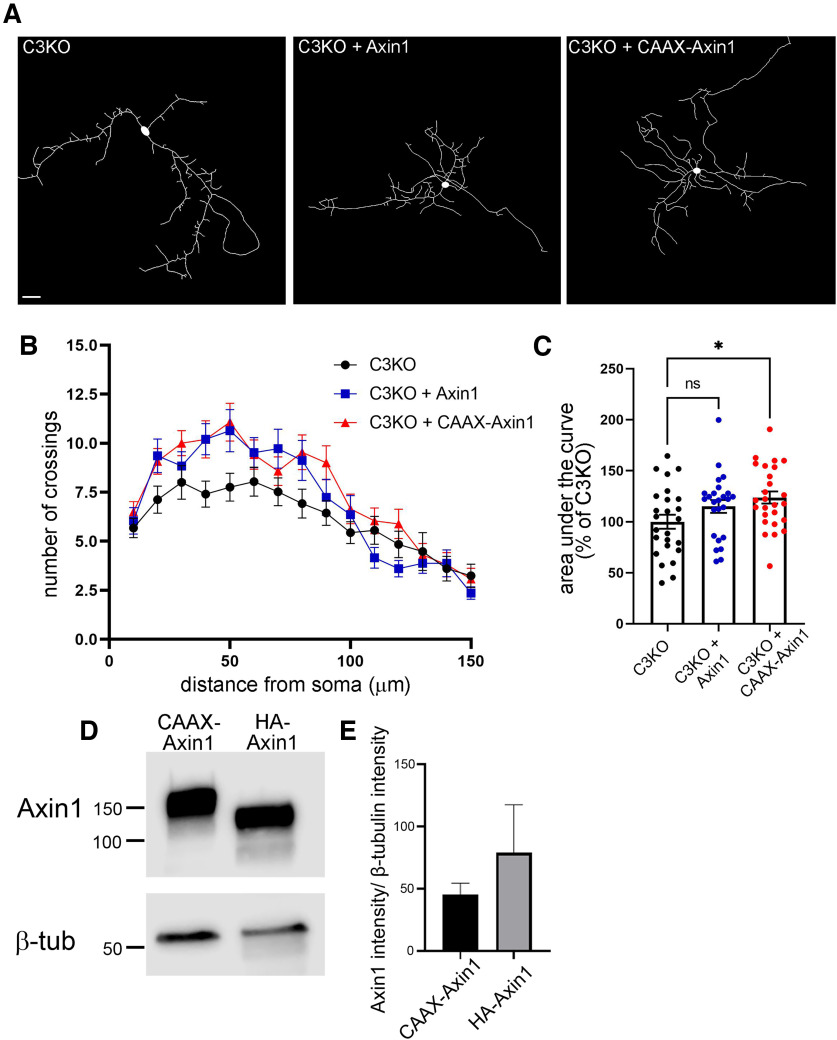
The establishment of a functional cerebral cortex depends on the proper execution of multiple developmental steps, culminating in dendritic and axonal outgrowth and the formation and maturation of synaptic connections. Dysregulation of these processes can result in improper neuronal connectivity, including that associated with various neurodevelopmental disorders. The γ-Protocadherins (γ-Pcdhs), a family of 22 distinct cell adhesion molecules that share a C-terminal cytoplasmic domain, are involved in multiple aspects of neurodevelopment including neuronal survival, dendrite arborization, and synapse development. The extent to which individual γ-Pcdh family members play unique versus common roles remains unclear. We demonstrated previously that the γ-Pcdh-C3 isoform (γC3), via its unique "variable" cytoplasmic domain (VCD), interacts in cultured cells with Axin1, a Wnt-pathway scaffold protein that regulates the differentiation and morphology of neurons. Here, we confirm that γC3 and Axin1 interact in the cortex in vivo and show that both male and female mice specifically lacking γC3 exhibit disrupted Axin1 localization to synaptic fractions, without obvious changes in dendritic spine density or morphology. However, both male and female γC3 knock-out mice exhibit severely decreased dendritic complexity of cortical pyramidal neurons that is not observed in mouse lines lacking several other γ-Pcdh isoforms. Combining knock-out with rescue constructs in cultured cortical neurons pooled from both male and female mice, we show that γC3 promotes dendritic arborization through an Axin1-dependent mechanism mediated through its VCD. Together, these data identify a novel mechanism through which γC3 uniquely regulates the formation of cortical circuitry.SIGNIFICANCE STATEMENT The complexity of a neuron's dendritic arbor is critical for its function. We showed previously that the γ-Protocadherin (γ-Pcdh) family of 22 cell adhesion molecules promotes arborization during development; it remained unclear whether individual family members played unique roles. Here, we show that one γ-Pcdh isoform, γC3, interacts in the brain with Axin1, a scaffolding protein known to influence dendrite development. A CRISPR/Cas9-generated mutant mouse line lacking γC3 (but not lines lacking other γ-Pcdhs) exhibits severely reduced dendritic complexity of cerebral cortex neurons. Using cultured γC3 knock-out neurons and a variety of rescue constructs, we confirm that the γC3 cytoplasmic domain promotes arborization through an Axin1-dependent mechanism. Thus, γ-Pcdh isoforms are not interchangeable, but rather can play unique neurodevelopmental roles.

Keywords: cell adhesion; dendritic arborization; signaling; synapse development; synaptic maturation.


Figures

Figure 1.
Figure 1.
Generation of the C3KO mouse. A, Schematic representation of: the mouse Pcdhg locus, comprising 22 variable exons (γA-types: blue, γB-types: cyan, and γC-types: dark blue) which are spliced to three constant exons (red); the PcdhgC3KO locus, with frameshift deletion in the C3 variable exon indicated by a red asterisk; and the γC3 protein domain structure, with six extracellular cadherin repeats, a transmembrane domain, and a short variable cytoplasmic domain (all encoded by the variable exon) followed by a shared cytoplasmic domain (encoded by the three constant exons). B, Start of the mouse Pcdhgc3 gene with protein translation. A 13-bp deletion in the C3KO allele results in a frameshift, leading to an early stop codon. C, Quantitative PCR performed on cortical cDNA for multiple cPcdh genes shows specific loss of the γC3 isoform (no signal using a forward primer spanning the deletion; reduced transcript levels using a forward primer at the 3′ end of the variable exon), while other cPcdh and overall Pcdhg expression remain unaltered. Data are presented as relative mRNA as compared with GAPDH. ±SEM from six individual animals. A two-way ANOVA with Bonferroni post hoc test was performed to assess statistical significance. ****p < 0.0001. D, Western blottings from cortical lysates show no γC3 expression in the C3KO, while expression of other γ-Pcdhs remain largely unaltered. E, F, Cryosections of adult C3KO, and control S1 cortices were stained with multiple layer-specific and cell type-specific markers, which indicate grossly normal cell types and morphology in C3KO mice, save for a somewhat thinner Layer I, suggesting a loss of apical dendritic tufts as was seen for complete Pcdhg cluster KO mice (Garrett et al., 2012). Scale bar: 100 µm.
Figure 2.
Figure 2.
C3KO mice do not exhibit increased apoptosis of spinal interneurons. A, Cryosections of P0 spinal cords from control and C3KO pups immunostained for the indicated markers (green) and counterstained with DAPI for nuclei (blue). Scale bar: 200 µm. No gross abnormalities of the spinal cord (previously observed in complete Pcdhg null mutants; Wang et al., 2002b; Prasad et al., 2008) were observed in C3KO mutants, nor were any alterations in the number of FoxP2-labeled ventral interneurons (B), CC3-labeled apoptotic cells (C), or Pax2-labeled dorsal and ventral interneurons (D). ns = not significant.
Figure 3.
Figure 3.
C3KO mice do not exhibit altered cell survival or dendrite self-avoidance in the retina. Whole-mount retinas were stained for (A, B) Vglut3 to label glutamatergic amacrine cells, (C, D) tyrosine hydroxylase (TH) to label dopaminergic amacrine cells (DA cells), and (E, F) melanopsin to label intrinsically photosensitive retinal ganglion cells (ipRGCs). For all cell types, there was no significant change in cell density in adult C3KO mutants (quantified in G). Self-avoidance in starburst amacrine cells (SACs) was assayed by analyzing interdendritic space in (H, I) the OFF strata and (J, K) the ON strata. T–V, No increase in the space between SAC dendrites was observed when quantified by Elo analysis (see Materials and Methods) or manual measurement. Projections of representative cell types within the inner plexiform layer (IPL) were analyzed to assess general retinal organization. L–M, SACs (ChAT+), Vglut3+ amacrine cells and rod bipolar cells (PKCa+) targeted normally, as did (N, O) DA cells (TH+) and ipRGCs (melanopsin+), (P, Q) bnos+ GABAergic amacrine cells and Glyt1+ glycinergic amacrine cells, and (R, S) Calbindin+ cells and type 3b cone bipolar cells (PKARIIb+). The expected organization of projections, which was observed in both control and C3KO retinae, is presented in W. For all experiments, six adult retinas per genotype were analyzed. ns = not significant.
Figure 4.
Figure 4.
γC3 interacts with Axin1 in vivo and is required for normal Axin1 subcellular localization. A, Endogenous Axin1 co-immunoprecipitates with γ-Pcdhs from P21 control cortical lysates; comparatively little Axin1 is co-immunoprecipitated from cortical lysates of C3KO mice. Representative Western blot images of (B) cytoplasmic and (C) synaptic fraction preparations from P21 cortex. Quantification of Axin1 (D), CDC42 (E), and β-actin (F) intensity normalized to total protein from cytoplasmic and synaptic fractions from C3KO and control mice. Levels of all three proteins are significantly reduced in synaptic, but not cytoplasmic fractions. n = 6 animals per genotype. Unpaired t test; *p <0.05, ***p < 0.001; ns = not significant.
Figure 5.
Figure 5.
C3KO mice show no alterations in dendritic spine density or morphology. A, Representative images showing dendritic spines of Thy1-YFPH-labeled Layer V pyramidal neurons from the S1 cortex of five-week-old mice. Scale bar: 5 µm. Quantification of overall dendritic spine density (B) and of the density of thin, mushroom, and stubby dendritic spine types (C). Quantification of average spine volume (D) and of average spine surface area (E) of all spines and of each dendritic spine class (thin, mushroom, and stubby). n ≥ 58 control dendritic segments across 7 mice, n = 54 C3KO dendritic segments across 9 mice. Error bars represent the SEM. No significant differences were observed between control and C3KO genotypes. ns = not significant.
Figure 6.
Figure 6.
C3KO mice exhibit decreased dendritic complexity. A, Representative YFP images (top) and tracings (bottom) of Thy1-YFPH-labeled Layer V pyramidal neurons of mice three weeks of age. B, Sholl analysis graphs show dendritic crossings at spheres of increasing 10-µm intervals of mice at three weeks of age. Graphs showing area under the curve of Sholl graphs (C), average number of branch points per neuron (D), total dendritic length (E), and average branch length (F) of mice at three weeks of age. G, Representative YFP images (top) and tracings (bottom) of Thy1-YFPH-labeled Layer V pyramidal neurons of mice six weeks of age. H, Sholl analysis graphs show dendritic crossings at spheres of increasing 10-µm intervals of mice at six weeks of age. Graphs showing area under the curve of Sholl graphs (I), average number of branch points per neuron (J), total dendritic length (K), and average branch length (L) of mice at six weeks of age. Significant reductions in dendritic complexity are seen in C3KO compared with control at both ages. Scale bar: 25 µm. n = ∼80 neurons from at least 4 mice per genotype. Unpaired t test. Error bars represent the SEM; *p < 0.05, **p < 0.01, ***p < 0.001, ****p < 0.0001; ns = not significant.
Figure 7.
Figure 7.
Dendritic arborization deficits found in C3KO mutants are not observed in other Pcdhg locus mutants. A, Schematic representation of the mouse Pcdhg locus (control), the PcdhgC4KO mutant mouse locus lacking only γC4, and the Pcdhg13R1 mutant locus lacking nine isoforms (mutations indicated by red asterisks). B, Representative images and tracings of Thy1-YFPH-labeled Layer V pyramidal neurons of mice six weeks of age. C, Sholl analysis graphs show dendritic crossings of spheres of increasing 10-µm intervals of mice at six weeks of age. D, Graphs showing area under the curve of Sholl graphs, (E) total branch points, (F) total dendritic length, and average branch length (G) of mice at six weeks of age. Although 13R1 neurons exhibit a slight trend toward lower complexity, neither they nor C4KO neurons were significantly different from controls on any measure. Scale bar: 25 µm. n ≥ 49 neurons per genotype and at least 3 animals from each genotype. One-way ANOVA; Dunnett's multiple comparisons test. Error bars represent the SEM; ns = not significant.
Figure 8.
Figure 8.
γC3-mediated dendritic arborization is dependent on Axin1. A, Cortical neurons cultured from P0 pups and nucleofected (∼50% efficiency) at plating with Axin1 shRNA (previously characterized by Chen et al., 2015) or its scrambled control. Neurons were lysed at day in vitro (DIV)8 and lysates analyzed by Western blotting using antibodies against Axin1. As expected, Axin1 levels are drastically reduced in neurons expressing the Axin1 shRNA knock-down construct. B, Quantification of area under the curve for Sholl analysis of control and C3KO cultured cortical neurons transfected with GFP, either Axin1 shRNA or the scrambled control shRNA, ± a full-length γC3 construct and analyzed at DIV8. Bars to the left indicate reduced complexity; bars to the right indicate increased complexity. C, Representative traces of neurons from each transfection condition. C3KO neurons exhibit significantly reduced dendrite complexity in culture as in vivo. Arbor complexity can be rescued by re-expression of γC3, but only if normal Axin1 levels are present. Scale bar: 50 µm. n ≥ 25 neurons per condition. One-way ANOVA; Dunnett's multiple comparisons test. Error bars represent the SEM; *p < 0.05, **p < 0.01, ***p < 0.001, ****p < 0.0001; ns = not significant.
Figure 9.
Figure 9.
The intracellular domain of γC3 is required for Axin1-mediated dendritic arborization. A, B, Quantification of area under the curve for Sholl analysis of control and C3KO cultured cortical neurons transfected with GFP, either Axin1 shRNA or the scrambled control shRNA, and a γC3 construct lacking either the cytoplasmic (C3Δcyto; A), or the extracellular domain (C3Δecto) and analyzed at DIV8. B, Bars to the left indicate reduced complexity; bars to the right indicate increased complexity. C, Schematic depiction of γC3 deletion transfection constructs transfected. D, Representative traces of neurons from each transfection condition. Scale bar: 50 µm. n ≥ 25 neurons per condition. One-way ANOVA; Dunnett's multiple comparisons test. Error bars represent the SEM; *p < 0.05, **p < 0.01, ***p < 0.001, ****p < 0.0001; ns = not significant.
Figure 10.
Figure 10.
The γC3 variable cytoplasmic domain is necessary and sufficient to rescue dendritic arborization in C3KO neurons. A, Schematic depiction of γC3 deletion transfection constructs transfected. Asterisk indicates palmitoylation sequence at the N terminus of the γC3-VCD construct. B, Quantification of area under the curve for Sholl analysis of C3KO cultured cortical neurons transfected with GFP (control), a full-length (FL) γC3 construct, constructs lacking either the extracellular domain (C3Δecto) or the constant domain (C3Δcon), or a construct encoding only a palmitoylated γC3 variable cytoplasmic domain (C3-VCD) and analyzed at DIV8. All constructs significantly rescued arborization toward wild-type levels (indicated by the dashed line), indicating that the C3 VCD is necessary (Fig. 9) and sufficient. n ≥ 25 neurons per condition. One-way ANOVA; Dunnett's multiple comparisons test. Error bars represent the SEM; ****p < 0.0001. C, Representative traces of neurons from each transfection condition. D, Representative images of transfected neurons immunostained for GFP (transfected to fill neuronal processes) and the HA tag on the indicated construct. All constructs localize in a punctate or patchy manner in neuronal dendrites. Scale bar: 50 µm (C) or 10 µm (D).
Figure 11.
Figure 11.
Reduced dendritic complexity of C3KO neurons can be rescued by targeted Axin1 overexpression in vitro. A, Representative images of DIV8 C3KO neurons transfected with GFP, and either an empty vector, Axin1 overexpression construct, or membrane-targeted CAAX-tagged Axin1. B, Sholl analysis graphs show dendritic crossings of spheres of increasing 10-µm intervals of neurons analyzed at DIV8. Expression of either Axin1 or CAAX-Axin1 rescues low arbor complexity proximal to the soma; only CAAX-Axin1 maintains that rescue at more distal Sholl spheres. C, Quantification of area under the curve of Sholl analysis of transfected C3KO neurons for all conditions. A significant rescue is observed with membrane-targeted CAAX-Axin1. n ≥ 25 neurons per transfection condition. One-way ANOVA; Dunnett's multiple comparisons test. Scale bar: 25 µm. Error bars represent the SEM; *p < 0.05. D, E, Western blottings of COS7 cells transfected with the indicated constructs and blotted using Axin1 and β-tubulin (loading control) antibodies. Quantification of three replicate experiments indicates no significant difference in expression levels between the two constructs.

Similar articles

Cited by

References

    1. Anderson RM, Johnson SB, Lingg RT, Hinz DC, Romig-Martin SA, Radley JJ (2020) Evidence for similar prefrontal structural and functional alterations in male and female rats following chronic stress or glucocorticoid exposure. Cereb Cortex 30:353–370. 10.1093/cercor/bhz092 - DOI - PMC - PubMed
    1. Ashby MC, Maier SR, Nishimune A, Henley JM (2006) Lateral diffusion drives constitutive exchange of AMPA receptors at dendritic spines and is regulated by spine morphology. J Neurosci 26:7046–7055. 10.1523/JNEUROSCI.1235-06.2006 - DOI - PMC - PubMed
    1. Carriere CH, Wang WX, Sing AD, Fekete A, Jones BE, Yee Y, Ellegood J, Maganti H, Awofala L, Marocha J, Aziz A, Wang LY, Lerch JP, Lefebvre JL (2020) The gamma-protocadherins regulate the survival of GABAergic interneurons during developmental cell death. J Neurosci 40:8652–8668. 10.1523/JNEUROSCI.1636-20.2020 - DOI - PMC - PubMed
    1. Chen J, Lu Y, Meng S, Han MH, Lin C, Wang X (2009) Alpha- and gamma-protocadherins negatively regulate PYK2. J Biol Chem 284:2880–2890. - PMC - PubMed
    1. Chen WV, Alvarez FJ, Lefebvre JL, Friedman B, Nwakeze C, Geiman E, Smith C, Thu CA, Tapia JC, Tasic B, Sanes JR, Maniatis T (2012) Functional significance of isoform diversification in the protocadherin gamma gene cluster. Neuron 75:402–409. 10.1016/j.neuron.2012.06.039 - DOI - PMC - PubMed

Publication types