. 2023 Jan 18;145(2):1389-1399.
doi: 10.1021/jacs.2c12086. Epub 2023 Jan 5.

Direct Expression of Fluorinated Proteins in Human Cells for 19F In-Cell NMR Spectroscopy

Affiliations

Direct Expression of Fluorinated Proteins in Human Cells for 19F In-Cell NMR Spectroscopy

Lan B T Pham et al. J Am Chem Soc. .

Abstract

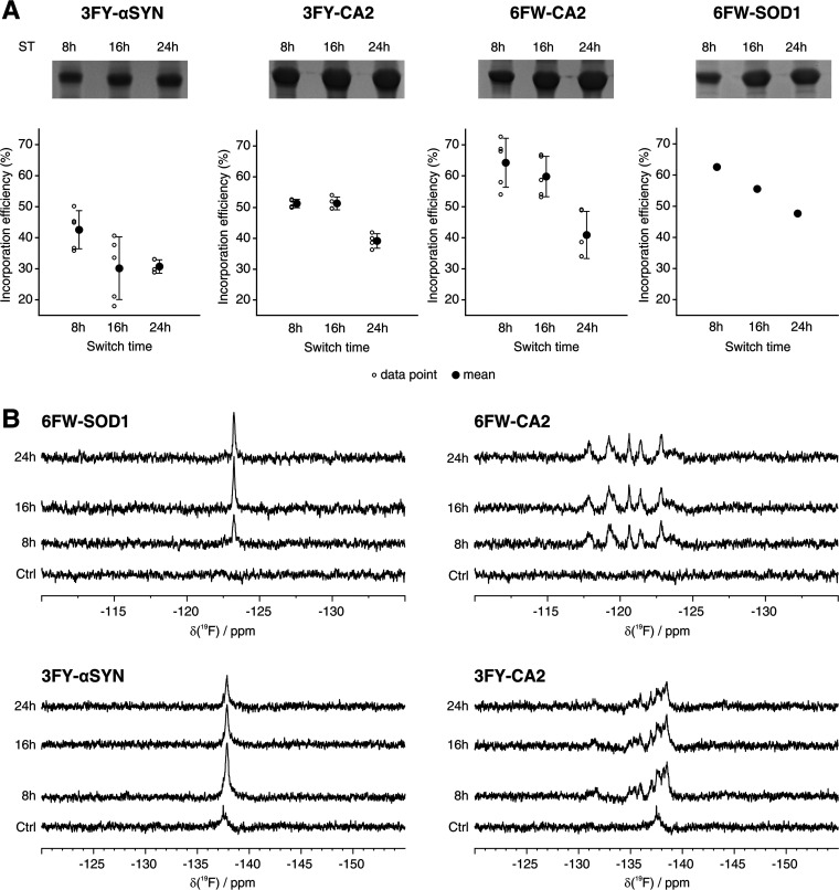
In-cell NMR spectroscopy is a powerful approach to study protein structure and function in the native cellular environment. It provides precious insights into the folding, maturation, interactions, and ligand binding of important pharmacological targets directly in human cells. However, its widespread application is hampered by the fact that soluble globular proteins often interact with large cellular components, causing severe line broadening in conventional heteronuclear NMR experiments. 19F NMR can overcome this issue, as fluorine atoms incorporated in proteins can be detected by simple background-free 1D NMR spectra. Here, we show that fluorinated amino acids can be easily incorporated in proteins expressed in human cells by employing a medium switch strategy. This straightforward approach allows the incorporation of different fluorinated amino acids in the protein of interest, reaching fluorination efficiencies up to 60%, as confirmed by mass spectrometry and X-ray crystallography. The versatility of the approach is shown by performing 19F in-cell NMR on several proteins, including those that would otherwise be invisible by 1H-15N in-cell NMR. We apply the approach to observe the interaction between an intracellular target, carbonic anhydrase 2, and its inhibitors, and to investigate how the formation of a complex between superoxide dismutase 1 and its chaperone CCS modulates the interaction of the chaperone subunit with the cellular environment.


Conflict of interest statement

The authors declare no competing financial interest.

Figures

Figure 1
Figure 1
In-cell and lysate 19F NMR of proteins incorporating different 19F-amino acids. 1D 19F NMR spectra of HEK293T cells (c, thick line) expressing different proteins in media supplemented with 3FY (top left), 4FF (top right), 5FW (bottom left), and 6FW (bottom right) with ST = 24 h, and the corresponding lysates (l, thin line). Ctrl: control spectra acquired on HEK293T cells transfected with pHL-empty and the corresponding lysates. The regions containing the background signal of 3FY and 4FF are shown as light gray bands; the chemical shift of each free FAA is marked with a dotted line (see also Figure S2).
Figure 2
Figure 2
Effect of different medium switch times on incorporation efficiency, protein expression levels, and 19F NMR signal intensities. (A) SDS-PAGE analysis of soluble cell lysates obtained from small-scale expression (top) of 3FY-αSYN, 3FY-CA2, 6FW-CA2, and 6FW-SOD1, and plots of LC-MS data of the corresponding gel bands (bottom). Each data point in the plots presents the percentage of fluorine incorporation in a peptide containing a single target residue (see also Table S2). The means and standard deviations of each set of data points are also shown. Fluorine incorporation in 6FW-SOD1 is estimated from a single peptide. ST: switch time. (B) 1D 19F NMR spectra of cells expressing 6FW-SOD1 (top left), 6FW-CA2 (top right), 3FY-αSYN (bottom left), and 3FY-CA2 (bottom right) at different ST. Ctrl: control spectrum acquired on HEK293T cells transfected with pHL-empty.
Figure 3
Figure 3
Structure of 6FW-CA2. (A) X-ray structure of 6FW-CA2 (PDB 8B29, cyan) superimposed to that of native, nonfluorinated CA2 (PDB 1CA2, yellow), showing negligible differences between the two structures. The inset shows the active site of the protein. Zn2+ is shown as sphere; Zn2+-coordinating histidines (cyan/yellow) and 6FW residues (red) are shown as sticks. (B–D) 2Fo-Fc maps of 6FW-CA2 contoured at 1σ (blue) showing the presence of fluorine atoms on Trp5 (B), Trp16 (C), and Trp123 (D). The polypeptide is shown as sticks, and water molecules are shown as spheres.
Figure 4
Figure 4
19F NMR of CA2 interacting with ligands in cells and soluble cell lysates. 1D 19F NMR spectra of cells (left) expressing 3FY-CA2 (ST = 8 h, top) and 6FW-CA2 (ST = 16 h, bottom) treated with different ligands, and of the corresponding lysates (right). No ligand: control spectra acquired on cells expressing CA2 with no ligand treatment, and corresponding lysates. ST: switch time. Peaks shifting upon binding are marked with dashed lines. *: disappearing signal upon ligand interaction.
Figure 5
Figure 5
Interaction of CCS-D2 and SOD1 observed by 19F in-cell NMR. 1D 19F NMR spectra of cells (left) expressing 3FY- (top), 4FF- (middle), and 6FW- (bottom) SOD1 and CCS-D2 either alone or together (ST = 24 h), and of the corresponding lysates (right). Signals attributed to CCS-D2 in the complex with SOD1 are marked with dashed lines. Ctrl: control spectra acquired on cells transfected with pHL-empty and corresponding lysates.

Similar articles

Cited by

References

    1. Luchinat E.; Cremonini M.; Banci L. Radio Signals from Live Cells: The Coming of Age of In-Cell Solution NMR. Chem. Rev. 2022, 122, 9267–9306. 10.1021/acs.chemrev.1c00790. - DOI - PMC - PubMed
    1. Theillet F.-X. In-Cell Structural Biology by NMR: The Benefits of the Atomic Scale. Chem. Rev. 2022, 122, 9497–9570. 10.1021/acs.chemrev.1c00937. - DOI - PubMed
    1. Banci L.; Barbieri L.; Bertini I.; et al. Atomic-resolution monitoring of protein maturation in live human cells by NMR. Nat. Chem. Biol. 2013, 9, 297–299. 10.1038/nchembio.1202. - DOI - PMC - PubMed
    1. Smith A. E.; Zhou L. Z.; Gorensek A. H.; Senske M.; Pielak G. J. In-cell thermodynamics and a new role for protein surfaces. Proc. Natl. Acad. Sci. U.S.A. 2016, 113, 1725–1730. 10.1073/pnas.1518620113. - DOI - PMC - PubMed
    1. Theillet F.-X.; Binolfi A.; Bekei B.; et al. Structural disorder of monomeric α-synuclein persists in mammalian cells. Nature 2016, 530, 45–50. 10.1038/nature16531. - DOI - PubMed

Publication types

MeSH terms