

Comparison between the CASIA SS-1000 and Pentacam in measuring corneal curvatures and corneal thickness maps
- PMID: 36604657
- PMCID: PMC9814456
- DOI: 10.1186/s12886-023-02768-w
Comparison between the CASIA SS-1000 and Pentacam in measuring corneal curvatures and corneal thickness maps
Abstract
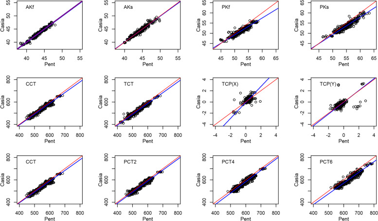
Purpose: To compare the intra-device repeatability and inter-device reproducibility between two anterior segment imaging instruments, the CASIA SS-1000 (Tomey Corp., Nagoya, Japan) and Pentacam (OCULUS, Arlington, WA) in measuring anterior segment parameters.
Methods: Single-center, prospective clinical trial. Participants ≥20 years of age were included. One eye was randomly selected, each imaged by three CASIA SS-1000 devices and three Pentacam devices by three different examiners. Each photographer operated a pair of devices, one CASIA SS-1000 and one Pentacam. The image order for each participant was determined by a random permutation table. Three images were taken from each device. A total of 18 images were taken for each eye. Ratios of the standard deviations, referenced as (CASIA/Pentacam), were calculated to compare the repeatability and reproducibility of the two imaging instruments.
Results: In all, 66 participants with a mean age of 46.4 years (±21.7) were enrolled in the study. All repeatability ratios and intra-device variability were less than 1 (anterior corneal curvature: flat = 0.86, steep = 0.85; posterior corneal curvature: flat = 0.43, steep = 0.61; and map: thinnest = 0.22; central = 0.24, 2 mm = 0.26, 4 mm = 0.27, and 6 mm = 0.30). All reproducibility ratios, which measure the inter-device variability, were less than 1 (anterior corneal curvature: flat = 0.58, steep = 0.73; posterior corneal curvature: flat = 0.25, steep = 0.31; and pachymetry map: thinnest = 0.20; central = 0.20; 2 mm = 0.20; 4 mm = 0.19; and 6 mm = 0.22). A ratio of less than 1 indicates that the CASIA SS-1000 has more consistent measurements.
Conclusions: The CASIA SS-1000 was found to have better repeatability and reproducibility compared to the Pentacam for both corneal curvature and pachymetry maps. This greater consistency may require further study to determine whether the decreased variability can be translated into improved clinical results.
Keywords: CASIA SS-1000; Corneal curvature; Laser refractive surgery; Pentacam.
© 2023. The Author(s).
Conflict of interest statement
KO and MT are employees of Tomey Corp. The other authors have no relevant conflicts of interest.
Figures

References
-
- Hurmeric V, Yoo SH, Mutlu FM. Optical coherence tomography in cornea and refractive surgery. Expert Rev Ophthalmol. 2012;7(3):241–250. doi: 10.1586/eop.12.28. - DOI
Publication types
MeSH terms
LinkOut - more resources
Full Text Sources
