

The Rise of Extracellular Vesicles as New Age Biomarkers in Cancer Diagnosis: Promises and Pitfalls
- PMID: 36604966
- PMCID: PMC9830000
- DOI: 10.1177/15330338221149266
The Rise of Extracellular Vesicles as New Age Biomarkers in Cancer Diagnosis: Promises and Pitfalls
Abstract
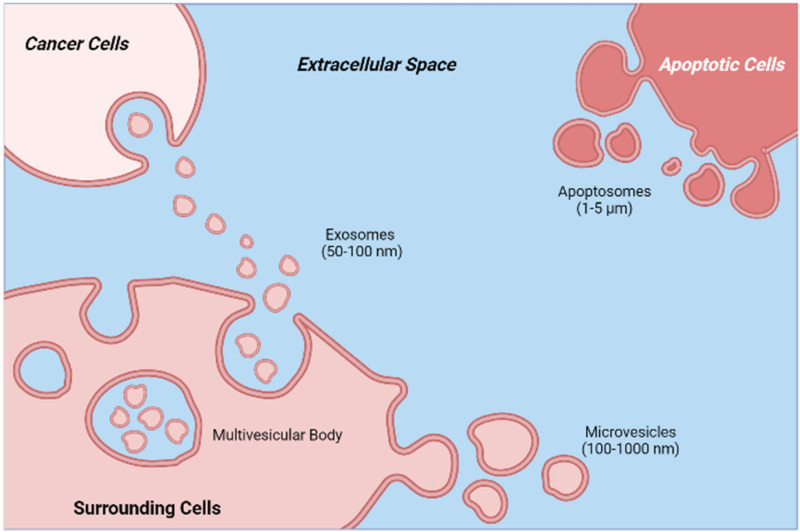
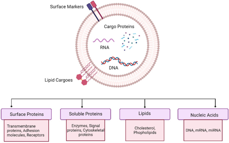
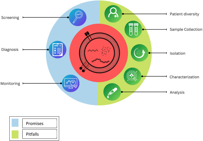
Cell-to-cell interactions in the intricate microenvironment of tissue have a significant impact on the progression of cancer at every stage. Both cancer cells and stromal cells are responsible for the secretion of soluble chemical compounds as well as membrane-encased components, which both influence and govern the cell-to-cell interactions within the micro-environment of tumor cells. These membrane structures are identified as extracellular vesicles (EVs), which include exosomes and microvesicles. These nanosized vesicles are made up of bilayered proteolipids and have dimensions ranging from 50 to 1000 nm. It has been speculated that extracellular vesicles that originate from cancer cells perform a variety of functions in the development and progression of cancer which may involve the transport of regulatory materials, such as oncogenic proteins between nearby cells and to distant biological locations. In addition, their level in the serum of cancer patients is noticeably higher than those of healthy controls. The release of extracellular vesicles into the extracellular space is a continual process in both healthy and diseased cells. These extracellular vesicles hold molecular signatures that are defining features of health as well as disease. And hence, the EVs present in biological fluids provide unparalleled and noninvasive access to the necessary molecular details about the health status of the cells. Recent discoveries about these complex extracellular organelles have accelerated the discovery of cancer-specific biological markers as well as the development of unique diagnostic tools based on extracellular vesicles. In this mini-review, we aim to highlight the hopes and hypes associated with the applications of extracellular vesicles as biomarkers for cancer diagnosis.
Keywords: EVs; biomarkers; cancer; diagnosis; extracellular vesicles.
Conflict of interest statement
The authors declared no potential conflicts of interest with respect to the research, authorship, and/or publication of this article.
Figures
References
-
- Cancer Research UK. Worldwide cancer statistics | Cancer Research UK [Internet]. Cancer Res. UK. 2016 [cited 2022 Apr 22]. p. 1–5. Available from: https://www.cancerresearchuk.org/health-professional/cancer-statistics/w....
-
- Siegel RL, Miller KD, Jemal A. Cancer statistics, 2019. CA Cancer J Clin. 2019;69:7‐34. - PubMed
-
- Kuth JC, Jones TS, Hanje J, Motl Moroney SE. Monoclonal antibodies in cancer. Pharm Biotechnol Fundam Appl Third Ed. Antibodies (Basel). 2016:339‐363.
-
- Hong M, Clubb JD, Chen YY. Engineering CAR-T cells for next-generation. Cancer Therapy. 2020:473‐488. - PubMed
Publication types
MeSH terms
Substances
LinkOut - more resources
Full Text Sources
Medical
