. 2022 Dec 20:13:1017780.
doi: 10.3389/fimmu.2022.1017780. eCollection 2022.

Attenuated Salmonella potentiate PD-L1 blockade immunotherapy in a preclinical model of colorectal cancer

Affiliations

Attenuated Salmonella potentiate PD-L1 blockade immunotherapy in a preclinical model of colorectal cancer

Besan H Al-Saafeen et al. Front Immunol. .

Abstract

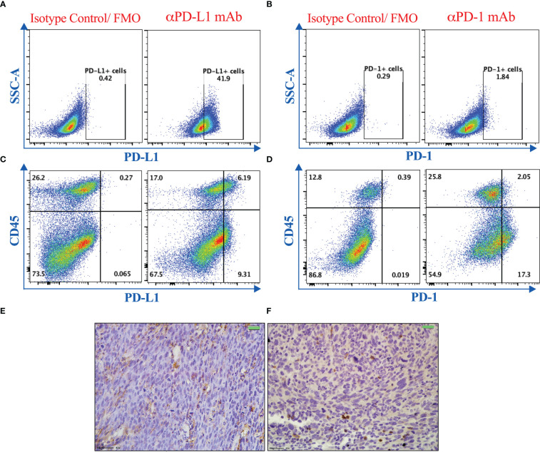
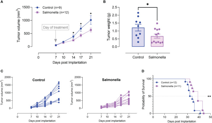
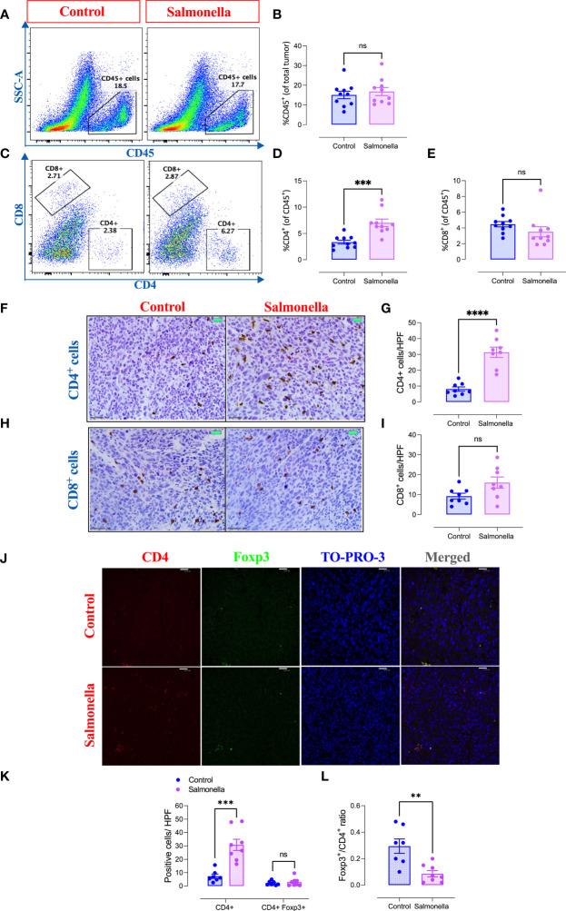
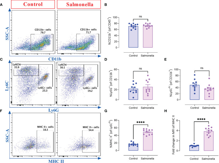
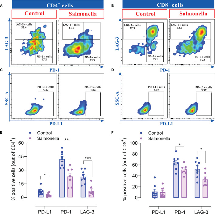
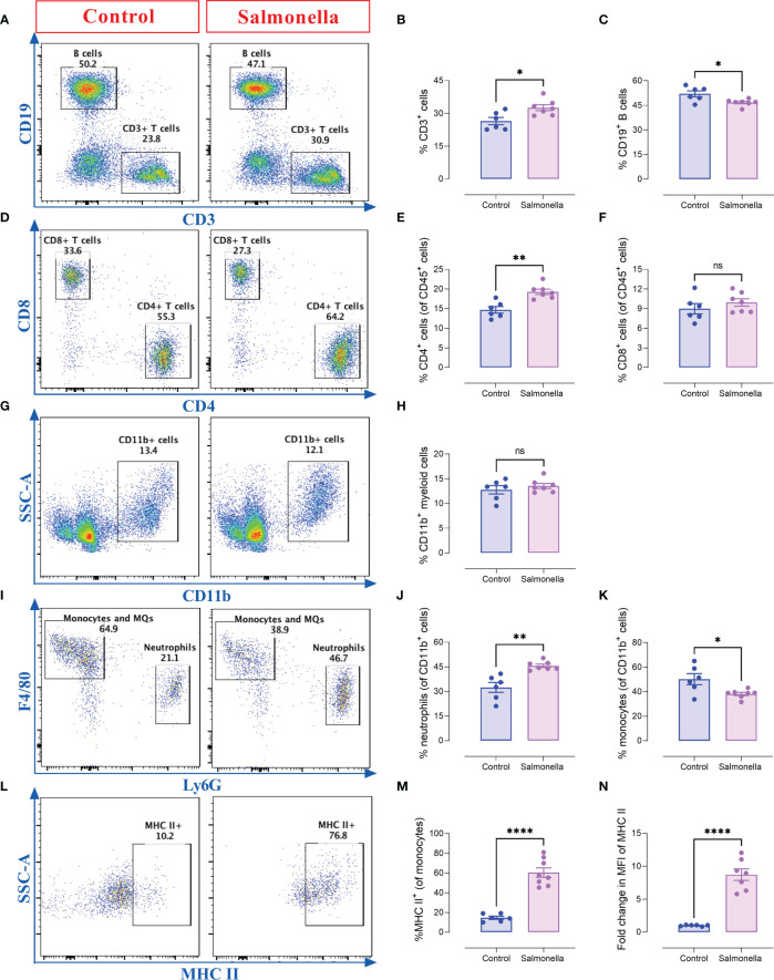
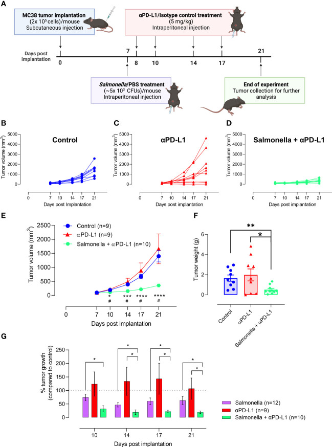
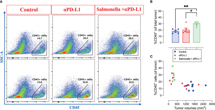
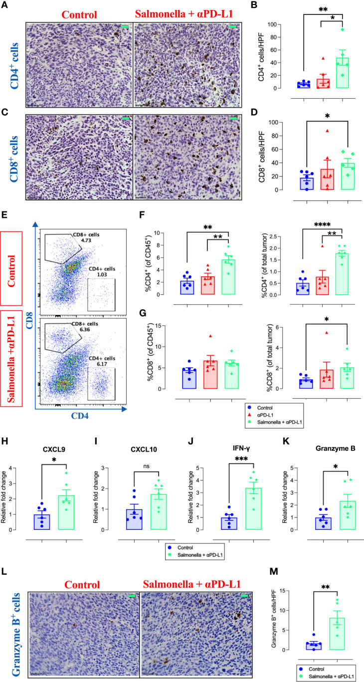
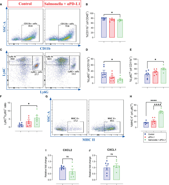
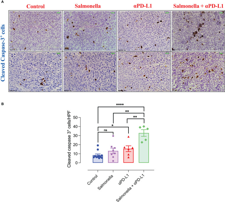
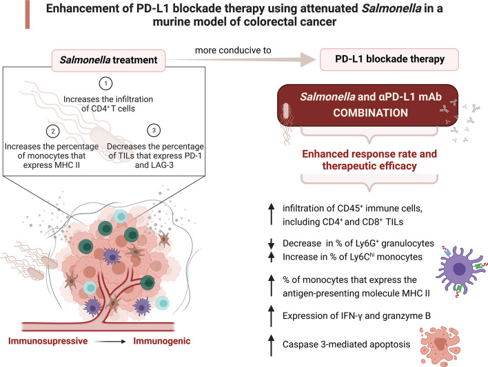
The use of immune checkpoint inhibitors to treat cancer resulted in unprecedented and durable clinical benefits. However, the response rate among patients remains rather modest. Previous work from our laboratory demonstrated the efficacy of using attenuated bacteria as immunomodulatory anti-cancer agents. The current study investigated the potential of utilizing a low dose of attenuated Salmonella typhimurium to enhance the efficacy of PD-L1 blockade in a relatively immunogenic model of colon cancer. The response of MC38 tumors to treatment with αPD-L1 monoclonal antibody (mAb) was variable, with only 30% of the mice being responsive. Combined treatment with αPD-L1 mAb and Salmonella resulted in 75% inhibition of tumor growth in 100% of animals. Mechanistically, the enhanced response correlated with a decrease in the percentage of tumor-associated granulocytic cells, upregulation in MHC class II expression by intratumoral monocytes and an increase in tumor infiltration by effector T cells. Collectively, these alterations resulted in improved anti-tumor effector responses and increased apoptosis within the tumor. Thus, our study demonstrates that a novel combination treatment utilizing attenuated Salmonella and αPD-L1 mAb could improve the outcome of immunotherapy in colorectal cancer.

Keywords: PD-L1 blockade; Salmonella typhimurium; colorectal cancer; immune checkpoints inhibitors; immunotherapy.


Conflict of interest statement

The authors declare that the research was conducted in the absence of any commercial or financial relationships that could be construed as a potential conflict of interest.

Figures

Figure 1
Figure 1
PD-1 and PD-L1 are expressed in MC38 tumors. Representative flow cytometric dot plots showing the expression of PD-L1 (A, C) and PD-1 (B, D) on MC38 tumor cells grown in vitro (A, B) or on single cell suspension of dissociated tumor tissues excised from mice 21 post subcutaneous implantation (C, D). Immunohistochemical staining was also used to illustrate the expression of PD-L1 (E) and PD-1 (F) on MC38 tumor tissue sections. Magnification 400×. Scale bar 20 μm.
Figure 2
Figure 2
Salmonella treatment retards MC38 tumor growth. MC38 tumor cells (2× 10⁵) were subcutaneously implanted into the right flank of C57BL/6 mice. Seven days post implantation, mice were injected intraperitoneally with either Salmonella strain BRD509E (~5× 10³ CFUs) or saline as control. (A) Tumor volumes were measured twice a week for up to 14 days post bacterial treatment. The data is shown as mean ± SEM of 9-12 mice per group, pooled from 2 independent experiments. (B) Tumor weights were recorded at the end of the observation period (day 21 post implantation). Each data point represents a single mouse, pooled from two independent experiments. (C) Tumor growth curves in each individual mouse in the control and BRD509E-treated groups are presented. (D) The effect of BRD509E treatment on the survival of MC38 tumor-bearing mice. Asterisks denote statistically significant differences from control group *(P ≤ 0.05) and ** (P ≤ 0.01).
Figure 3
Figure 3
Treatment with Salmonella enhances the infiltration of CD4+ T cells and decreases the ratio of Tregs/CD4+ T cells in MC38 tumors. MC38 tumor-bearing mice were treated with Salmonella, or saline as control, on day 7 post tumor implantation. Mice were sacrificed 2 weeks later and tumors were collected for further analysis. The percentages of different intratumoral immune cells were determined using flow cytometry. Representative dot plots and the combined results analyses for the percentages of CD45+ immune cells (A, B), CD4+ T cells (C, D) and CD8+ T cells (C, E) were shown for each group. Each data point represents a single mouse, pooled from 3 independent experiments. Tumor sections were stained with anti CD4 and anti CD8 antibodies as described in the material and methods section. Representative images and graphs depict the number of CD4+ (F, G) and CD8+ cells (H, I)/HPF (high-power field) are presented for each group. Magnification 400×. Each data point represents a single mouse pooled from two independent experiments. Representative immunofluorescent images of CD4, Foxp3, TO-PRO-3 nuclear staining and the merge picture from the control and Salmonella-treated groups (J), and the number of CD4+ and CD4+ Foxp3+ cells were quantified/HPF (K). Scale bar 25 μm. The ratio of Tregs (CD4+ Foxp3+)/CD4+ cells were also determined (L). Each data point represents a single mouse pooled from two independent experiments. Asterisks denote statistically significant differences from control group, ** (P ≤ 0.01), *** (P ≤ 0.001), **** (P ≤ 0.0001) and ns (no statistical significance, ≥ 0.05).
Figure 4
Figure 4
Treating MC38 tumor-bearing mice with Salmonella increases the antigen presentation potential of CD11b+ Ly6Chi intratumoral myeloid cells. MC38 tumors were collected from control and Salmonella-treated mice on day 21 post tumor implantation and the percentages of different intratumoral immune cells were determined using flow cytometry. Representative dot plots and the combined results analyses for the percentages of CD11b+ (A, B), Ly6G+ (C, D), Ly6Chi (C, E) intratumoral myeloid cells and MHC II-expressing cells (gated on CD11b+ Ly6Chi cells) (F, G) are shown for each group. The expression of MHC II was evaluated using MFI (Median Fluorescence Intensity). (H). Each data point represents a single mouse, pooled from 3 independent experiments. Asterisks denote statistically significant differences from control group, **** (P ≤ 0.0001) and ns (no statistical significance, ≥ 0.05).
Figure 5
Figure 5
Salmonella treatment decreases the percentage of TILs that express inhibitory checkpoint ligands. MC38 tumors were harvested from tumor-bearing mice on day 14 post bacterial treatment and tumor-infiltrating T cells were analyzed for their expression of PD-1, PD-L1 and LAG-3 using flow cytometry. Dot plots representing the percentage of CD4+ T cells that express PD-1 and LAG-3 (A) or PD-L1 (C) are illustrated. Dot plots showing the percentage of CD8+ T cells that express PD-1 and LAG-3 (B) or PD-L1 (D) are also presented. The percentage of CD4+ (E) and CD8+ (F) cells that express different inhibitory checkpoint molecules are illustrated. The data is presented using mean ± SEM of 7-8 mice per group pooled from two independent experiments. Asterisks denote statistically significant differences from control group, * (P ≤ 0.05), ** (P ≤ 0.01) and *** (P ≤ 0.001).
Figure 6
Figure 6
Salmonella treatment increases the percentages of splenic CD4+ T cells and Ly6G+ neutrophils in MC38-tumor bearing mice. Tumor-bearing mice were treated with ~5× 10³ CFUs of Salmonella on day 7 post tumor implantation. Mice were sacrificed on day 14 post bacterial treatment and spleens were collected for flow cytometric analysis. Representative dot plots and the percentage of T cells (A, B), B cells (A, C), CD4+ T cells (D, E), CD8+ T cells (D, F), CD11b+ myeloid cells (G, H), neutrophils (Ly6G+) (I, J) and monocytes/macrophages (Ly6G- F4/80+) (I, K) are illustrated. The percentage of macrophages that express MHC class II was also determined (L, M). The expression of MHC II on monocytes/macrophages was evaluated (N). Each data point represents a single mouse, pooled from two independent experiments. Asterisks denote statistically significant differences from control group, * (P ≤ 0.05), ** (P ≤ 0.01), **** (P ≤ 0.0001) and ns (no statistical significance, ≥ 0.05).
Figure 7
Figure 7
Treatment with Salmonella improves the response and anti-tumor efficacy of PD-L1 blockade in MC38 tumors. (A) Schematic diagram representing the treatment protocol. MC38 tumor cells were subcutaneously implanted into male C57BL/6 mice. αPD-L1-treated group received 100 μg of αPD-L1 mAb (5 mg/kg) on days 8, 10, 14 and 17 post implantation. Combined αPD-L1 and Salmonella-treated group received ~5 × 10³ CFUs of BRD509 on day 7 post implantation followed by 4 doses of 5 mg/kg of αPD-L1 mAb on days 8, 10, 14 and 17 post implantation. MC38 tumor growth curve for each mouse in the control (B) or αPD-L1-treated (C) or αPD-L1 and Salmonella-treated (D) groups are displayed. The mean tumor volume (E) and tumor weights (F) are presented for each group. In (E), each data point is presented using mean ± SEM of 9-10 mice per group, pooled from two independent experiments. Each data point in (F) represents a single mouse, pooled from 2 independent experiments. (G) The percent tumor growth rate in mice treated with Salmonella alone, αPD-L1 alone or combination of αPD-L1 and Salmonella at different time points. The data is presented using mean ± SEM of 9-12 mice per group. Asterisks denote statistically significant differences between control and combination groups, * (P ≤ 0.05) and ** (P ≤ 0.01). Number signs in (E) denote statistically significant differences between control and αPD-L1 groups, # (P ≤ 0.05).
Figure 8
Figure 8
The combination of αPD-L1 and Salmonella enhances the infiltration of CD45+ immune cells into MC38 tumors. MC38 tumor-bearing mice were treated with αPD-L1 or combination of Salmonella and αPD-L1 or isotype control. On day 21 post tumor implantation, mice were sacrificed, tumors were collected and the percentage of CD45+ cells were determined using flow cytometry. (A) Representative dot plots showing the percentage of CD45+ cells out of MC38 tumors in control, αPD-L1 and combination-treated groups. (B) Quantification of the percentage of CD45+ immune cells in the three groups. The correlation between tumor volumes and the infiltration of CD45+ cells is illustrated (C). Each data point in (B) represents a single mouse, pooled from 2 independent experiments. Each data point in (C) represents a single mouse, pooled from 3 independent experiments for control and αPD-L1-treated groups and from 2 independent experiments for combination-treated group. Asterisks denote statistically significant differences from control group, * (P ≤ 0.05) and ** (P ≤ 0.01).
Figure 9
Figure 9
Combination treatment increases MC38 tumor infiltration by CD4+ and CD8+ T cells and the expression of their effector molecules. MC38 tumor tissues were resected from non-treated, αPD-L1 and combination-treated mice on day 21 post implantation for further analyses using immunohistochemistry, flow cytometry and qRT-PCR. Representative immunohistochemical images for CD4 (A) and CD8 (C) in tumor tissues are presented for the control and combination-treated groups. Magnification 400×. Scale bar 20 μm. CD4+ (B) and CD8+ (D) cells were quantified in 15 HPF for control, αPD-L1 and combination-treated groups. Each data point represents the average of positive cells/HPF from a single mouse, pooled from 2 independent experiments. Representative flow cytometric dot plots and the combined result analyses for the percentages of CD4+ (E, F) and CD8+ (E, G) cells in MC38 tumors are illustrated. RNA was extracted from total MC38 tumor tissues and gene expression levels were determined using qRT-PCR. The effect of combination treatment on the expression levels of CXCL9 (H), CXCL10 (I), IFN-γ (J) and granzyme B (K) was assessed. Representative images for granzyme B staining in tumor tissues are presented for each group (L). Graph depicts the number of cells/HPF (M). Magnification 400×. Scale bar 20 μm. Each data point represents a single mouse pooled from two independent experiments. Asterisks denote statistically significant differences from control group, * (P ≤ 0.05), ** (P ≤ 0.01), *** (P ≤ 0.001) and **** (P ≤ 0.0001).
Figure 10
Figure 10
Combined treatment alters the intratumoral myeloid cells compartment in MC38 tumor-bearing mice. MC38 tumor tissues were resected from non-treated, αPD-L1 and combination-treated mice on day 21 post implantation for flow cytometric analysis. Representative dot plots and the combined result analyses for the percentages of CD11b+ myeloid cells (A, B), Ly6G+ (C, D), Ly6G- Ly6Chi (C, E) cells in MC38 tumors are illustrated. The expression of MHC II on Ly6Chi cells was also assessed (G, H). The levels of CXCL2 (I) and CXCL1 (J) expression in total MC38 tumor tissues were determined using qRT-PCR. Each data point represents a single mouse pooled from two independent experiments. Asterisks denote statistically significant differences from control group, * (P ≤ 0.05), **** (P ≤ 0.0001) and ns (no statistical significance, ≥ 0.05).
Figure 11
Figure 11
Combination treatment of Salmonella and αPD-L1 induces apoptosis more efficiently than monotherapy. Representative immunohistochemical images for cleaved caspase-3 in tumor tissues are presented for control, Salmonella, αPD-L1 and combination-treated groups (A). Magnification 400×. Scale bar 20 μm. Cleaved caspase 3+ cells were quantified in 15 HPF for the different groups (B). Each data point represents the average of positive cells from a single mouse, pooled from 2 independent experiments for all groups except for the control group, from three independent experiments. Asterisks denote statistically significant differences from control group, * (P ≤ 0.05), ** (P ≤ 0.01), *** (P ≤ 0.001) and ns (no statistical significance, ≥ 0.05).
Figure 12
Figure 12
Attenuated Salmonella enhances the therapeutic efficacy of PD-L1 blockade in a murine colon adenocarcinoma model. A low dose of attenuated Salmonella transforms the tumor microenvironment from being immunosuppressive to become immunogenic. Salmonella-induced alterations in the intratumoral immune system components enhances the response rate and therapeutic outcome of PD-L1 blockade. The potential underlying mechanisms of the improved effect of combination treatment are illustrated. Created with BioRender.com.

References

    1. Cancer. Available at: https://www.who.int/news-room/fact-sheets/detail/cancer.
    1. Yang Y. Cancer immunotherapy: Harnessing the immune system to battle cancer. J Clin Invest (2015) 125(9):3335–7. doi: 10.1172/JCI83871 - DOI - PMC - PubMed
    1. Sharma P, Allison JP. The future of immune checkpoint therapy. Sci (1979) (2015) 348(6230):56–61. doi: 10.1126/science.aaa8172 - DOI - PubMed
    1. Pardoll DM. The blockade of immune checkpoints in cancer immunotherapy. Nat Rev Cancer (2012) 12(4):252–64. doi: 10.1038/nrc3239 - DOI - PMC - PubMed
    1. Fourcade J, Sun Z, Benallaoua M, Guillaume P, Luescher IF, Sander C, et al. . Upregulation of Tim-3 and PD-1 expression is associated with tumor antigen-specific CD8+ T cell dysfunction in melanoma patients. J Exp Med (2010) 207(10):2175–86. doi: 10.1084/jem.20100637 - DOI - PMC - PubMed

Publication types