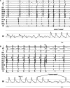
The Frog Sign Revisited
- PMID: 36605297
- PMCID: PMC9635569
- DOI: 10.19102/icrm.2022.13101
The Frog Sign Revisited
Abstract
The frog sign is a classic physical examination finding of typical atrioventricular nodal re-entrant tachycardia. We present the case of a 78-year-old man with recurrent, symptomatic supraventricular tachycardia referred for ablation in whom the frog sign was observed during physical examination.
Keywords: Frog sign; right atrial pressure; typical atrioventricular nodal re-entrant tachycardia.
Copyright: © 2022 Innovations in Cardiac Rhythm Management.
Conflict of interest statement
The authors report no conflicts of interest for the published content. No funding information was provided.
Figures


References
-
- Gursoy S, Steurer G, Brugada J, Andries E, Brugada P. Brief report: The hemodynamic mechanism of pounding in the neck in atrioventricular nodal reentrant tachycardia. N Engl J Med. 1992;327(11):772–774. [CrossRef] [PubMed] - DOI - PubMed
-
- Padanilam BJ, Manfredi JA, Steinberg LA, Olson JA, Fogel RI, Prystowsky EN. Differentiating junctional tachycardia and atrioventricular node re-entry tachycardia based on response to atrial extrastimulus pacing. J Am Coll Cardiol. 2008;52(21):1711–1717. [CrossRef] [PubMed] - DOI - PubMed
-
- Al Mahammeed ST, Buxton AE, Michaud GF. New criteria during right ventricular pacing to determine the mechanism of supraventricular tachycardia. Circ Arrhythm Electrophysiol. 2010;3(6):578–584. [CrossRef] [PubMed] - DOI - PubMed
-
- Dandamudi G, Mokabberi R, Assal C, et al. A novel approach to differentiating orthodromic reciprocating tachycardia from atrioventricular nodal reentrant tachycardia. Heart Rhythm. 2010;7(9):1326–1329. [CrossRef] [PubMed] - DOI - PubMed
-
- Knight B, Zivin A, Souza J, et al. A technique for the rapid diagnosis of atrial tachycardia in the electrophysiology laboratory. J Am Coll Cardiol. 1999;33(3):775–781. [CrossRef] [PubMed] - DOI - PubMed
Publication types
LinkOut - more resources
Full Text Sources
