Review
. 2022 Dec 20:16:1083159.
doi: 10.3389/fncel.2022.1083159. eCollection 2022.

Functions and dysfunctions of oligodendrocytes in neurodegenerative diseases

Affiliations
Review

Functions and dysfunctions of oligodendrocytes in neurodegenerative diseases

Seungwan Han et al. Front Cell Neurosci. .

Abstract

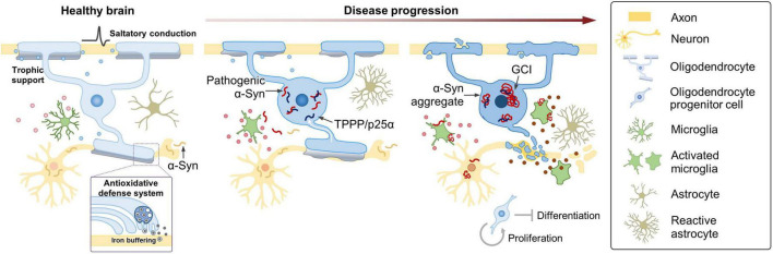
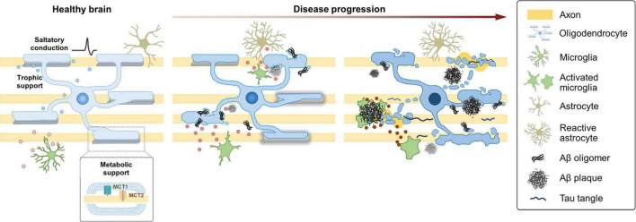
Neurodegenerative diseases (NDDs) are characterized by the progressive loss of selectively vulnerable populations of neurons, which is responsible for the clinical symptoms. Although degeneration of neurons is a prominent feature that undoubtedly contributes to and defines NDD pathology, it is now clear that neuronal cell death is by no means mediated solely by cell-autonomous mechanisms. Oligodendrocytes (OLs), the myelinating cells of the central nervous system (CNS), enable rapid transmission of electrical signals and provide metabolic and trophic support to neurons. Recent evidence suggests that OLs and their progenitor population play a role in the onset and progression of NDDs. In this review, we discuss emerging evidence suggesting a role of OL lineage cells in the pathogenesis of age-related NDDs. We start with multiple system atrophy, an NDD with a well-known oligodendroglial pathology, and then discuss Alzheimer's disease (AD) and Parkinson's disease (PD), NDDs which have been thought of as neuronal origins. Understanding the functions and dysfunctions of OLs might lead to the advent of disease-modifying strategies against NDDs.

Keywords: Alzheimer’s disease; Parkinson’s disease; multiple system atrophy; neurodegenerative disease; oligodendrocyte.


Conflict of interest statement

The authors declare that the research was conducted in the absence of any commercial or financial relationships that could be construed as a potential conflict of interest.

Figures

FIGURE 1
FIGURE 1
Oligodendroglial pathology and possible mechanisms contributing to MSA. Under healthy conditions, OLs enable saltatory conduction of electrical signals, provide metabolic and trophic support, and play a role in providing an antioxidant defense system to neurons. In MSA, TPPP/p25α relocates from the myelin sheath to the cell body, prior to α-Syn aggregation. Redistribution of TPPP/p25α causes cellular enlargement, and α-Syn starts to accumulate in the enlarged cytoplasm. Metabolic and trophic support of OLs to neurons is compromised, iron overload increases, and oxidative stress becomes aggravated. At later stages, α-Syn aggregates form GCIs in OLs. α-Syn is transformed into a toxic form in the intracellular environment of OL, where α-Syn seems to acquire distinct characteristics of MSA. MSA-type α-Syn might be transferred to neurons, and pathogenic α-Syn may induce neurodegeneration by directly interfering with essential neuronal functions or indirectly via aberrant activation of micro- and astrogliosis. As the disease progresses, microglia become destructive by releasing neurotoxic inflammatory mediators, such as cytokines, chemokines, and reactive oxygen and nitrogen species. The changing state of microglia, reflected in part, by their secretory profile, might play a crucial role in the progression of MSA. Enhanced proliferation of OPCs occurs, perhaps in an attempt to compensate for the myelin loss, but under the burden of α-Syn, immature oligodendroglia fail to differentiate and undergo cell death.
FIGURE 2
FIGURE 2
Dynamic changes of oligodendroglia during the progression of AD. Under healthy conditions, OLs enable saltatory conduction of electrical signals and provide metabolic and trophic support to neurons. In the AD brain, OLs are vulnerable to neurodegenerative conditions and acquire a distinct cellular state in response to the changing microenvironment. Under mild Aβ exposure, OLs alter gene expression and start to exhibit myelin defects. As the disease progresses, defects in OL function and myelin loss become more evident in correlation with the increased Aβ burden. Perturbation of myelin-axon coupling affects signal propagation as well as metabolic and trophic support. At later stages with higher Aβ burden, a specific subgroup of OLs express C4b and Serpina3n. Some aspects of the late-stage OLs might be shared across several NDDs. As AD progresses, microglia also become destructive by releasing neurotoxic inflammatory mediators, such as cytokines, chemokines, and reactive oxygen and nitrogen species. The changing state of microglia, reflected in part, by their secretory profile, might play a crucial role in the progression of AD.

Similar articles

Cited by

References

    1. Agarwal D., Sandor C., Volpato V., Caffrey T. M., Monzón-Sandoval J., Bowden R., et al. (2020). A single-cell atlas of the human substantia nigra reveals cell-specific pathways associated with neurological disorders. Nat. Commun. 11:4183. 10.1038/s41467-020-17876-0 - DOI - PMC - PubMed
    1. Agosta F., Canu E., Stefanova E., Sarro L., Tomiæ A., Špica V., et al. (2014). Mild cognitive impairment in Parkinson’s disease is associated with a distributed pattern of brain white matter damage. Hum. Brain Mapp. 35 1921–1929. 10.1002/hbm.22302 - DOI - PMC - PubMed
    1. Ahmed Z., Asi Y. T., Lees A. J., Revesz T., Holton J. L. (2013). Identification and quantification of oligodendrocyte precursor cells in multiple system atrophy, progressive supranuclear palsy and Parkinson’s disease. Brain Pathol. 23 263–273. 10.1111/j.1750-3639.2012.00637.x - DOI - PMC - PubMed
    1. Akay L. A., Effenberger A. H., Tsai L. H. (2021). Cell of all trades: oligodendrocyte precursor cells in synaptic, vascular, and immune function. Genes Dev. 35 180–198. 10.1101/gad.344218.120 - DOI - PMC - PubMed
    1. Allen N. J., Lyons D. A. (2018). Glia as architects of central nervous system formation and function. Science 362 181–185. 10.1126/science.aat0473 - DOI - PMC - PubMed

LinkOut - more resources