Review
. 2022 Aug 1;17(3):9-21.
doi: 10.51866/rv.37. eCollection 2022 Nov 30.

Overview of common oral lesions

Affiliations
Review

Overview of common oral lesions

Esha Zahid et al. Malays Fam Physician. .

Abstract

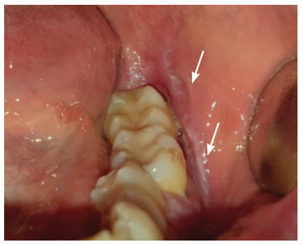
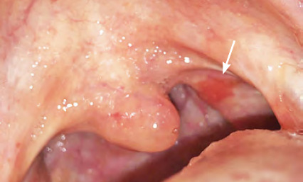
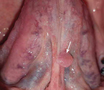
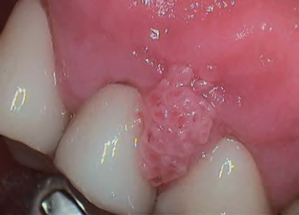
This article summarises common oral lesions that clinicians may face in everyday practice by categorising them by clinical presentation: ulcerated lesions, white or mixed white-red lesions, lumps and bumps, and pigmented lesions. The pathologies covered include recurrent aphthous stomatitis, herpes simplex virus, oral squamous cell carcinoma, geographic tongue, oral candidosis, oral lichen planus, pre-malignant disorders, pyogenic granuloma, mucocele and squamous cell papilloma, oral melanoma, hairy tongue and amalgam tattoo. The objective of this review is to improve clinician knowledge and confidence in assessing and managing common oral lesions presenting in the primary care setting.

Keywords: Dentistry; Oral lesions; Oral medicine; Primary health care.


Figures

Figure 1
Figure 1. Minor recurrent aphthous ulcer on right floor of mouth
Figure 2
Figure 2. Major recurrent aphthous ulcer on the palate
Figure 3
Figure 3. Primary herpetic stomatitis — lesions on upper and lower lip
Figure 4
Figure 4. Primary herpetic stomatitis - lesion on tongue
Figure 5
Figure 5. Herpes labialis of the lower left lip (crusted lesion)
Figure 6
Figure 6. Squamous cell carcinoma of the right pterygomandibular region
Figure 7
Figure 7. Squamous cell carcinoma of the left ventral surface of the tongue
Figure 8
Figure 8. Pseudomembranous candidosis of the tongue
Figure 9
Figure 9. Pseudomembranous candidosis of the hard palate (confirmed by oral swab)
Figure 10
Figure 10. Erythematous candidosis of the hard palate
Figure 11
Figure 11. Hyperplastic candidosis associated within the edentulous ridge 47-48-retromolar region in a diabetic with poor denture hygiene
Figure 12
Figure 12. Denture-induced stomatitis
Figures 13 and 14
Figures 13 and 14. Reticular form of oral lichen planus on buccal mucosa
Figures 13 and 14
Figures 13 and 14. Reticular form of oral lichen planus on buccal mucosa
Figure 15
Figure 15. Erosive oral lichen planus
Figures 16 and 17
Figures 16 and 17. Gingival oral lichen planus areas of desquamation or loss of the epithelial surface appearing intensely red — desquamative gingivitis
Figures 16 and 17
Figures 16 and 17. Gingival oral lichen planus areas of desquamation or loss of the epithelial surface appearing intensely red — desquamative gingivitis
Figures 18 and 19
Figures 18 and 19. Geographic tongue on dorsal and lateral surfaces of the tongue
Figures 18 and 19
Figures 18 and 19. Geographic tongue on dorsal and lateral surfaces of the tongue
Figure 20
Figure 20. Leukoplakia (biopsied with diagnosis of oral lichen planus and secondary oral candidosis)
Figure 21
Figure 21. Erythroplakia
Figure 22
Figure 22. Submucosal fibrosis in a patient who used betel leaf frequently (the pallor of the soft palate is an extensive fibrous change within the submucosa)
Figure 23
Figure 23. Dry and fissured lips (called cheilitis) secondary to oral candidosis with solar elastosis (actinic cheilitis)
Figure 24
Figure 24. Mucocele on lower lip
Figure 25
Figure 25. Squamous papilloma of the lingual frenum
Figure 26
Figure 26. Pyogenic granuloma
Figure 27
Figure 27. Oral melanoma
Figure 28
Figure 28. Amalgam tattoo biopsied of left buccal mucosa
Figure 29
Figure 29. Hairy tongue on dorsal surface of tongue

Similar articles

Cited by

References

    1. Akintoye SO, Greenberg MS. Recurrent aphthous stomatitis. Dent Clin North Am. 2014;58(2):281–297. doi: 10.1016/j.cden.2013.12.002. - DOI - PMC - PubMed
    1. Tarakji B, Gazal G, Al-Maweri S, Azzeghaiby SN, Alaizari N. Guideline for the diagnosis and treatment of recurrent aphthous stomatitis for dental practitioners. J In Oral Health. 2015 May;7(5):74–80. - PMC - PubMed
    1. Edgar N, Saleh D, Miller R. Recurrent aphthous stomatitis: a review. J Clin Aesthet Dermatol. 2017;10(3):26–36. - PMC - PubMed
    1. Montgomery-Cranny JA, Wallace A, Rogers HJ, Hughes SC, Hegarty AM, Zaitoun H. Management of Recurrent Aphthous Stomatitis in Children. Dent Update. 2015;42(6):564–572. doi: 10.12968/denu.2015.42.6.564. - DOI - PubMed
    1. Xu F, Schillinger JA, Sternberg MR, et al. Seroprevalence and coinfection with herpes simplex virus type 1 and type 2 in the United States, 1988-1994. J Infect Dis. 2002;185(8):1019–1024. doi: 10.1086/340041. - DOI - PubMed

LinkOut - more resources