. 2023 Jan 6;80(1):29.
doi: 10.1007/s00018-022-04666-8.

Galactic cosmic radiation exposure causes multifaceted neurocognitive impairments

Affiliations

Galactic cosmic radiation exposure causes multifaceted neurocognitive impairments

Yasaman Alaghband et al. Cell Mol Life Sci. .

Abstract

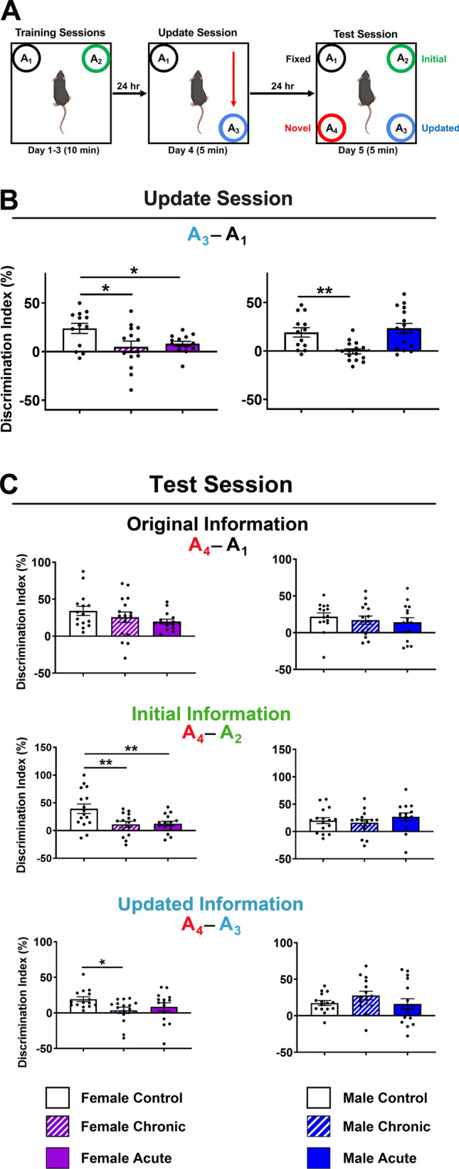
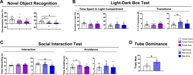
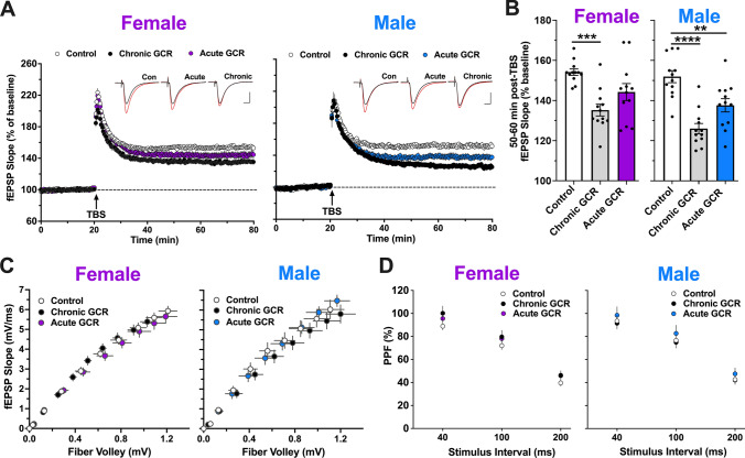
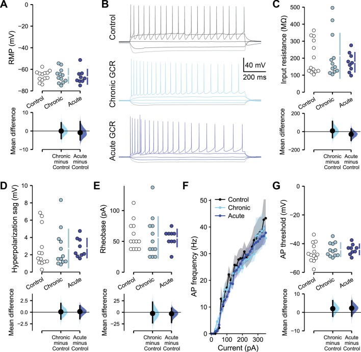
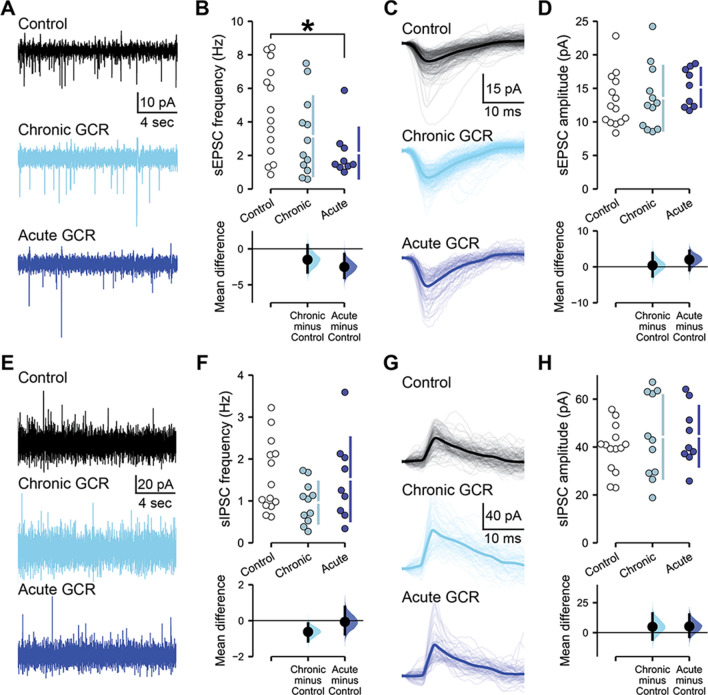
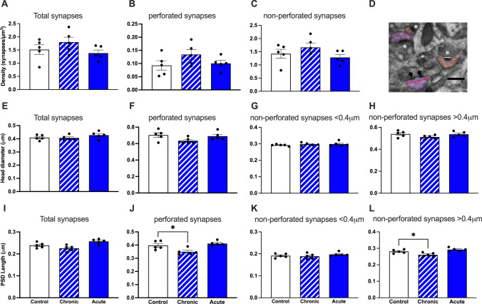
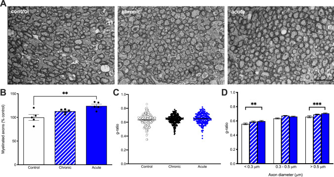
Technological advancements have facilitated the implementation of realistic, terrestrial-based complex 33-beam galactic cosmic radiation simulations (GCR Sim) to now probe central nervous system functionality. This work expands considerably on prior, simplified GCR simulations, yielding new insights into responses of male and female mice exposed to 40-50 cGy acute or chronic radiations relevant to deep space travel. Results of the object in updated location task suggested that exposure to acute or chronic GCR Sim induced persistent impairments in hippocampus-dependent memory formation and reconsolidation in female mice that did not manifest robustly in irradiated male mice. Interestingly, irradiated male mice, but not females, were impaired in novel object recognition and chronically irradiated males exhibited increased aggressive behavior on the tube dominance test. Electrophysiology studies used to evaluate synaptic plasticity in the hippocampal CA1 region revealed significant reductions in long-term potentiation after each irradiation paradigm in both sexes. Interestingly, network-level disruptions did not translate to altered intrinsic electrophysiological properties of CA1 pyramidal cells, whereas acute exposures caused modest drops in excitatory synaptic signaling in males. Ultrastructural analyses of CA1 synapses found smaller postsynaptic densities in larger spines of chronically exposed mice compared to controls and acutely exposed mice. Myelination was also affected by GCR Sim with acutely exposed mice exhibiting an increase in the percent of myelinated axons; however, the myelin sheathes on small calibur (< 0.3 mm) and larger (> 0.5 mm) axons were thinner when compared to controls. Present findings might have been predicted based on previous studies using single and mixed beam exposures and provide further evidence that space-relevant radiation exposures disrupt critical cognitive processes and underlying neuronal network-level plasticity, albeit not to the extent that might have been previously predicted.

Keywords: Cognitive dysfunction; Electrophysiology; Space radiation; Synaptic plasticity.


Conflict of interest statement

The authors have no conflict of interest or competing interests to disclose.

Figures

Fig. 1
Fig. 1
Study design. Single cohorts of 178 wild-type and 91 female C57BL/6J were randomly divided into 3 experimental groups: sham irradiated controls, and acutely or chronically irradiated using the 33-beam GCR Sim protocol [59–60 male mice and 30–31 female mice for each irradiation paradigm]. Chronically irradiated animals received a GCR Sim dose of 2.08 cGy/day, 6 days/week for 4 weeks (24 irradiation days; total accumulated dose ~ 50 cGy). Acutely irradiated animals received a single total GCR Sim dose on same day that the chronically irradiated mice received their final exposure (~ 40 cGy over ~ 2 h). Within 5 days post-irradiation mice were shipped to their respective institutions (i.e., Harvard, UC Irvine, Stanford). Animals were acclimated at least 2 months prior to behavioral, electrophysiological or structural analyses. (IRR, irradiation; OFT, open-field testing; EM, electron microscopy; USUHS, Uniformed Services University of Health Sciences; LTP, long-term potentiation; OUL, object in updated location; NOR, novel object recognition; LDB, light–dark box; FE, fear extinction)
Fig. 2
Fig. 2
Exposure to simulated GCR elicits impairments in memory formation and updating. A Experimental design. All objects were identical aside from location. B Female mice exposed to chronic or acute GCR Sim exhibited significantly lower discrimination indices (DI) relative to controls during the update session, demonstrating no preference for the object in the updated location (A3) as compared to the fixed location object (A1). Only chronically irradiated male mice were impaired in update session performance. C During the test session, female and male mice exposed to either GCR Sim paradigm retained the memory of the fixed location (A1) relative to the novel location (A4), exhibiting DI scores similar to control animals (top panels). The irradiated male mice also retained the memory of the initial location (A2) relative to the novel location (A4), but both chronically and acutely irradiated females showed significantly lower DIs relative to control animals (middle panels). Similarly, irradiated male mice retained the memory of the updated location (A3) relative to the novel location (A4), but the chronically irradiated female mice showed significantly lower DIs relative to control animals. Data are mean ± SEM (N = 13–16 per group); P values derived from one-way ANOVA followed by a Bonferroni’s multiple comparison test. *P < 0.05, **P < 0.01
Fig. 3
Fig. 3
GCR Sim exposures induce memory impairments and changes in social behaviors. A Novel Object Recognition (NOR) testing indicated that chronically and acutely exposed males show significantly reduced discrimination index scores relative to controls, indicating no preference for the novel object. Acutely irradiated GCR females showed a trend toward reduced discrimination index scores compared to controls. B Using time spent in the light compartment of the light–dark box test (LDB) as a measure of anxiety-like behavior, none of the GCR-exposed animals were affected (left panel). Evaluation of numbers of transitions between the light and dark compartments on the LDB could suggest that chronic GCR males showed elevated number of transitions possibly suggesting increased frantic, anxiety-like behavior although analysis of time spent in each compartment do not support this conclusion (right panel). C During social interaction test (SIT), only acutely irradiated males showed reductions in social interactions with a novel mouse (left panel), while chronically irradiated females exhibited avoidance behavior (right panel). D Male mice exposed to chronic GCR Sim showed an increase in trials won compared with unirradiated control mice when tested on the Tube Dominance behavioral task, suggesting an increase in aggressive behavior. For NOR, LDB and SIT, data are the mean ± SEM (N = 13–16 mice/group); P values derived from one-way ANOVA followed by a Bonferroni’s multiple comparison test. For the Tube Dominance test, data are the mean ± SEM (N = 8 per group); P values derived from unpaired t-test. *P < 0.05, **P < 0.01
Fig. 4
Fig. 4
Hippocampal long-term synaptic plasticity is perturbed by GCR Sim exposure. Extracellular field recordings of CA1 dorsal hippocampus apical dendrite responses to Schaffer collateral stimulation at 5 months following completion of chronic and acute GCR Sim exposures. A Following a stable 20 min baseline recording, a single train of theta burst stimulation (TBS; arrow) was applied, and then, recordings were continued for an additional 60 min. The time course shows that TBS-induced long-term potentiation (LTP) was markedly reduced in slices from chronically irradiated female and both groups of GCR Sim-exposed male mice compared with slices from respective control animals. Representative traces collected during baseline (inset; black line) and 60 min post-TBS (red line). Scale bars indicate 0.4 mV/5 ms. B Chronically GCR Sim-exposed female mice showed a marked reduction in LTP at 60 min post-TBS relative to control mice (left). Field excitatory postsynaptic potential (fEPSP) slope was significantly reduced 60 min post-TBS in slices from both chronically and acutely GCR-exposed male mice (right). C The relationships between stimulation current and fEPSP slope were not detectably different between groups. D Transmitter release kinetics, as assessed with paired pulse facilitation (PPF), were also comparable among all animals. Data are mean ± SEM (total N = 6 mice per group; 1 slice/hemisphere per mouse); P values for mean potentiation and fEPSP slope derived from one-way ANOVA followed by a Bonferroni’s multiple comparison test. P values for PPF derived from two-way ANOVA. **P < 0.01, ***P < 0.001. ****P < 0.0001
Fig. 5
Fig. 5
GCR Sim exposure does not alter the intrinsic electrophysiological properties of CA1 pyramidal neurons in male mice. All data are from whole cell current clamp recordings of CA1 pyramidal neurons from the superficial layer of the dorsal hippocampus, 2–4 months following either chronic or acute exposures of male mice to GCR Sim. A Resting membrane potential (RMP) was unchanged between groups. B Representative examples of responses to a range of brief current injections in control and irradiated neurons. There was no alteration in the input resistance (C), sag during a − 100 pA hyperpolarizing current injection (D) or rheobase current required to evoke an action potential (E) between groups. F Action potential (AP) frequency remained equivalent across a range of current injections and G the threshold potential for action potential initiation remained unchanged. Data are Control: 5 animals, 13 cells; Chronic: 5 animals, 12 cells; acute: 4 animals, 9 cells. A, C–E, G Cumming estimation plots show raw data on the top axis and a bootstrapped sampling distribution on the bottom axis; black dots depict the mean difference between groups and the 95% confidence interval is indicated by the ends of the vertical black bars. F Data are mean ± SEM. P values derived from linear mixed-effect model regression or two-way ANOVA
Fig. 6
Fig. 6
Acute exposure to GCR Sim preferentially suppresses excitatory synaptic signaling in male mice. All data are from whole cell voltage clamp recordings of CA1 pyramidal neurons from the superficial layer of the dorsal hippocampus, 2–4 months following either chronic or acute irradiations of male mice. A Representative examples of spontaneous excitatory postsynaptic current (sEPSC) recordings from control and GCR Sim-exposed neurons. B The frequency of sEPSCs was reduced in male mice following acute irradiation. C Aligned examples of sEPSCs in representative control and irradiated neurons. Light lines show individual sEPSCs, while darker lines display the average sEPSC during a 200 s recording from that neuron. D sEPSC amplitude remained similar between groups. E Representative examples of spontaneous inhibitory postsynaptic current (sIPSC) recordings from control and GCR Sim-exposed neurons. F Frequency and G, H amplitude of sIPSCs was equivalent between groups. Data are Control: 5 animals, 14 cells; Chronic: 5 animals, 12 cells (11 cells for sIPSCs); Acute: 4 animals, 9 cells. B, D, F, G Cumming estimation plots show raw data on the top axis and a bootstrapped sampling distribution on the bottom axis; black dots depict the mean difference between groups and the 95% confidence interval is indicated by the ends of the vertical black bars. P values derived from linear mixed-effect model regression. *P < 0.05
Fig. 7
Fig. 7
GCR Sim exposure alters PSD length in large, > 0.4 mm diameter synapses in CA1 pyramidal neurons of male mice. There were no significant changes in total synapse density, perforated synapse density or non-perforated synapse density compared to controls. A total synapse density, B perforated synapse density and C non-perforated synapse density. D Representative electron micrograph depicting non-perforated synapses (white asterisks), perforated synapses (arrow heads) and measurements of PSD length (white line) and head diameter (red line). Scale bar = 500 nm. E–H HD measurements for all synapses, perforated synapses, non-perforated synapses < 0.4 mm and non-perforated synapses > 0.4 mm, respectively. I PSD length in all synapses showed smaller PSDs in chronic GCR mice. J Perforated synapse PSD lengths were significantly reduced following chronic GCR exposure. K PSD lengths in non-perforated synapses < 0.4 mm in HD. L Non-perforated synapses > 0.4 mm in HD were significantly reduces in chronically exposed GCR mice. N = 5 mice per group. Data are mean ± SEM, one-way ANOVA *P < 0.05, **P < 0.01
Fig. 8
Fig. 8
Acute exposure to GCR Sim results in myelin degeneration in male mice. A Representative images from the corpus callosum from control (N = 5 mice; N = 4303 axons), chronic (N = 5 mice; N = 4868 axons) and acute (N = 5 mice; N = 5343 axons) GCR-exposed mice. Scale bar = 2 mm. B There was an increase in the percent of myelinated axons following acute irradiation with no differences between chronic irradiation and controls. C There is no significant difference in overall g-ratios in chronic or acute irradiated mice compared to controls. D Acute irradiation results in less myelin in the smallest (< 0.3 mm) and largest axons (> 0.5 mm). N = 5 mice per group. P values derived from linear mixed-effect model regression. *P < 0.05

References

    1. Cucinotta F, Alp M, Sulzman F, Wang M. Space radiation risks to the central nervous system. Life Sci Space Res. 2014;2:54–69. doi: 10.1016/j.lssr.2014.06.003. - DOI
    1. Ehresmann B, et al. Charged particle spectra measured during the transit to mars with the mars science laboratory radiation assessment detector (MSL/RAD) Life Sci Space Res (Amst) 2016;10:29–37. doi: 10.1016/j.lssr.2016.07.001. - DOI - PubMed
    1. Klein PM, et al. Detrimental impacts of mixed-ion radiation on nervous system function. Neurobiol Dis. 2021;151:105252. doi: 10.1016/j.nbd.2021.105252. - DOI - PubMed
    1. Keiser AA, et al. Systemic HDAC3 inhibition ameliorates impairments in synaptic plasticity caused by simulated galactic cosmic radiation exposure in male mice. Neurobiol Learn Mem. 2021;178:107367. doi: 10.1016/j.nlm.2020.107367. - DOI - PMC - PubMed
    1. Parihar VK et al (2015) What happens to your brain on the way to Mars. Sci Adv 1 - PMC - PubMed