. 2023 Jan 5;186(1):80-97.e26.
doi: 10.1016/j.cell.2022.12.004.

Glucose dissociates DDX21 dimers to regulate mRNA splicing and tissue differentiation

Affiliations

Glucose dissociates DDX21 dimers to regulate mRNA splicing and tissue differentiation

Weili Miao et al. Cell. .

Abstract

Glucose is a universal bioenergy source; however, its role in controlling protein interactions is unappreciated, as are its actions during differentiation-associated intracellular glucose elevation. Azido-glucose click chemistry identified glucose binding to a variety of RNA binding proteins (RBPs), including the DDX21 RNA helicase, which was found to be essential for epidermal differentiation. Glucose bound the ATP-binding domain of DDX21, altering protein conformation, inhibiting helicase activity, and dissociating DDX21 dimers. Glucose elevation during differentiation was associated with DDX21 re-localization from the nucleolus to the nucleoplasm where DDX21 assembled into larger protein complexes containing RNA splicing factors. DDX21 localized to specific SCUGSDGC motif in mRNA introns in a glucose-dependent manner and promoted the splicing of key pro-differentiation genes, including GRHL3, KLF4, OVOL1, and RBPJ. These findings uncover a biochemical mechanism of action for glucose in modulating the dimerization and function of an RNA helicase essential for tissue differentiation.

Keywords: DDX21; RNA helicases; glucose; mRNA splicing; tissue differentiation.


Conflict of interest statement

Declaration of interests The authors declare no competing interests.

Figures
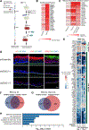
Figure 1.
Figure 1.. Glucose binds to DDX21 and DDX21 is essential for epidermal differentiation.
(A) Workflow for the discovery of glucose binding proteins by MS using either dextran column (left) or azido glucose (right). (B) Heatmap of the top enriched proteins among the 91 commonly enriched proteins. (C) Heatmap of the top enriched helicases in the dextran column. (D) Keratin 1 (K1), Keratin 10 (K10), DDX21 and Collagen VII (Col7) staining in skin tissue organoids comparing DDX21 shRNA with control shRNA; dotted line, basement membrane. (E) Changes in progenitor and differentiation markers upon DDX21 loss in differentiated cells and skin tissue organoids (RNA-seq); differentiation genes shown include all genes with a GO term of keratinocyte differentiation, regulation of keratinocyte differentiation, or keratinization, with an FDR<0.05 in ≥4 biological replicates. Venn diagram showing the overlap of down-regulated genes (FDR<0.05) between the two shDDX21 in RNA-seq of differentiated cells (F) and organotypic human epidermal tissue (G). (H) Adjusted p-values of biological process GO terms for RNAs down-regulated in RNA-seq of organotypic human epidermal tissue (178 genes from Figure 1G).
Figure 2.
Figure 2.. Glucose binds to the ATP binding site of DDX21 and changes DDX21 protein conformation.
(A) MST of DDX21’s binding affinity for glucose ±1 mM ATP. (B) MST of DDX21 binding to ATP ±350 μM glucose or galactose. (C) MST of glucose binding to DDX21 WT, K236R and K236G mutants. (D) MST of ATP-binding affinities of DDX21 WT, K236R and K236G mutants. (E) Helicase activity of DDX21 ±galactose or ±glucose. (F) Molecular docking by UCSF Chimera shows the predicted binding site of glucose to DDX21 (PDB: 6L5N). (G) Relative abundance of the peptides with no glucose modification, calculated from the intensity obtained from 350 μM glucose/no-glucose, both with UVC crosslinking (top 15 most abundant peptides). (H) Selected ion chromatography (SIC) and MS of the targeted peptide of DDX21 (ELANQVSK) with (red) or without (black) glucose incubation during UVC crosslinking. 10 ppm mass tolerance was applied to plot SIC. *, MS peak for the glucose crosslinked peptide. (I) MST analysis of glucose binding to DDX21 WT and V276W mutant. (J)-(K) BS3-induced protein sites crosslinked in PBS, 1 mM ATP or 350 μM glucose. Crosslinking sites within 5 amino acids are marked as the same site. The black lines and boxes in the diagram represent BS3-induced crosslinks within DDX21 and the red lines and boxes highlight the crosslinks near ATP-binding domain of DDX21. Error bars, standard deviations from ≥3 biological replicates. p-values, two-tailed student’s t-test or Welch’s t-test.
Figure 3.
Figure 3.. Glucose inhibits DDX21 dimerization and re-localizes DDX21 to the nucleoplasm.
(A) MST of DDX21 dimerization ±350 μM glucose or galactose. (B) MST of the effects of DDX21 mutations on DDX21 dimerization. (C) The dimer/monomer ratio of DDX21 ±350 μM glucose or galactose, as obtained from HA Western blot analysis of blue native gels. (D) PLA signal for the interaction between FHH-DDX21 and V5-DDX21 (dimerization) in progenitor, differentiated cells maintained in low or normal glucose. (E) MST of dimerization mutant DDX21611R binding to ATP ±350 μM glucose. (F) Percentage of cells with DDX21 WT or dimerization mutants localized in the nucleoplasm in HEK293T or keratinocyte progenitors. (G) Immunofluorescence of HA-DDX21 WT and dimerization mutants in HEK293T cells. (H) Immunofluorescence of HA-DDX21 in keratinocyte progenitor, differentiated cells grown in normal or low glucose. Green, anti-HA signal. (I) Western blot of nucleolus fractionation in progenitor, differentiated cells grown in normal or low glucose. Cp, cytoplasm; Np, nucleoplasm; No, nucleolus. (J) Relative FHH-DDX21 abundance in the nucleoplasm (Np)/nucleolus (No) from Western blot. (K) Percentage of cells with nucleoplasmic FHH-DDX21 in three conditions: progenitor keratinocytes, differentiated cells grown in low or normal glucose. Error bars, standard deviations from ≥3 biological replicates. p-values, two-tailed student’s t-test or Welch’s t-test.
Figure 4.
Figure 4.. DDX21-enabled differentiation is ATPase-independent and requires glucose binding.
(A) NanoSIMS of 13C and 2H in progenitor and differentiated keratinocytes cultured with 13C6 glucose and 2H4 ATP (top two rows) or light glucose and ATP (bottom two rows). Intensities represent the relative counts normalized to total carbon counts (12C+13C). (B) Relative counts normalized to total carbon counts (12C+13C) for 2H and 13C in progenitor and differentiated cells. The nucleus was defined by 31P signal that was collected simultaneously. (C) Keratin 1 (K1, red) and Keratin 10 (K10, green) staining in organotypic human epidermal tissue, comparing DDX21 shRNA with control shRNA, and DDX21 shRNA with WT or mutant DDX21 rescue; dotted line, basement membrane. (D) qPCR of progenitor and differentiation markers in control shRNA, DDX21 shRNA, and DDX21 shRNA with WT or mutant DDX21 rescue in organotypic human epidermal tissue. Data is normalized to DDX21 shRNA with GFP control. (E) qPCR of progenitor and differentiation markers in primary keratinocytes after gene editing at the endogenous DDX21 gene locus with K236R, K236G and 611R mutations, grown as organotypic human epidermal tissue. Data represents the mean from ≥2 replicates. Mutants are normalized to their respective control edit; that is, K236K for K236R and K236G, and 611LAAALA for 611RRRRRR.
Figure 5.
Figure 5.. Glucose transitions DDX21 into a larger complex containing RNA splicing factors.
(A) Western blot of HA-DDX21 and HNRNPC in FPLC from 13.75 to 19 mL ±350 μM glucose incubation in the high molecular weight (HMW) fraction. (B) Distribution of DDX21 protein in FPLC, ±350 μM glucose in the HMW fraction from Western blot. (C) Overlap of enriched genes in progenitor, differentiated cell maintained in low or normal glucose in BioID hits, each normalized to their corresponding GFP control. (D) Cell components enrichment in proteins from DDX21 proximal proteomes. P & L, proteins enriched only in progenitor and low glucose cells (n=194); N, proteins enriched only in normal glucose cells (n=67). (E) Correlation between the DDX21/GFP enrichment ratios obtained via LC-MS/MS from low and normal glucose in differentiated cells among 472 total enriched genes. Adjusted p-values of top biological process GO terms enriched in DDX21 proximal proteomes in low glucose compared to normal glucose (F), or normal glucose compared to low glucose (G). (H) Western blot of HNRNPUL1 after HA-DDX21 immunoprecipitation in progenitor, differentiated cell grown in normal or low glucose. (I) Relative enrichment of HNRNPUL1 in normal glucose compared to low glucose from Western blot. (J) Distribution of DDX21 FPLC from 13.75 to 17.25 mL in the HMW fraction in differentiated cells grown in normal or low glucose from Western blot. Error bars, standard deviations from ≥3 biological replicates. p-values, two-tailed student’s t-test.
Figure 6.
Figure 6.. Glucose regulates DDX21 to bind to RNA introns.
(A) Motif identified by HOMER de novo searching of DDX21 CLIP-seq peaks in differentiated keratinocytes grown in normal glucose. (B) MST analysis of DDX21 binding to the SCUGSDGC (D: A, G or U; S: C or G) motif RNA (“CLIP RNA”), G4 RNA or a random RNA sequence. (C) Percentage of the indicated RNA binding to DDX21 in progenitor, differentiated cells grown in normal or low glucose in easyCLIP-seq. (D) Percentage of DDX21 CLIP-seq reads at RNA introns in progenitor cells, differentiated cells grown in low or normal glucose. (E) Binding of DDX21 to the JUP and GRHL3 RNAs in differentiated keratinocytes grown in low (red) or normal (black) glucose. The SCUGSDGC motif and %GC are plotted as smoothed lines below in green and blue, respectively. (F) Binding of DDX21 to the indicated RNA in progenitor, low or normal glucose in differentiated cells. (G) Percentage of the indicated RNA binding for DDX21 WT and mutants in HEK293T cells by easyCLIP-seq. (H) Binding of DDX21 WT and mutants to the indicated RNA in HEK293T cells. Error bars, standard deviations from ≥3 biological replicates. p-values, two-tailed student’s t-test. FOX2 is a splicing factor control.
Figure 7.
Figure 7.. DDX21 regulates mRNA splicing.
Traces of easyCLIP signal (A) and box plot of total signal (B) around exons differentially expressed between differentiated cells grown in low or normal glucose (top, FDR<0.1, ≥10% difference) or around exons differentially skipped between shScramble- and shDDX21-treated cells (bottom, FDR<0.15, ≥10% difference). The p-values at ±1 introns in (A) are relative to ±2 introns. (C) The top two forms of altered splicing upon shDDX21 loss. The number of differential events at FDR<0.2 is given for shDDX21 knockdown, followed by the percentage of those events rescued (FDR<0.2) by WT or K236R. (D) Averaged coverage of the SCUGSDGC motif around exons differentially skipped upon differentiation (FDR<0.05, top), or upon glucose restriction (FDR<0.05, bottom). The x-axis denotes the last 50 nt of the upstream exon, the first and last 250 nt of the upstream intron, the first and last 50 nt of the skipped exon. Traces are the average number of nucleotides covered by the motif in a 50 nt window excluding the first and last 2 bases of introns. Densities of 100 control motifs with nucleotide-frequency/length/degeneracy matched to the SCUGSDGC motif were calculated, and the 95% confidence interval (CI) fit independently at each point along the transcript, with the bounds of the 95% CI plotted in grey. The log10 p-value is the enrichment of motif coverage in the 50 nt window for the blue line cases vs non-differential exons; horizontal red line, p=0.05. (E) %Exon inclusion in ECM1 mRNA. All conditions except “Progenitor” are from differentiated cells. Numbers, average percentage from two biological replicates. (F) DDX21 binding across ECM1 mRNA in differentiated keratinocytes grown in low (red) or normal (black) glucose. Green lines, the SCUGSDGC motif (smoothed). (G) Exon usage changes between treatment with a control shRNA vs DDX21 loss (x-axis) and rescue with WT (black) or K236R (red) DDX21 vs DDX21 loss (y-axis). (H) Intron retention changes of progenitor and differentiation markers upon DDX21 loss or upon WT or K236R DDX21 rescue in differentiated cells. (I) qPCR of progenitor and differentiation markers in progenitor and differentiated cells with overexpressing FHH-GFP, FHH-GRHL3 or FHH-GRHL3-intron 13-retained. Data is normalized to FHH-GFP. Error bars, standard deviations from ≥3 biological replicates. p-values, Mann-Whitney U (two-sided).

Comment in

  • Sweet splicing.
    Carmo-Fonseca M. Carmo-Fonseca M. Cell. 2023 Jan 5;186(1):10-11. doi: 10.1016/j.cell.2022.11.025. Cell. 2023. PMID: 36608648

References

    1. Lopez-Pajares V, Yan K, Zarnegar BJ, Jameson KL, and Khavari PA (2013). Genetic pathways in disorders of epidermal differentiation. Trends in genetics : TIG 29, 31–40. 10.1016/j.tig.2012.10.005. - DOI - PMC - PubMed
    1. Bikle DD, Xie Z, and Tu C-L (2012). Calcium regulation of keratinocyte differentiation. Expert review of endocrinology & metabolism 7, 461–472. 10.1586/eem.12.34. - DOI - PMC - PubMed
    1. Fuchs E (2007). Scratching the surface of skin development. Nature 445, 834–842. 10.1038/nature05659. - DOI - PMC - PubMed
    1. Moreci RS, and Lechler T (2020). Epidermal structure and differentiation. Current Biology 30, R144–R149. 10.1016/j.cub.2020.01.004. - DOI - PubMed
    1. Hay RJ, Johns NE, Williams HC, Bolliger IW, Dellavalle RP, Margolis DJ, Marks R, Naldi L, Weinstock MA, Wulf SK, et al. (2014). The Global Burden of Skin Disease in 2010: An Analysis of the Prevalence and Impact of Skin Conditions. Journal of Investigative Dermatology 134, 1527–1534. 10.1038/jid.2013.446. - DOI - PubMed

Publication types

LinkOut - more resources