

Application of 3D printed pelvic fracture related urethra and surrounding tissue as preoperative planning model
- PMID: 36609237
- PMCID: PMC9824970
- DOI: 10.1186/s12894-022-01165-7
Application of 3D printed pelvic fracture related urethra and surrounding tissue as preoperative planning model
Abstract
Objective: Urethral stenosis caused by pelvic fracture urethral injury (PFUI) is a complex urological disease, especially for the redo cased. However, to find the proximal end of the posterior urethra, and to avoid injury to the rectum and to forecast to remove the inferior pubic margin are two key points for a successful surgery. These steps can be challenging for even the most experienced urologists. This study is to describe a new technique for understanding the three-dimensional (3D) anatomy of the urethra, which will also aid in surgical planning and simplify urethroplasty.
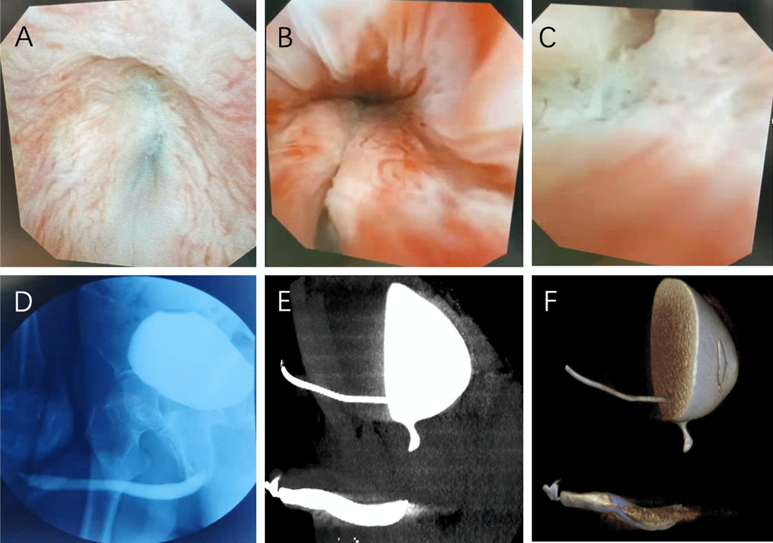
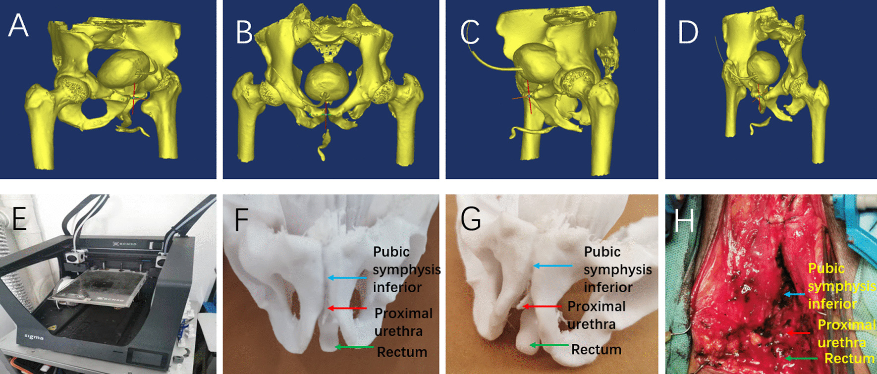
Materials and methods: Three patients underwent routine urethroscopy, X ray urethrography and contrast CT urethrography. The 3D images were then reconstructed, and the data were transmitted to a 3D printer. 3D models were printed with polyacrylic acid to simulate the anatomical structure and relationship of urethral stenosis with pubic symphysis and rectum. Various diagnosis methods were compared with the condition in surgery. The patients and trainee questionnaires were performed.
Results: Three models of urethral CT were obtained. These models were presented to patients and trainee doctors along with routine urethroscopy, urethrography, and urethral CT. The scores of patients and trainee question forms demonstrated that the 3D printed urethral stenosis model of pelvic fracture has obvious advantages in urethral adjacency and ease of understanding. The 3D printed urethras were easy to show the pubic symphysis and simulate its excision and exposure of urethra. The model could show the precise distance from urethra to rectum to prevent the rectum injury in surgery.
Conclusions: 3D printing technology can be applied to the preoperative evaluation of urethral stenosis caused by PFUI. It can be auxiliary to understand the anatomical structure of the posterior urethra, the direction of urethral displacement, protecting the rectum and the forecasting for pubectomy. It is especially helpful for the accurate preoperative planning of some complex urethral stenosis and redo cases.
Keywords: 3D printing; Pelvic fracture urethral injury; Urethral plasty; Urethral stenosis.
© 2023. The Author(s).
Conflict of interest statement
The authors declares that they have no competing interests.
Figures
Similar articles
-
The effects of primary realignment or suprapubic cystostomy on prostatic displacement in patients with pelvic fracture urethral injury: a clinical study based on MR urethrography.Injury. 2022 Feb;53(2):534-538. doi: 10.1016/j.injury.2021.09.050. Epub 2021 Oct 2. Injury. 2022. PMID: 34645564
-
Management of male pelvic fracture urethral injuries: Review and current topics.Int J Urol. 2019 Jun;26(6):596-607. doi: 10.1111/iju.13947. Epub 2019 Mar 20. Int J Urol. 2019. PMID: 30895658 Review.
-
Management of long-term functional sequelae of pelvic fracture urethral injury.Fr J Urol. 2024 Nov;34(10):102711. doi: 10.1016/j.fjurol.2024.102711. Epub 2024 Jul 27. Fr J Urol. 2024. PMID: 39074537 Review.
-
Predictors of elaborated perineal or a combined abdominoperineal approach during repair for pelvic fracture urethral injury.World J Urol. 2024 Jan 20;42(1):40. doi: 10.1007/s00345-023-04733-0. World J Urol. 2024. PMID: 38244107
-
[A CASE OF PELVIC FRACTURE URETHRAL INJURY RECONSTRUCTED BY DEFERRED URETHROPLASTY].Nihon Hinyokika Gakkai Zasshi. 2017;108(1):52-55. doi: 10.5980/jpnjurol.108.52. Nihon Hinyokika Gakkai Zasshi. 2017. PMID: 29367512 Japanese.
Cited by
-
Advances in urethral stricture diagnostics and urethral reconstruction beyond traditional imaging: a scoping review.Cent European J Urol. 2024;77(3):528-537. doi: 10.5173/ceju.2024.121. Epub 2024 Sep 30. Cent European J Urol. 2024. PMID: 40115474 Free PMC article. Review.
References
MeSH terms
Grants and funding
LinkOut - more resources
Full Text Sources
Medical
