. 2023 Jan 7;42(1):10.
doi: 10.1186/s13046-022-02581-1.

The N6-methyladenosine METTL3 regulates tumorigenesis and glycolysis by mediating m6A methylation of the tumor suppressor LATS1 in breast cancer

Affiliations

The N6-methyladenosine METTL3 regulates tumorigenesis and glycolysis by mediating m6A methylation of the tumor suppressor LATS1 in breast cancer

Youqin Xu et al. J Exp Clin Cancer Res. .

Abstract

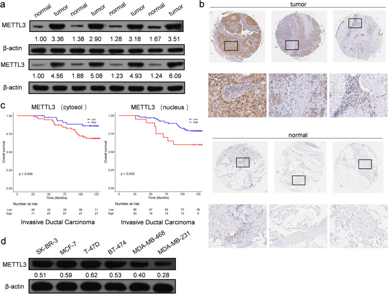
Background: Posttranscriptional modification of tumor-associated factors plays a pivotal role in breast cancer progression. However, the underlying mechanism remains unknown. M6A modifications in cancer cells are dynamic and reversible and have been found to impact tumor initiation and progression through various mechanisms. In this study, we explored the regulatory mechanism of breast cancer cell proliferation and metabolism through m6A methylation in the Hippo pathway. METHODS: A combination of MeRIP-seq, RNA-seq and metabolomics-seq was utilized to reveal a map of m6A modifications in breast cancer tissues and cells. We conducted RNA pull-down assays, RIP-qPCR, MeRIP-qPCR, and RNA stability analysis to identify the relationship between m6A proteins and LATS1 in m6A regulation in breast cancer cells. The expression and biological functions of m6A proteins were confirmed in breast cancer cells in vitro and in vivo. Furthermore, we investigated the phosphorylation levels and localization of YAP/TAZ to reveal that the activity of the Hippo pathway was affected by m6A regulation of LATS1 in breast cancer cells. RESULTS: We demonstrated that m6A regulation plays an important role in proliferation and glycolytic metabolism in breast cancer through the Hippo pathway factor, LATS1. METTL3 was identified as the m6A writer, with YTHDF2 as the reader protein of LATS1 mRNA, which plays a positive role in promoting both tumorigenesis and glycolysis in breast cancer. High levels of m6A modification were induced by METTL3 in LATS1 mRNA. YTHDF2 identified m6A sites in LATS1 mRNA and reduced its stability. Knockout of the protein expression of METTL3 or YTHDF2 increased the expression of LATS1 mRNA and suppressed breast cancer tumorigenesis by activating YAP/TAZ in the Hippo pathway.

Conclusions: In summary, we discovered that the METTL3-LATS1-YTHDF2 pathway plays an important role in the progression of breast cancer by activating YAP/TAZ in the Hippo pathway.

Keywords: Breast cancer; Hippo-YAP/TAZ signaling pathway; LATS1; METTL3.


Conflict of interest statement

Youqin Xu, Mu Song, Ziyang Hong, Wancheng Chen, Qianbing Zhang, Jianlong Zhou, Chao Yang, Zilong He, Juanjuan Yu, Xiaolin Peng, Qiuhong Zhu, Shaotian Li, Kaiyuan Ji, Minfeng Liu and Qiang Zuo declare that they have no conflict of interest.

Figures

Fig. 1
Fig. 1
Abnormal levels of m6A modified mRNA are related to multiple pathways in breast cancer. a The volcano plot of differential methylation sites of breast cancer tissues compared with adjacent normal tissues, n = 2, |log2(fold change)|> 1 and p value < 0.05. b Venn diagram (left panel) of common (intersection region) and unique peaks (non-intersection region) of breast cancer tissue compared with adjacent normal tissue, |log2(fold change)|> 1 and p value < 0.05. The gene region (right panel) annotated with different peaks of breast cancer tissue compared with adjacent normal tissue, |log2(fold change)|> 1 and p value < 0.05. c Differential m6A and differential gene expression combined analysis diagram. Blue dots represent transcripts with significantly downregulated and upregulated m6A expression, red dots represent transcripts with significantly upregulated expression and upregulated m6A expression, green dots represent transcripts with significantly downregulated and downregulated m6A expression, and yellow dots represent transcripts with significantly upregulated and downregulated m6A expression. d Functional enrichment of differentially expressed genes with different m6A methylation sites (left panel). The KEGG pathway enrichment bubble map of differential m6A methylation and differentially expressed genes (right panel). e List of LATS1 containing transcripts of differentially methylated sites (upper panel) in breast cancer tissues compared with adjacent normal tissues, n = 2, |log2(fold change)|> 1 and p value < 0.05. SRAMP prediction results of m6A sites on LATS1 (lower panel). f m6A2Target prediction results of m6A proteins related to LATS1
Fig. 2
Fig. 2
Changes in METTL3 expression were found in breast cancer tissues. a The protein level of METTL3 in breast cancer tissues and adjacent normal tissues, n = 8. b The expression of METTL3 protein in breast cancer tissues and normal tissues by IHC in the TMA (129 breast cancer tissue samples and 3 normal breast tissue samples). Strong staining of mettl3 was detected in breast cancer tissues (upper panel) and weak staining was detected in normal tissues (lower panel). c High METTL3 protein expression was significantly associated with poor prognosis of invasive ductal carcinoma, p < 0.05. d The protein level of METTL3 in 6 different breast cancer cell lines
Fig. 3
Fig. 3
METTL3 promotes tumorigenesis of breast cancer cell MCF-7 both in vitro and in vivo. a The proliferation of MCF-7 cells was suppressed when the expression of METTL3 was altered in cells. The cell number was determined with EdU cellular proliferation assay with flow detection, n = 3, *** p < 0.001. b An apoptosis assay was performed after knocking out or overexpressing METTL3 in MCF-7 cells, and the apoptotic cell number was counted with a fluorescein assay, n = 3, ** p < 0.01, *** p < 0.001. c Blocking METTL3 inhibited the migration of MCF-7 cells, as detected by wound healing tests at 0 h, 12 h, 24 h and 36 h. d The invasion ability of MCF-7 cells, as revealed by the trans-well assay, was significantly suppressed by knocking out METTL3, n = 3, * p < 0.05, ** p < 0.01. e The tumor growth of MCF-7 cells in nude mice was delayed by METTL3 knockout, n = 7, * p < 0.05, *** p < 0.001
Fig. 4
Fig. 4
METTL3 affects the Hippo pathway and glycolysis in breast cancer cells. a Knocking out METTL3 remarkably affected transcriptome expression in MCF-7 cells. b KEGG enrichment analysis indicated that the Hippo pathway was upregulated, whereas glycolysis progression was downregulated by MTEEL3 knockout. c RNA-seq results showed that multiple differentially expressed genes involved in different pathways were significantly related to METTL3, n = 3. d KEGG enrichment of differential metabolites from metabolomic-seq analysis demonstrated that metabolites of glycolysis progression could be largely affected by METTL3, n = 6
Fig. 5
Fig. 5
METTL3 promotes glycolysis and the colony-forming ability of breast cancer cells. a The ECAR in the METTL3 KO group and control group of MCF-7 cells in response to glucose, oligomycin and 2-DG, * p < 0.05. b The levels of glycolysis, glycolysis reserve and glycolysis capacity in MCF-7 cells compared with the control cells after deleting the expression of METTL3, n = 3, *** p < 0.001. c RNA-pull down assay showing the interaction between METTL3 and LATS1 mRNA. d The m6A level of LATS1 mRNA when the expression of METTL3 was altered in MCF-7 cells, as detected by MeRIP-qPCR, n = 3, ** p < 0.01. e The m6A level and METTL3 binding level of LATS1 mRNA when the m6A methylation sites on LATS1 mRNA were mutated in MCF-7 cells detected by MeRIP-qPCR, n = 3, *** p < 0.001. (f) Rescue of the expression of LATS1 by knocking out the expression of METTL3 and siRNA of LATS1 remarkably affected the proliferation of MCF-7 cells, n = 3, ** p < 0.01, *** p < 0.001. g Rescue of the expression of LATS1 by METTL3 significantly restored glycolysis in MCF-7 cells, *** p < 0.001
Fig. 6
Fig. 6
M6A regulation of LATS1 mRNA was identified by ythdf2 in breast cancer cells. a Deletion of METTL3 improved the stability of LATS1 mRNA, n = 3, * p < 0.05, *** p < 0.001. b Conducting the m6A methylation sites mutation on LATS1 mRNA helped enhance the stability of LATS1 mRNA, n = 3, * p < 0.05. c RNA-pull down assay showed the interaction between YTHDF2 and LATS1 mRNA. d Altering the expression of METTL3 and mutating the LATS1 mRNA binding site of METTL3 affected only the protein level of LATS1. e YTHDF2 negatively regulated the protein level of LATS1 in breast cancer cells. f Rescue of the expression of LATS1 by altering the protein level of METTL3 together with YTHDF2 affected the phosphorylation level of YAP/TAZ in the nucleus
Fig. 7
Fig. 7
Rescuing the protein level of LATS1 by altering the expression of YTHDF2 in breast cancer cells could help suppress tumorigenesis. a YTHDF2 deletion remarkably suppressed glycolysis progression in MCF-7 cells, *** p < 0.001. b Knocking out the expression of YTHDF2 significantly inhibited tumor growth in vivo, n = 7, * p < 0.05, ** p < 0.01. c The colony formation ability of MCF-7 cells was suppressed by knocking out the expression of YTHDF2 and reversed by inhibiting the expression of LATS1 at the same time. YTHDF2 overexpression led to the rapid proliferation of MCF-7 cells and such stimulation could be rescued by LATS1 knockdown, n = 3, ** p < 0.01. e YTHDF2 deletion raised the apoptosis level of MCF-7 cells, while LATS1 knockdown rescued this stimulation, n = 3, ** p < 0.01, *** p < 0.001. f The suppressed invasion ability of MCF-7 cells affected by YTHDF2 knockout could be rescued by LATS1 knockdown, n = 3, ** p < 0.01, *** p < 0.001

Similar articles

Cited by

References

    1. Sung H, Ferlay J, Siegel RL, Laversanne M, Soerjomataram I, Jemal A, Bray F. Global Cancer Statistics 2020: GLOBOCAN Estimates of Incidence and Mortality Worldwide for 36 Cancers in 185 Countries. CA Cancer J Clin. 2021;71(3):209–249. doi: 10.3322/caac.21660. - DOI - PubMed
    1. Shieh Y, Tice JA. Medications for Primary Prevention of Breast Cancer. JAMA. 2020;324(3):291–292. doi: 10.1001/jama.2020.9246. - DOI - PMC - PubMed
    1. Denkert C, von Minckwitz G, Darb-Esfahani S, Lederer B, Heppner BI, Weber KE, Budczies J, Huober J, Klauschen F, Furlanetto J, et al. Tumour-infiltrating lymphocytes and prognosis in different subtypes of breast cancer: a pooled analysis of 3771 patients treated with neoadjuvant therapy. Lancet Oncol. 2018;19(1):40–50. doi: 10.1016/S1470-2045(17)30904-X. - DOI - PubMed
    1. Harbeck N, Gnant M. Breast cancer. Lancet. 2017;389(10074):1134–1150. doi: 10.1016/S0140-6736(16)31891-8. - DOI - PubMed
    1. Hanahan D. Hallmarks of Cancer: New Dimensions. Cancer Discov. 2022;12(1):31–46. doi: 10.1158/2159-8290.CD-21-1059. - DOI - PubMed

MeSH terms