. 2023 Jan;11(1):e005627.
doi: 10.1136/jitc-2022-005627.

STING agonism overcomes STAT3-mediated immunosuppression and adaptive resistance to PARP inhibition in ovarian cancer

Affiliations

STING agonism overcomes STAT3-mediated immunosuppression and adaptive resistance to PARP inhibition in ovarian cancer

Liya Ding et al. J Immunother Cancer. 2023 Jan.

Abstract

Background: Poly (ADP-ribose) polymerase (PARP) inhibition (PARPi) has demonstrated potent therapeutic efficacy in patients with BRCA-mutant ovarian cancer. However, acquired resistance to PARPi remains a major challenge in the clinic.

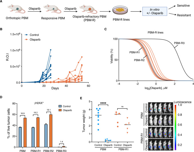
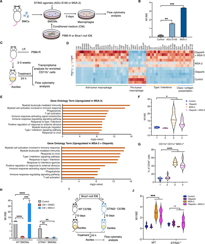
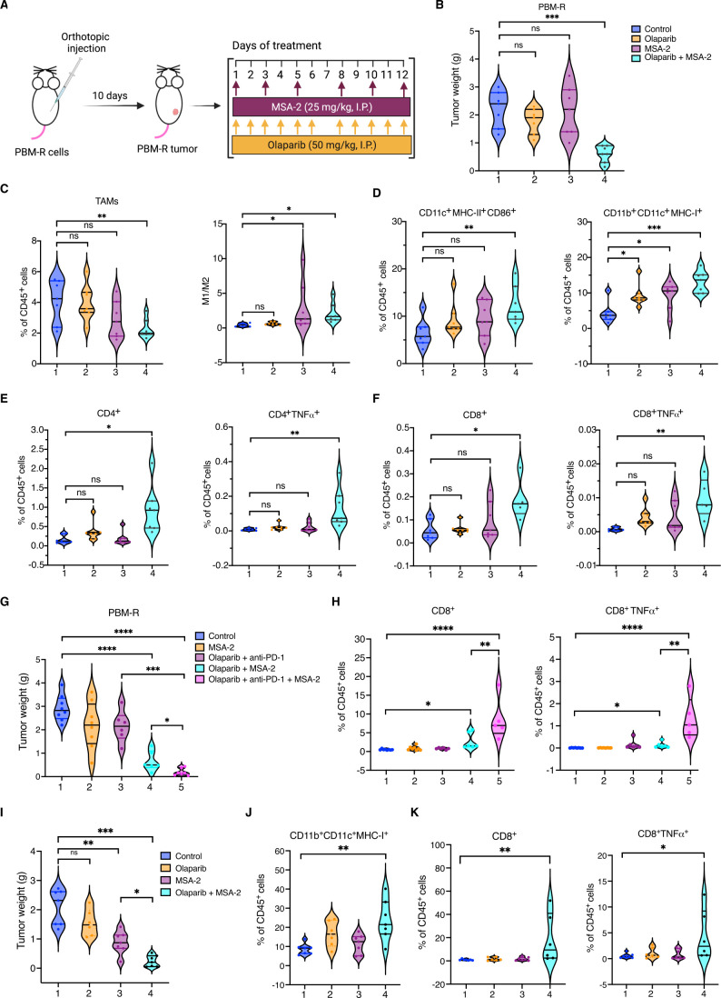
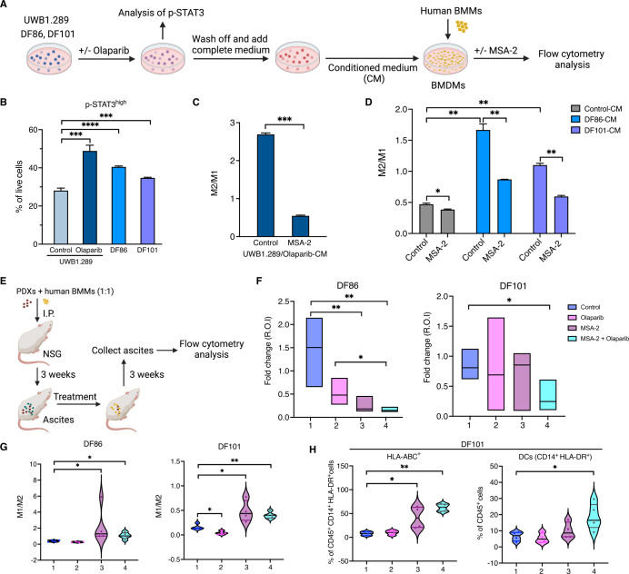
Methods: PARPi-resistant ovarian cancer mouse models were generated by long-term treatment of olaparib in syngeneic Brca1-deficient ovarian tumors. Signal transducer and activator of transcription 3 (STAT3)-mediated immunosuppression was investigated in vitro by co-culture experiments and in vivo by analysis of immune cells in the tumor microenvironment (TME) of human and mouse PARPi-resistant tumors. Whole genome transcriptome analysis was performed to assess the antitumor immunomodulatory effect of STING (stimulator of interferon genes) agonists on myeloid cells in the TME of PARPi-resistant ovarian tumors. A STING agonist was used to overcome STAT3-mediated immunosuppression and acquired PARPi resistance in syngeneic and patient-derived xenografts models of ovarian cancer.

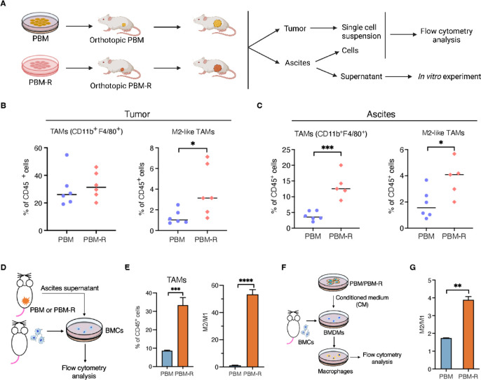
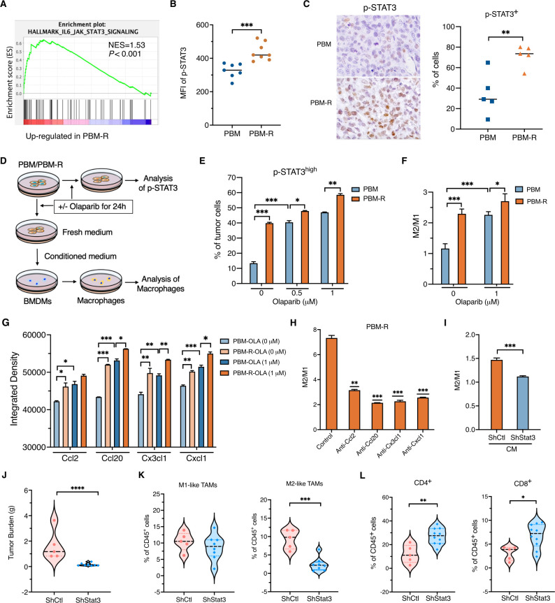
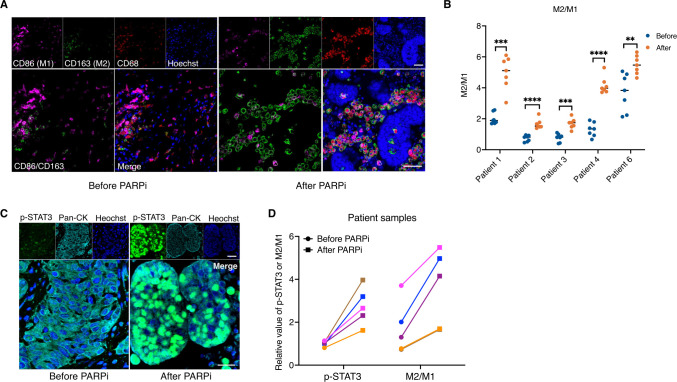
Results: In this study, we uncover an adaptive resistance mechanism to PARP inhibition mediated by tumor-associated macrophages (TAMs) in the TME. Markedly increased populations of protumor macrophages are found in BRCA-deficient ovarian tumors that rendered resistance to PARPi in both murine models and patients. Mechanistically, PARP inhibition elevates the STAT3 signaling pathway in tumor cells, which in turn promotes protumor polarization of TAMs. STAT3 ablation in tumor cells mitigates polarization of protumor macrophages and increases tumor-infiltrating T cells on PARP inhibition. These findings are corroborated in patient-derived, PARPi-resistant BRCA1-mutant ovarian tumors. Importantly, STING agonists reshape the immunosuppressive TME by reprogramming myeloid cells and overcome the TME-dependent adaptive resistance to PARPi in ovarian cancer. This effect is further enhanced by addition of the programmed cell death protein-1 blockade.

Conclusions: We elucidate an adaptive immunosuppression mechanism rendering resistance to PARPi in BRCA1-mutant ovarian tumors. This is mediated by enrichment of protumor TAMs propelled by PARPi-induced STAT3 activation in tumor cells. We also provide a new strategy to reshape the immunosuppressive TME with STING agonists and overcome PARPi resistance in ovarian cancer.

Keywords: Drug Therapy, Combination; Immunomodulation; Immunotherapy; Macrophages; Tumor Microenvironment.


Conflict of interest statement

Competing interests: LD, QW and JJZ are co-inventors of the patent related to this study (US application 63/148426). QW is a scientific consultant for Crimson Biopharm. PAK is a scientific consultant for Alkermes, AstraZeneca, Bayer, GSK, Merck, Pfizer, Tesaro, Mersana, Repare, Kadmon and served as the principal investigator (PI) of institutional support clinical trial for AstraZeneca, Bayer, GSK, Merck, Lilly, and BMS. TMR is cofounder of Crimson Biopharm and Geode Therapeutics, and a SAB member for Shiftbio and K2B Therapeutics. GJF has patents/pending royalties on the PD-L1/PD-1 pathway from Roche, Merck MSD, Bristol Myers Squibb, Merck KGA, Boehringer Ingelheim, AstraZeneca, Dako, Leica, Mayo Clinic, Eli Lilly, and Novartis. GJF has served on advisory boards for Roche, Bristol Myers Squibb, Origimed, Triursus, iTeos, NextPoint, IgM, Jubilant, Trillium, GV20, IOME, and Geode. GJF has equity in Nextpoint, Triursus, Xios, iTeos, IgM, Trillium, Invaria, GV20, and Geode. JFL has served as a scientific consultant for Genentech/Roche and Bristol Myers Squibb, and has been on advisory boards for Clovis Oncology, Genentech/Roche, GlaxoSmithKline, Regeneron, AstraZeneca, and Eisai. JFL has served as the institutional PI (support to the institution) of clinical trials by Genentech/Roche, AstraZeneca, Boston Biomedical, Acetylon Pharmaceuticals, Bristol Myers Squibb, Agenus, CytomX Therapeutics, Regeneron, Tesaro/GSK, Clovis Oncology, Surface Oncology, 2X Oncology, Vigeo Therapeutics, Aravive, Arch Oncology and Zentalis Pharmaceuticals. UM has served on the advisory boards of 2X Oncology, Fujifilm, Immunogen, Mersana, Geneos, and Merck. JJZ is cofounder and director of Crimson Biopharm and Geode Therapeutics. The remaining authors declare no competing interests.

Figures

Figure 1
Figure 1
Characterization of protumor macrophages in Brca1-deficient ovarian tumors that acquired resistance to PARP inhibition (A) Generation of PARPi-resistant ovarian tumor models: PARPi-responsive Brca1-null/p53-null/Myc-high ovarian tumors (PBM) tumors were orthotopically allografted into syngeneic host mice. Over a course of olaparib treatment, tumors that had eventually progressed were harvested as PBM-R tumors for further characterization. (B) Tumor growth curve of PBM-tumor bearing mice treated with olaparib and vehicle control (control, n=10; olaparib, n=12). (C) Measurement of IC50 value of PARPi-naïve PBM cells and PBM-R tumor cell lines from PBM-R tumors (blue, PBM; orange, PBM-R; black, PBM-R3). (D) PBM and PBM-R cells were treated with 1 µM olaparib or vehicle control for 24 hours and subsequently subjected to analysis of γ-H2AX by flow cytometry analysis (n=3). (E) Tumor burden and representative bioluminescence-imaging analysis of PBM or PBM-R1 tumor-bearing mice treated with vehicle control or olaparib. Data are presented as mean±SD. One-way analysis of variance (ANOVA). ****p<0.0001. PARP, poly (ADP-ribose) polymerase; PARPi, PARP inhibitors; PBM, Trp53-/-Brca1-/-Myc; PBM-R, PARPi-resistant PBM; ROI, region of interest.
Figure 2
Figure 2
PBM-R tumors have increased M2-like macrophages in tumors and ascites of tumor-bearing mice (A) Diagram of workflow for B–D. (B) Flow cytometric analysis of tumor-infiltrating total and protumor (M2-like) TAMs in PBM and PBM-R tumor-bearing mice (n=6). (C) Flow cytometric analysis of total and M2-like TAMs in the ascites of PBM and PBM-R tumor-bearing mice (n=5 or 6). (D) Diagram of workflow for E. (E) Flow cytometric analysis of bone marrow cells (BMCs) cultured in 50% complete medium and 50% ascites supernatant harvested from PBM and PBM-R tumor-bearing mice for 5 days (n=3). (F) Diagram of workflow for G. (G) Flow cytometric analysis of bone marrow-derived macrophages (BMDMs) cultured in 50% complete medium and 50% PBM-CM or PBM-R-CM for 3 days (n=3). Data are presented as mean±SD. One-way analysis of variance (ANOVA). *p<0.05, **p<0.01, ***p<0.001, ****p<0.0001. MHC, major histocompatibility complex; TAM, tumor-associated macrophages; PBM, Trp53-/-Brca1-/-Myc; PBM-R, PARPi-resistant PBM; M2/M1, M2-like macrophages/M1-like macrophages.
Figure 3
Figure 3
PARPi-induced STAT3 signaling activation in tumors is important for M2-like macrophage polarization (A) GSEA analysis of RNA sequencing data revealed an upregulated STAT3 signaling pathway enriched in PBM-R tumors (n=6 for each group). (B) Flow cytometric analysis of phosphorylation level of STAT3 (Y705) in PBM and PBM-R tumor cells (n=7 or 8). (C) Representative images of immunohistochemistry staining and quantification data for p-STAT3 (Y705) in PBM and PBM-R tumors, each dot represents one mouse. (D) Diagram of workflow for (E) and (F). (E) Flow cytometric analysis of p-STAT3 in PBM and PBM-R tumor cells treated with indicated concentration of olaparib or vehicle control (n=3). (F) Flow cytometric analysis of BMDMs cultured in CM from PBM and PBM-R with or without olaparib treatment (n=3). (G) Analysis of cytokines in the medium of PBM and PBM-R cells treated with olaparib or vehicle control (n=2). (H) Analysis of mouse BMDMs cultured with or without 50% PBM-R-CM in the presence or absence of indicated neutralizing antibodies for 3 days (n=3). (I) Flow cytometric analysis of BMDMs cultured in CM from PBM-R with or without knockdown of STAT3. (J) Tumor burden of mice transplanted with PBM-R tumor cells expressing control or STAT3 shRNAs and treated with olaparib or vehicle control. (K, L) Flow cytometric analysis of total TAMs and the ratio of M1-like and M2-like macrophages (K), CD4+ and CD8+ effector T cells (L) in olaparib-treated PBM-R tumors expressing control or STAT3 shRNAs in (J). CM, conditioned medium; BMDMs, bone marrow-derived macrophages; PARP, PARP, poly (ADP-ribose) polymerase; PARPi, PARP inhibitors; p-STAT3, phosphorylated STAT3; STAT3, signal transducer and activator of transcription 3; TAM, tumor-associated macrophages; PBM, Trp53-/-Brca1-/-Myc; PBM-R, PARPi-resistant PBM; MFI, median fluorescence intensity; GSEA, Gene Set Enrichment Analysis.
Figure 4
Figure 4
M2-like macrophages and tumor cell-intrinsic phosphorylated STAT3 increased after PARPi treatment in patient ovarian cancer samples with BRCA-mutations. (A) Representative immunofluorescence images of a pair of matched tumor specimens before and after PARPi treatment from a patient with ovarian cancer with germline BRCA mutation were stained for CD86 (magenta), CD163 (green), CD68 (red) and Hoechst (blue). Scale bars=50 µm. (B) Quantification of the ratios of M2/M1 in five matched pairs of tumor specimens before and after PARPi treatment collected from BRCA-mutant ovarian patients. Each dot represents the quantification data of a single image. (C) Representative immunofluorescence images of p-STAT3 (Y705) (green), Pan-CK (Pan-Cytokeratin, cyan) and Hoechst (blue) in the tumors of patients with ovarian cancer with germline BRCA mutations. Scale bars=20 µm. (D) Quantification of p-STAT3 and the ratio of M2/M1 before and after PARPi treatment in each of these BRCA-mutant ovarian patients. Each dot represents the median value of p-STAT3 or M2/M1 from one patient. Data are presented as mean±SD, or median with quartiles (violin plots). One-way analysis of variance. *p<0.05, **p<0.01, ***p<0.001, ****p<0.0001. PARP, poly (ADP-ribose) polymerase; PARPi, PARP inhibitors; p-STAT3, phosphorylated STAT3; STAT3, signal transducer and activator of transcription 3.
Figure 5
Figure 5
STING agonists reprogram myeloid cells in vitro and in vivo in a STING-dependent manner. (A) Diagram of workflow for (B) and (H). Bone marrow-derived macrophages (BMDMs) were cultured in 50% PBM-R-CM or control medium for 72 hours with or without STING agonists (10 µM ADU-S100 or 5 µg/mL MSA-2) treatment. (B) Flow cytometric analysis of macrophage phenotypes in (A). (C) Diagram of workflow for (D). (D) Heat map of differentially expressed genes in myeloid cells (CD45+CD11b+) collected from the ascites of PBM-R tumor-bearing mice with indicated treatment. (E) Top-ranked upregulated gene ontology terms in myeloid cells treated MSA-2 or MSA-2 in combination with olaparib. (F, G) Flow cytometric analysis of the ratio of M1/M2 (F) and major histocompatibility complex-I+ myeloid DCs (CD11b+ CD11c+) (G) in the ascites of PBM-R tumor-bearing mice treated with control, olaparib, MSA-2 and olaparib in combination with MSA-2 for 24 hours. (H) Analysis of the ratio of M1/M2 in WT BMDMs and STING–/– BMDMS cultured in CM from Brca1-null ID8 cells treated with olaparib or vehicle control (n=3). (I) Diagram of workflow for (J). (J) Analysis of macrophages (M1/M2) in WT and STING–/– mice injected with Brca1-null ID8 cells and treated with indicated drugs for 24 hours. ns, not significant. Data are presented as mean±SD, or median with quartiles (violin plots). One-way analysis of variance (ANOVA) (B, F, G, H). *p<0.05, **p<0.01, ***p<0.001, ****p<0.0001. BMC, bone marrow cells; DCs, dendritic cells; i.p., intraperitoneally; STING, stimulator of interferon genes; PBM-R, PARPi-resistant PBM; M1/M2, M1-like macrophages/M2-like macrophages.
Figure 6
Figure 6
Treatment of PBM-R tumors with a stimulator of interferon genes agonist changes immune profile of TME and improves tumor’s response to PARP inhibition. (A) Experiment design and treatment scheme of (B). (B) Tumor burden of PBM-R tumor-bearing mice treated with control, olaparib, MSA-2 and olaparib in combination with MSA-2 for 14 days (n=7). (B–F) Flow cytometric analysis of tumor-infiltrating immune cells in PBM-R tumor-bearing mice as described in (A) and (B): (C) TAMs+ (left), M1/M2 (right); (D) activated DCs (CD11c+MHC-II+CD86+) (left) and MHC-I+ myeloid DCs (CD11b+ CD11c+ MHC-I+) (right), (E) total and TNF-α+ CD4+ T cells and (F) total and TNF-α+ CD8+ T cells. (G) Tumor burden of PBM-R tumor-bearing mice treated with indicated agents for 14 days (n=6~8). (H) Flow cytometric analysis of tumor-infiltrating total and TNF-α+ CD8+ T cells in PBM-R tumor-bearing mice as described in (G). (I) Tumor burden of PBM-R3 tumor-bearing mice treated with control, olaparib, MSA-2 and MSA-2 in combination with olaparib for 14 days (n=6 or 7). (J and K) Flow cytometric analysis of tumor-infiltrating immune cells in PBM-R3 tumor-bearing mice as described in (I): (J) MHC-I+ myeloid DCs (CD11b+ CD11c+ MHC-I+); (K) total and TNF-α-producing CD8+ T cells. Data are presented as median with quartiles. One-way analysis of variance (ANOVA). ns, not significant; *p<0.05, **p<0.01, ***p<0.001, ****p<0.0001. DCs, dendritic cells; i.p., intraperitoneally; PD-1, programmed cell death protein-1; MHC, major histocompatibility complex; TAM,; TAM, tumor-associated macrophage; TNF, tumor necrosis factor; PBM-R, PARPi-resistant PBM.
Figure 7
Figure 7
Overcoming PARPi-resistance in ovarian PDXs with a stimulator of interferon genes agonist treatment. (A) Diagram of workflow for (B–D). (B) Detection of phosphorylation level of STAT3 (Y705) by flow cytometry in UWB1.289 cells with or without olaparib treatment and PARPi-resistant ovarian cancer patient-derived PDXs (DF86 and DF101) (n=3). (C) Flow cytometric analysis of human BMDMs cultured in CM from olaparib-treated UWB1.289 cells with or without MSA-2 treatment as described in (A) for 3 days (n=3). (D) Analysis of human BMDMs cultured in CM from PDXs in the presence or absence of MSA-2 (n=3). (E) Diagram of workflow for (F). (F) Tumor burden of DF86 and DF101 PDX-bearing mice treated with control, MSA-2, olaparib and MSA-2 in combination with olaparib for 2 weeks (n=4 or 5). (G) Analysis of the phenotypes of macrophages (M1/M2) in the ascites of DF86 and DF101PDX-bearing mice as described in (F). (H) Analysis of the proportion and HLA expression in myeloid DCs (CD14+HLA-DR+) in DF101 PDX-bearing mice with indicated treatment. Data are presented as mean±SD, or median with quartiles (violin plots). One-way analysis of variance (ANOVA) (C–H). *p<0.05, **p<0.01, ***p<0.001, ****p<0.0001. BMDM, bone marrow-derived macrophage; BMM, bone marrow monocyte; DCs, dendritic cells; HLA, human leukocyte antigen; i.p., intraperitoneally; PARP, poly (ADP-ribose) polymerase; PARPi, PARP inhibitors; p-STAT3, phosphorylated STAT3; STAT3, signal transducer and activator of transcription 3.

Similar articles

Cited by

References

    1. Farmer H, McCabe N, Lord CJ, et al. Targeting the DNA repair defect in BRCA mutant cells as a therapeutic strategy. Nature 2005;434:917–21. 10.1038/nature03445 - DOI - PubMed
    1. Chan CY, Tan KV, Cornelissen B. Parp inhibitors in cancer diagnosis and therapy. Clin Cancer Res 2021;27:1585–94. 10.1158/1078-0432.CCR-20-2766 - DOI - PubMed
    1. Mullard A. Parp inhibitors plough on. Nat Rev Drug Discov 2017;16:229. 10.1038/nrd.2017.61 - DOI - PubMed
    1. González-Martín A, Pothuri B, Vergote I, et al. Niraparib in patients with newly diagnosed advanced ovarian cancer. N Engl J Med 2019;381:2391–402. 10.1056/NEJMoa1910962 - DOI - PubMed
    1. Robson M, Im S-A, Senkus E, et al. Olaparib for metastatic breast cancer in patients with a germline BRCA mutation. N Engl J Med 2017;377:523–33. 10.1056/NEJMoa1706450 - DOI - PubMed

Publication types

Substances