. 2023 Jan 6;13(1):270.
doi: 10.1038/s41598-023-27531-5.

Expression of LIM domain-binding 3 (LDB3), a striated muscle Z-band alternatively spliced PDZ-motif protein in the nervous system

Affiliations

Expression of LIM domain-binding 3 (LDB3), a striated muscle Z-band alternatively spliced PDZ-motif protein in the nervous system

Yotam Blech-Hermoni et al. Sci Rep. .

Abstract

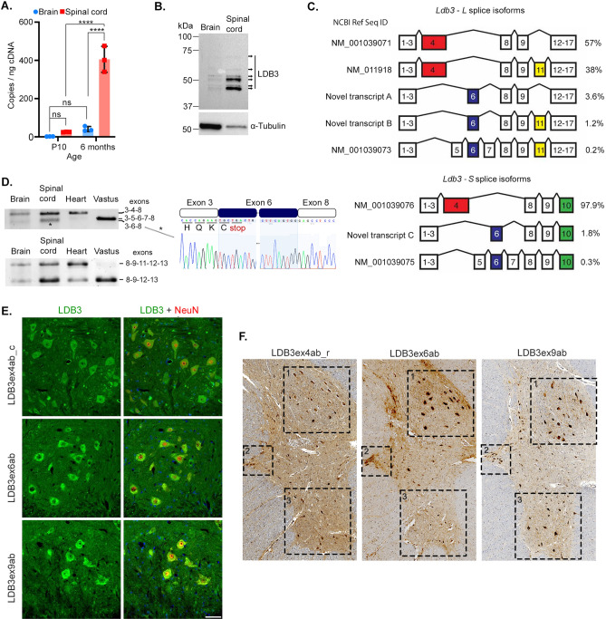
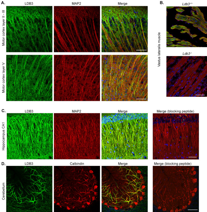
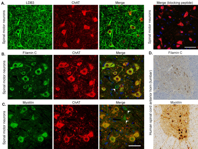
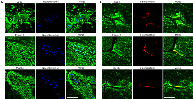
LIM domain-binding 3 (LDB3) is a member of the Enigma family of PDZ-LIM proteins. LDB3 has been reported as a striated muscle-specific Z-band alternatively spliced protein that plays an important role in mechanosensory actin cytoskeleton remodeling. This study shows that LDB3 is broadly expressed in the central and peripheral nervous system of human and mouse. LDB3 is predominantly expressed in the adult stages compared to early development and at a significantly higher level in the spinal cord than in the brain. As in skeletal muscle and heart, LDB3 is extensively alternatively spliced in the neurons. Three novel splice isoforms were identified suggesting splicing-dependent regulation of LDB3 expression in the nervous system. Expression of LDB3 in the motor cortex, cerebellum, spinal motor neuron, peripheral nerve, and neuromuscular junction in addition to skeletal muscle indicates important roles for this PDZ-LIM family protein in motor planning and execution. Moreover, expression in the hippocampal neurons suggests roles for LDB3 in learning and memory. LDB3 interactors filamin C and myotilin are also expressed in the spinal motor neuron, nerve, and neuromuscular junction, thereby providing the basis for neurogenic manifestations in myopathies associated with mutations in these so-called muscle proteins.


Conflict of interest statement

The authors declare no competing interests.

Figures

Figure 1
Figure 1
Expression of LDB3 splice isoforms in brain and spinal cord. (A) Expression levels of Ldb3 mRNA in brain (blue circle) and spinal cord (red square) of 10-day (P10)- and 6 month-old mice measured with ddPCR assays using equal amounts of input cDNA per reaction. The bar graph with scatter plot represents mean ± S.D.; n = 3 mice; triplicate assay; a two-way ANOVA with the Bonferroni correction ****p < 0.0001. (B) A representative immunoblot of LDB3 isoforms in human motor cortex and spinal cord detected by a rabbit monoclonal antibody (Abcam; ab171936). Multiple bands are seen between 37 and 75 kDa (arrows). Two-fold higher amount of proteins was loaded from the brain lysate compared to the spinal cord; α-tubulin served as a loading control. (C) Spectrum of LDB3-L and LDB3-S isoforms detected by SMRT sequencing in the brain of six month old mice (n = 3). Relative percentages of total reads are shown. (D) Representative gel electrophoresis of Ldb3 splice isoforms in different mouse tissues in a RT-PCR assay (n = 6 mice). Exon composition identified by Sanger sequencing is indicated. Chromatogram of a cloned amplicon from the lower band (*) in the spinal cord validates the novel splicing events joining Ldb3 exon 3 to 6 and exon 6 to 8. Predicted reading frame including premature stop codon in exon 6 is shown. Dotted line indicates continuity of exon 6 sequence in the chromatogram. See Supplementary Table S2 for all primer sequences. (E) Representative immunofluorescence on lumbar spinal cord cross sections of six month old mice (n = 6) show staining for the exon domain-specific LDB3 antibodies (green) in NeuN (red)-positive neurons of the anterior horn. Scale bar: 20 µm. (F) Representative immunohistochemistry on serial cross sections of human L1-L2 spinal segments (n = 3 autopsies) show staining for the exon-specific antibodies in neurons of Clarke’s dorsal nucleus (1), the lateral column (2), and the anterior horn (3). Individual tiles were acquired at 20 × magnification and stitched together into a composite image. Original blots/gels for the image B and D are presented in Supplementary Fig. 2 of supplementary file 1.
Figure 2
Figure 2
Immunolocalization of LDB3 in different regions of mouse brain. (A) Representative immunofluorescence using GTX115593 antibody on motor cortex coronal section shows LDB3 signal (green) in the apical dendrite bundles probably from layers III and V stained by MAP2 (red) and in thinner dendrites not labeled by MAP2 which are likely from superficial neurons in six-month old mice (n = 6). Bottom panel shows LDB3 signal (green) in MAP2 (red) stained cell body and dendrites of pyramidal neurons in motor cortex layer V in the same coronal section. (B) Representative immunofluroscence shows the same LDB3 antibody signal (green) co-localized with α-actinin-2 signal (red) at the Z-discs in the vastus lateralis muscle fibers of 18 day old Ldb3+/+ embryo but not Ldb3−/− littermates (n = 3). Note Ldb3−/− mice do not survive beyond birth. (C, D) Representative immunofluorescence using the same antibody shows LDB3 (green) in MAP2 (red) stained dendrites from the hippocampal CA1 neurons (C) and in calbindin (red) stained dendrites from the cerebellar Purkinje cells (D) in 6 month old mice (n = 6). LDB3 signal is absent in parallel sections incubated with LDB3 antibody plus a blocking peptide. Scale bar in all images 50 µm.
Figure 3
Figure 3
Immunolocalization of LDB3 in human motor cortex and the L1-L2 spinal cord anterior horn. (A) Immunohistochemistry staining with GTX115593 antibody shows LDB3 immunoreactivity in neurons of the motor cortex layers. (B) Images show neurons of layers II, III, and V at a higher resolution. The LDB3 signal is seen in the soma and processes of the neurons. (C) Immunofluorescent staining with the same antibody shows LDB3 (green) in the soma and dendrites of MAP2 co-stained (red) pyramidal neurons of layer V in the motor cortex. (D) Immunohistochemistry staining with the same antibody shows LDB3 immunoreactivity in the soma and processes of the anterior horn neurons in lumbar segments of the spinal cord. Scale bar 250 µm (A and D), 100 µm (B), 50 µm (B inset and C), and 20 µm (D inset).
Figure 4
Figure 4
Immunolocalization of LDB3, filamin C and myotilin in spinal motor neurons. (A) Representative immunofluorescence on spinal cord lumbar cross section stained by GTX115593 antibody show LDB3 signal (green) in the cell body and processes of ChAT-positive (red) spinal motor neurons of six month old mice (n = 6). Addition of a blocking peptide results in loss of the LDB3 signal. (B, C) Immunostaining of parallel sections with antibodies against filamin C (B) and myotilin (C) detect both proteins in cholinergic neurons in the anterior horn of the spinal cord in mice (n = 6). Arrow heads indicate myotilin- and filamin C -antibody stained cells which are not ChAT positive. (D) Representative immunohostochemistry on human spinal cord lumbar segments (n = 3; 20 × magnification) show that neurons in the anterior horn react to the filamin C and myotilin antibody. Scale bar: 50 µm.
Figure 5
Figure 5
Immunolocalization of LDB3, filamin C, and myotilin in peripheral nerve and NMJ. (A, B) Representative immunofluorescence on tibialis anterior muscle cross-sections from six-month old mice (n = 6) show LDB3, filamin C, and myotilin staining (green) localized to intramuscular nerve fibers (A) and NMJs (B) marked by expression of neurofilament-M (blue) and α-Bungarotoxin (red), respectively. Scale bar (25 µm).

References

    1. Boden SD, et al. LMP-1, a LIM-domain protein, mediates BMP-6 effects on bone formation. Endocrinology. 1998;139:5125–5134. doi: 10.1210/endo.139.12.6392. - DOI - PubMed
    1. Gardezi SR, Weber AM, Li Q, Wong FK, Stanley EF. PDLIM5 is not a neuronal CaV2.2 adaptor protein. Nat. Neurosci. 2009;12:957–958. doi: 10.1038/nn0809-957a. - DOI - PubMed
    1. Ueki N, et al. Isolation, tissue expression, and chromosomal assignment of a human LIM protein gene, showing homology to rat enigma homologue (ENH) J. Hum. Genet. 1999;44:256–260. doi: 10.1007/s100380050155. - DOI - PubMed
    1. Faulkner G, et al. ZASP: A new Z-band alternatively spliced PDZ-motif protein. J. Cell Biol. 1999;146:465–475. doi: 10.1083/jcb.146.2.465. - DOI - PMC - PubMed
    1. Zhou Q, Ruiz-Lozano P, Martone ME, Chen J. Cypher, a striated muscle-restricted PDZ and LIM domain-containing protein, binds to alpha-actinin-2 and protein kinase C. J. Biol. Chem. 1999;274:19807–19813. doi: 10.1074/jbc.274.28.19807. - DOI - PubMed

Publication types

MeSH terms

LinkOut - more resources