. 2023 Jan;55(1):132-142.
doi: 10.1038/s12276-022-00917-7. Epub 2023 Jan 6.

PRPF8 increases the aggressiveness of hepatocellular carcinoma by regulating FAK/AKT pathway via fibronectin 1 splicing

Affiliations

PRPF8 increases the aggressiveness of hepatocellular carcinoma by regulating FAK/AKT pathway via fibronectin 1 splicing

Juan L López-Cánovas et al. Exp Mol Med. 2023 Jan.

Abstract

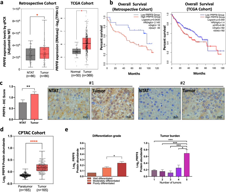
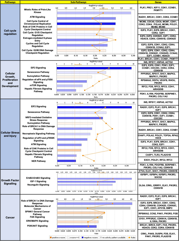
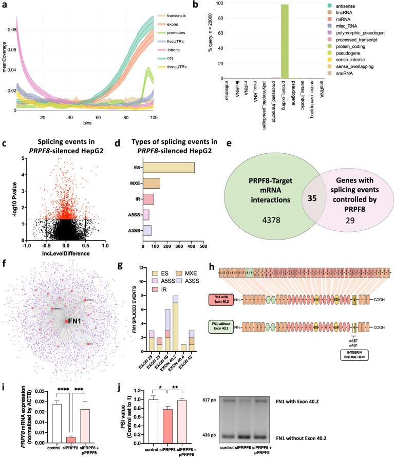
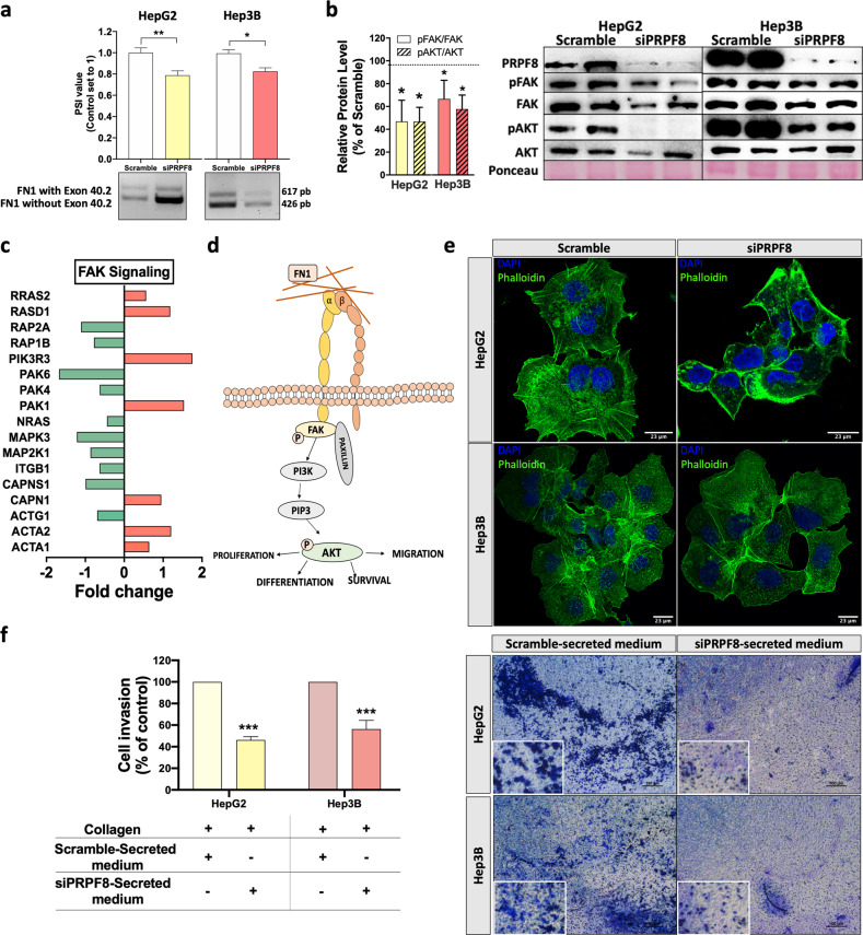
Hepatocellular carcinoma (HCC) pathogenesis is associated with alterations in splicing machinery components (spliceosome and splicing factors) and aberrant expression of oncogenic splice variants. We aimed to analyze the expression and potential role of the spliceosome component PRPF8 (pre-mRNA processing factor 8) in HCC. PRPF8 expression (mRNA/protein) was analyzed in a retrospective cohort of HCC patients (n = 172 HCC and nontumor tissues) and validated in two in silico cohorts (TCGA and CPTAC). PRPF8 expression was silenced in liver cancer cell lines and in xenograft tumors to understand the functional and mechanistic consequences. In silico RNAseq and CLIPseq data were also analyzed. Our results indicate that PRPF8 is overexpressed in HCC and associated with increased tumor aggressiveness (patient survival, etc.), expression of HCC-related splice variants, and modulation of critical genes implicated in cancer-related pathways. PRPF8 silencing ameliorated aggressiveness in vitro and decreased tumor growth in vivo. Analysis of in silico CLIPseq data in HepG2 cells demonstrated that PRPF8 binds preferentially to exons of protein-coding genes, and RNAseq analysis showed that PRPF8 silencing alters splicing events in multiple genes. Integrated and in vitro analyses revealed that PRPF8 silencing modulates fibronectin (FN1) splicing, promoting the exclusion of exon 40.2, which is paramount for binding to integrins. Consistent with this finding, PRPF8 silencing reduced FAK/AKT phosphorylation and blunted stress fiber formation. Indeed, HepG2 and Hep3B cells exhibited a lower invasive capacity in membranes treated with conditioned medium from PRPF8-silenced cells compared to medium from scramble-treated cells. This study demonstrates that PRPF8 is overexpressed and associated with aggressiveness in HCC and plays important roles in hepatocarcinogenesis by altering FN1 splicing, FAK/AKT activation and stress fiber formation.


Conflict of interest statement

The authors declare no competing interests.

Figures

Fig. 1
Fig. 1. PRPF8 is overexpressed in HCC and is associated with clinical aggressiveness and poor survival in HCC patients.
a Expression level of PRPF8 in HCC [retrospective cohort (determined by qPCR): n = 86 patients and TCGA cohort (obtained from RNAseq data): n = 419 samples]. b Overall survival in the retrospective and TCGA cohorts, categorized into the high and low PRPF8 expression groups based on the median PRPF8 expression level, and analyzed by determining the log-rank-p-value. c Immunohistochemical (IHC) score of PRPF8 in NTAT and tumor samples (n = 14 patients from the retrospective cohort). Representative images (60X) from two patients are shown. d Protein level of PRPF8 in paratumor and tumor tissues from CPTAC data. e Correlations between the PRPF8 protein level and clinical aggressiveness in the CPTAC cohort. The data are presented as the means ± SEMs. The asterisks (*p < 0,05; **p < 0,01; ***p < 0,001; ****p < 0,0001) indicate statistically significant differences. NTAT Nontumor adjacent tissue, HR Hazard ratio.
Fig. 2
Fig. 2. PRPF8 silencing alters the expression of several genes involved in key cancer-related pathways.
RNAseq data from PRPF8-silenced HepG2 cells vs. control cells were processed and analyzed by Ingenuity Pathway Analysis. Key pathways and subpathways altered in response to PRPF8 silencing are shown. The bars indicate the -log(B-H p values), and the significance threshold was calculated as <0.05. The most representative altered genes in each subpathway are shown.
Fig. 3
Fig. 3. PRPF8 modulation by a specific siRNA decreases the aggressive features of liver cancer cell lines.
Validation of siRNA-mediated PRPF8 silencing at the mRNA (a) and protein (b) levels. c Proliferation of PRPF8-silenced cells compared to scramble-treated cells. d mRNA expression levels of key cell cycle-related genes (CDK2 and CDK4) in PRPF8-silenced vs. scramble-treated cells. e Migration of PRPF8-silenced cells compared to scramble-treated cells. Representative images at 0 h and after 24 h are shown. f Number of colonies formed in PRPF8-silenced vs. scramble-treated cells. Representative images of colonies formed after 10 days are shown. g Mean size of tumorspheres formed from PRPF8-silenced vs. scramble-treated cells. Representative images of tumorspheres formed after 10 days are shown. h Ratios of mRNA expression levels of key oncogenic splice variants and the corresponding full-length transcripts (CCDC50S/CCDC50 and KLF6SV1/KLF6) in PRPF8-silenced vs. scramble-treated cells. The data are presented as the mean ± SEM of n = 3–5 independent experiments. The asterisks (*p < 0,05, **p < 0,01; ***p < 0,001, ****p < 0,0001) indicate statistically significant differences.
Fig. 4
Fig. 4. PRPF8 silencing decreases HCC cell growth in vivo.
a Diagram showing the in vivo experimental design. At the third week post grafting, each tumor was transfected with scramble siRNA or siPRPF8 (n = 6 mice). b The growth rate of tumors was determined throughout the first 6 days after transfection. Representative images of scramble- and siPRPF8-treated tumors are shown. c The final tumor weight was calculated after sacrifice. d Tumor tissue hematoxylin and eosin (H&E) staining. e Validation of the siRNA-mediated reduction in PRPF8 mRNA expression. f Validation of the siRNA-mediated reduction in PRPF8 protein expression. g mRNA expression levels of key cell cycle-related genes (CDK2 and CDK4) in PRPF8-silenced vs. scramble-treated cells. h Ratios of mRNA expression levels between key oncogenic splice variants and the corresponding full-length transcripts (CCDC50S/CCDC50 and KLF6SV1/KLF6) in PRPF8-silenced vs. scramble-treated xenografts. The asterisks (*p < 0,05; **p < 0,01) indicate statistically significant differences.
Fig. 5
Fig. 5. PRPF8 binds to exonic regions in protein-coding transcripts to modulate splicing.
Coverage profile of the preferred binding location of PRPF8 (a) and transcript features (b) determined from in silico analysis of eCLIP data for PRPF8 in HepG2 cells. c Volcano plot of splicing events altered by silencing of PRPF8 in HepG2 cells. Data were obtained from ENCODE and analyzed with rmats. d Number of events of each type of splicing induced by PRPF8 silencing in HepG2 cells. e Venn diagram of PRPF8-target mRNA interactions (CLIPseq data) and splicing events induced by PRPF8 silencing (RNAseq data) showing that 35 genes whose mRNA is targeted by PRPF8 exhibit altered splicing events in response to PRPF8 silencing. f Protein‒protein interaction analysis identified fibronectin 1 (FN1) as a hub gene. g Splicing events found in the FN1 gene in response to PRPF8 silencing, among which exon 40.2 skipping was the most common altered event. RNAseq data from HepG2 cells were obtained and analyzed. h Diagram showing the skipping of FN1 exon 40.2, which encodes an integrin interaction region. i Validation of PRPF8 expression levels by qPCR in silencing (siPRPF8) and rescue (siPRPF8 + pPRPF8) experiments in Hep3B cells, with comparison to those in mock transfected cells. j Evaluation of the FN1 exon 40.2 inclusion PSI value by PCR in response to PRPF8 silencing and PRPF8 silencing/overexpression compared with that in scramble-treated/mock Hep3B cells. PSI (percent spliced in) indicates the inclusion of an exon. ES Exon skipping, MXE Mutually exclusive exons, A5SS Alternative 5´ splice site, A3SS Alternative 3´ splice site, IR Intron retention. The asterisks (*p < 0,05; **p < 0,01, ***p < 0,001, ****p < 0,0001) indicate statistically significant differences.
Fig. 6
Fig. 6. PRPF8 modulates the splicing of FN1, activation of FAK/AKT, remodeling of the cytoskeleton and invasion capacity.
a In vitro validation of the implication of PRPF8 in exon 40.2 skipping in HepG2 and Hep3B cells, as demonstrated by PCR. PSI (percent spliced in) indicates the inclusion of an exon. b Phosphorylation levels of FAK and AKT in response to PRPF8 silencing in HepG2 and Hep3B cells. The images show representative western blots. c Expression of FAK signaling-related genes in response to PRPF8 silencing. RNAseq data from HepG2 cells were obtained and analyzed. d Schematic representation of integrin-mediated FAK/AKT signaling. e Confocal fluorescence microscopy analysis of stress fibers (using phalloidin dye) in response to PRPF8 silencing in HepG2 and Hep3B cells. Nuclei were stained with DAPI (blue). f Invasion capacity of HepG2 and Hep3B cells using collagen membranes incubated with conditioned medium from scramble- or siPRPF8-treated cells. The data are presented as the mean ± SEM of n = 3–5 independent experiments. The asterisks (*p < 0,05; **p < 0,01) indicate statistically significant differences.

References

    1. Villanueva A. Hepatocellular Carcinoma. N. Engl. J. Med. 2019;380:1450–1462. doi: 10.1056/NEJMra1713263. - DOI - PubMed
    1. Forner A, Reig M, Bruix J. Hepatocellular carcinoma. Lancet. 2018;391:1301–1314. doi: 10.1016/S0140-6736(18)30010-2. - DOI - PubMed
    1. Sveen A, Kilpinen S, Ruusulehto A, Lothe RA, Skotheim RI. Aberrant RNA splicing in cancer; expression changes and driver mutations of splicing factor genes. Oncogene. 2016;35:2413–2427. doi: 10.1038/onc.2015.318. - DOI - PubMed
    1. Wang H, et al. A Coiled-Coil Domain Containing 50 Splice Variant Is Modulated by Serine/Arginine-Rich Splicing Factor 3 and Promotes Hepatocellular Carcinoma in Mice by the Ras Signaling Pathway. Hepatology. 2019;69:179–195. doi: 10.1002/hep.30147. - DOI - PubMed
    1. Vetter D, et al. Enhanced hepatocarcinogenesis in mouse models and human hepatocellular carcinoma by coordinate KLF6 depletion and increased messenger RNA splicing. Hepatology. 2012;56:1361–1370. doi: 10.1002/hep.25810. - DOI - PMC - PubMed

Publication types

MeSH terms