. 2023 Jan 6;14(1):102.
doi: 10.1038/s41467-022-35763-8.

Brucella effectors NyxA and NyxB target SENP3 to modulate the subcellular localisation of nucleolar proteins

Affiliations

Brucella effectors NyxA and NyxB target SENP3 to modulate the subcellular localisation of nucleolar proteins

Arthur Louche et al. Nat Commun. .

Abstract

The cell nucleus is a primary target for intracellular bacterial pathogens to counteract immune responses and hijack host signalling pathways to cause disease. Here we identify two Brucella abortus effectors, NyxA and NyxB, that interfere with host protease SENP3, and this facilitates intracellular replication of the pathogen. The translocated Nyx effectors directly interact with SENP3 via a defined acidic patch (identified from the crystal structure of NyxB), preventing nucleolar localisation of SENP3 at late stages of infection. By sequestering SENP3, the effectors promote cytoplasmic accumulation of nucleolar AAA-ATPase NVL and ribosomal protein L5 (RPL5) in effector-enriched structures in the vicinity of replicating bacteria. The shuttling of ribosomal biogenesis-associated nucleolar proteins is inhibited by SENP3 and requires the autophagy-initiation protein Beclin1 and the SUMO-E3 ligase PIAS3. Our results highlight a nucleomodulatory function of two Brucella effectors and reveal that SENP3 is a crucial regulator of the subcellular localisation of nucleolar proteins during Brucella infection, promoting intracellular replication of the pathogen.


Conflict of interest statement

The authors declare no competing interests.

Figures

Fig. 1
Fig. 1. The Brucella NyxA and NyxB proteins are translocated into host cells during infection, accumulating in punctate or filament-like cytoplasmic and nuclear structures and can interact with each other.
a RAW macrophage-like cells were infected for 24 h with either B. abortus wild-type expressing TEM1 (encoded by the bla gene) fused with NyxA, NyxB or BAB1_0466. The percentage of cells with coumarin emission, which is indicative of translocation, was quantified after incubation with the CCF2-AM substrate. Data represent means ± 95% confidence intervals from five independent experiments, with more than 500 cells counted for each condition. b Representative images for B. abortus wild-type carrying pbla:nyxA, pbla:nyxB or pbla:BAB1_0466 to exemplify the presence of effector translocation visible in coumarin-positive cells (violet) or absence of translocation. Scale bar corresponds to 25 µm. c Accumulation of 4HA-tagged NyxA (top) or 4HA-tagged NyxB (bottom) in cytoplasmic punctate or filament-like structures in HeLa cells infected for 48 h with ∆nyxA or ∆nyxB strains expressing DSRed and the corresponding 4HA-tagged effector. The cell nucleus is visible with DAPI. A fluorescence intensity profile along a defined straight line across the 4HA-positive structures is included for each image, with the HA signal represented in green and the bacterial signal in red. d Representative confocal microscopy images showing accumulation of 4HA-tagged NyxA (top) or 4HA-tagged NyxB (bottom) in punctate nuclear structures in HeLa cells infected for 48 h with ∆nyxA or ∆nyxB strains expressing DSRed and the corresponding 4HA-tagged effector. e Representative confocal microscopy images of HA-tagged NyxA and NyxB (red) ectopically expressed in HeLa cells. Examples of predominant cytosolic localisation are on the left panels and nuclear localisation on the right panels. The nucleus of the cells is labelled with DAPI. Scale bars are 5 μm. f Confocal imaging showing co-localisation of HA-NyxA (green) and myc-NyxB (red) aggregates in the nucleus (white). All scale bars correspond to 5 µm.
Fig. 2
Fig. 2. The Nyx effectors interact with host protease SENP3, essential for efficient B. abortus intracellular multiplication.
a Pull-down assay with the N-terminal region of SENP3 from amino acid 7 until 159 (SENP37-159) against His-V5-NyxA or His-V5-NyxB immobilised on Ni NTA resins. An empty column was used as a control for non-specific binding and purified His-NyxA and His-Nyx-B inputs are shown. Interactions were visualised by western blotting using an anti-SENP3 antibody and column binding with anti-V5 (lower blot). Non-bound fractions (F1 and F2), last wash (W) and elution (E) are shown for each sample and the molecular weights indicated (kDa). b Co-immunoprecipitation (co-IP) assay from cells expressing GFP-SENP3 and Myc-NyxA or NyxB. GFP was used as a control for non-specific binding. The co-IP was revealed using an anti-Myc antibody, the fraction bound to GFP-trapping beads with an anti-GFP antibody, and the inputs (shown on the bottom two images) with anti-Myc and anti-GFP antibodies. Molecular weights are indicated (kDa). c Western blot of HeLa cell lysate treated with siRNA control (siControl) or siRNA SENP3 (siSENP3(a)) for 72 h. The membrane was probed with an anti-SENP3 antibody followed by anti-actin for loading control. Depletion was also verified by microscopy, showing a predominant nucleolar localisation of SENP3 in control cells which is strongly reduced in siSENP3(a) treated cells. Scale bar is 5 μm. d HeLa cells depleted for SENP3 or treated with the control siRNA for 72 h were infected with wild-type B. abortus expressing DSRed and the percentage of cells with either 1– 5, 6–10 or more than 10 bacteria per cell were quantified by microscopy at 2, 24 or 48 h post-infection. Data correspond to means ± 95% confidence intervals from three independent experiments, with more than 500 cells being counted for each siRNA treatment at each time point. A two-way ANOVA with Bonferroni correction was used to compare the bacterial counts obtained in siControl-treated cells with siSENP3 depleted cells, for each subgroup (1–5, 6–10 or >10 bacteria/cell) at each time-point. e Same as c but using an alternative siRNA mix (siSENP3(b)). Depletion was achieved after 48 h treatment. f Same as in d with the alternative siRNA mix treatment for 48 h and quantification of intracellular replication 48 h after. Data correspond to means ± 95% confidence intervals from five independent experiments. The p values are indicated in graphs (d) and (f), and not significant (ns) corresponds to p > 0.05.
Fig. 3
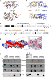
Fig. 3. The NyxB structure defines a novel family of effectors allowing the identification of the SENP3 interacting groove.
a Two views of the NyxB monomer depicted in ribbon with helices coloured in wheat, strands in blue and loops in pink. b Two views of the NyxB dimer. c Structure-based sequence alignment of NyxB and NyxA. Secondary structure elements are indicated above the sequences. Identical residues are not shaded, residues shaded in black and grey is non-conserved and conserved, respectively. Dots indicate residues identified in the acidic patch and cyan dots indicate acidic groove mutants (MAG). d Surface representation of NyxB dimer coloured according to electrostatic potential (red negative, blue positive) showing the extended acidic patch. The inset shows a close-up view of the area with residues’ side chains displayed as ball-and-sticks and mutated residues (E82, Y66 and D80) coloured in cyan. e Pull-down assay with His-NyxA, His-NyxB or the specific catalytic mutants (His-NyxAMAG or His-NyxBMAG) immobilised on Ni NTA resins that were incubated with a HeLa cell extract. An empty column was used as a control for non-specific binding. Interactions with endogenous SENP3, NPM1 or Histone 3 (H3) were visualised by western blotting using the corresponding antibody and column binding with anti-His (lower blot). Non-bound fractions (F1 and F2), last wash (W) and elution (E) are shown for each sample and the molecular weights indicated (kDa). The cell extract and the different purified Nyx inputs are also shown.
Fig. 4
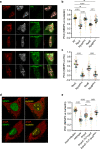
Fig. 4. The Brucella Nyx effectors directly perturb the SENP3 nucleolar localisation in host cells, including during infection.
a Representative confocal microscopy images of HeLa cells expressing the HA empty vector, HA-NyxA, HA-NyxAMAG, HA-NyxB and HA-NyxBMAG. Nucleolin (red), SENP3 (white) and HA (green) were revealed with specific antibodies. b Quantification of the Pearson correlation coefficient of SENP3 versus nucleolin (see methods for plugin description). Data are represented as means ± 95% confidence intervals from four independent experiments. Each experiment is colour coded and all events counted are shown. Data were analysed using one-way ANOVA by including all comparisons with Tukey’s correction. Not all comparisons are shown. All the cells quantified are shown in the format of SuperPlots, with each colour representing an independent experiment and its corresponding mean (N = 4). c The same data set as in b was used for quantification of the Pearson correlation coefficient of SENP3 versus HA to assess recruitment and data represented and statistical comparison are as described in (b). d Representative confocal microscopy images of HeLa cells infected for 48 h with wild-type DSRed expressing B. abortus or ΔnyxA, with nucleolin (yellow) and SENP3 (green). e Quantification of the Pearson’s coefficient of SENP3 versus nucleolin in HeLa cells infected for 48 h with either B. abortus wild-type or ΔnyxA, its complemented strain ΔnyxA::Tn7-nyxA or a complementing strain expressing the mutated acidic groove responsible for interaction with SENP3 (ΔnyxA::Tn7-nyxAMAG). Data are represented and analysed as in (b). Not all comparisons are shown. All microscopy images displayed have scale bars corresponding to 5 μm. The p values are indicated in graphs b, c and e, and not significant (ns) correspond to p > 0.05.
Fig. 5
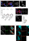
Fig. 5. B. abortus NyxA and NyxB induce cytoplasmic punctate accumulation of NVL and RPL5 in Brucella-induced foci (Bif).
a Confocal microscopy image of wild-type B. abortus (expressing DSRed) infected HeLa cells in comparison with non-infected cells or c immortalised bone-marrow derived macrophages (iBMDM) labelled with an anti-NVL antibody (white). b Quantification of the number of NVL-positive cytoplasmic structures in mock-infected control HeLa cells in comparison to wild-type or a mutant strain lacking each or both nyxA and nyxB, complemented strains or expressing MAG mutants. Data are represented as means ± 95% confidence intervals from three independent experiments. Each experiment is colour coded and all events counted are shown. Data were analysed using one-way ANOVA by including all comparisons with Tukey’s correction. Not all comparisons are shown but are available in Supplementary Table 4. d Same as in b in iBMDMs, focusing on control mock-infected cells in comparison to iBMDMs infected with wild-type or a mutant lacking NyxA/B. e Representative confocal microscopy images of HeLa cells infected with either ΔnyxA expressing DSRed and 4HA-NyxA (top) or ΔnyxB expressing DSRed and 4HA-NyxB for 48 h and labelled for NVL (white) and DAPI (cyan). f Proximity ligation assay (PLA) in HeLa cells infected for 48 h with ∆nyxA expressing 3Flag-NyxA, with either NVL (antibody from M. Nagahama, top panel), RPL5 (antibody from M. Nagahama, middle panel) or SENP3 (bottom panel). Zoom inlets correspond to positive PLA signal observed surrounding individual bacteria. All scale bars correspond to 5 µm. The p values are indicated in graphs (b) and (d), and not significant (ns) corresponds to p > 0.05.
Fig. 6
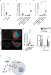
Fig. 6. Formation of Bif is dependent on Beclin1 and negatively regulated by SENP3.
a Quantification of the number of NVL-positive structures in cells infected by the wild-type B. abortus and depleted for Beclin1 with siRNA or treated with scrambled siRNA (siControl). Data are represented as means ± 95% confidence intervals from three independent experiments. Data were analysed using one-way ANOVA by including all comparisons with Tukey’s correction. b Quantification of the number of NVL-positive cytoplasmic structures in HeLa cells pre-treated with control siRNA or siSENP3(a) and infected for an additional 24 h with wild-type B. abortus or c 48 h. Mock-infected cells are included as controls. Data are represented as means ± 95% confidence intervals from four independent experiments. Each experiment is colour coded and all events counted are shown. Data were analysed using one-way ANOVA by including all comparisons with Tukey’s correction. Not all comparisons are shown. d Representative images of NVL cytoplasmic punctate accumulation (white) in HeLa cells treated with control siRNA (top) or depleted for SENP3 (bottom) followed by infection for 48 h with B. abortus expressing DSRed. Scale bars correspond to 5 μm. e Quantification of the number of NVL-positive Bif in HeLa cells pre-treated with control siRNA or an alternative siSENP3(b) mix, siBeclin or siPIAS3 and infected for an additional 24 h with wild-type B. abortus. Data correspond to means ± 95% confidence intervals from five independent experiments. Data were analysed using one-way ANOVA by including all comparisons with Tukey’s correction. f HeLa cells depleted for Beclin or PIAS3 or treated with the control siRNA for 48 h were infected with wild-type B. abortus expressing DSRed and the percentages of cells with either 1–5, 6–10 or more than 10 bacteria per cell were quantified by microscopy at 48 h post-infection. Data correspond to means ± 95% confidence intervals from five independent experiments. A two-way ANOVA with Bonferroni correction was used to compare each condition to the siControl-treated cells. The p values are indicated in the graphs (af), and not significant (ns) corresponds to p > 0.05. g NyxA/B are translocated into host cells, being detected on the vacuolar membrane, in the vicinity of replicating bacteria and in the nucleus. Within the nucleus, NyxA/B interact with SENP3, retaining it in the nucleoplasm and preventing its accumulation in the nucleoli. This sequestration promotes the formation of Bif, which are novel cytosolic structures induced upon Brucella infection, enriched in the Nyx effectors and the ribosomal proteins RPL5 and NVL. SENP3 acts as a negative regulator of Bif whereas Beclin and PIAS3 activities are required for Bif formation. The effect of NyxA/B sequestration of SENP3 on the SUMOylation levels of Beclin remains to be determined. Created with Biorender.com.

References

    1. Hegde RS, Zavodszky E. Recognition and degradation of mislocalized proteins in health and disease. Cold Spring Harb. Perspect. Biol. 2019;11:a033902. - PMC - PubMed
    1. Long X, et al. The critical roles of the SUMO-specific protease SENP3 in human diseases and clinical implications. Front. Physiol. 2020;11:558220. - PMC - PubMed
    1. Yan S, et al. Redox regulation of the stability of the SUMO protease SENP3 via interactions with CHIP and Hsp90. EMBO J. 2010;29:3773–3786. - PMC - PubMed
    1. Kunz K, Piller T, Müller S. SUMO-specific proteases and isopeptidases of the SENP family at a glance. J. Cell Sci. 2018;131:jcs211904. - PubMed
    1. Liu K, et al. A fine-tuning mechanism underlying self-control for autophagy: deSUMOylation of BECN1 by SENP3. Autophagy. 2020;16:975–990. - PMC - PubMed

Publication types

MeSH terms