. 2023 Jan 6;13(1):293.
doi: 10.1038/s41598-022-26899-0.

Ubiquilin-2 regulates pathological alpha-synuclein

Affiliations

Ubiquilin-2 regulates pathological alpha-synuclein

Stephanie S Sandoval-Pistorius et al. Sci Rep. .

Abstract

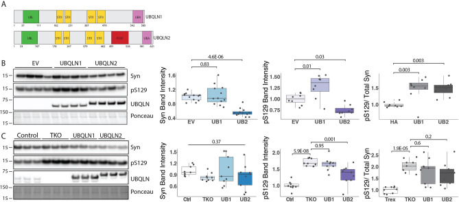
The key protein implicated in Parkinson's disease and other synucleinopathies is α-synuclein, and a post-translationally modified form of the protein, phosphorylated at serine 129 (pS129), is a principal component in Lewy bodies, a pathological hallmark of PD. While altered proteostasis has been implicated in the etiology of Parkinson's disease, we still have a limited understanding of how α-synuclein is regulated in the nervous system. The protein quality control protein Ubiquilin-2 (UBQLN2) is known to accumulate in synucleinopathies, but whether it directly regulates α-synuclein is unknown. Using cellular and mouse models, we find that UBQLN2 decreases levels of α-synuclein, including the pS129 phosphorylated isoform. Pharmacological inhibition of the proteasome revealed that, while α-synuclein may be cleared by parallel and redundant quality control pathways, UBQLN2 preferentially targets pS129 for proteasomal degradation. Moreover, in brain tissue from human PD and transgenic mice expressing pathogenic α-synuclein (A53T), native UBQLN2 becomes more insoluble. Collectively, our studies support a role for UBQLN2 in directly regulating pathological forms of α-synuclein and indicate that UBQLN2 dysregulation in disease may contribute to α-synuclein-mediated toxicity.


Conflict of interest statement

The authors declare no competing interests.

Figures

Figure 1
Figure 1
UBQLN2 insolubility is increased in Parkinson’s disease human tissue and mouse models. (A,B) Western blots of lysates from post-mortem cingulate cortex of PD and DLB patients show an increase in insoluble UBQLN2 in (A) PD (n = 8) and (B) DLB (n = 10) compared to age-matched controls. S PBS soluble, I PBS insoluble/sarkosyl soluble. (C) Representative western blot of whole brain lysate from mice overexpressing human mutant α-syn A53T solubilized in sarkosyl (n = 8) shows elevated levels of insoluble UBQLN2 compared to non-transgenic controls (n = 3). Western blots were cropped to focus on protein(s) of interest. Uncropped Western blots are shown in Supplemental Figure S1.
Figure 2
Figure 2
UBQLN KO leads to pS129 accumulation while UBQLN2 overexpression decreases levels of pS129 in vitro. (A) Diagram of UBQLN2 and UBQLN1 protein structure. (B) Representative western blot of UBQLN 1,2, 4 Total Knock out (TKO) cells transfected to express WT α-syn alone or together with UBQLN1 (UB1) or UBQLN2 (UB2). Total α-syn and pS129 levels are decreased by co-expression of UBQLN2 but not UBQLN1. The ratio of pS129/total Syn is increased in both UBQLN1 and UBQLN2 co-expression (n = 8). (C) Representative western blot of TKO, UBQLN1 rescue, UBQLN2 rescue, and control cells were transfected with α-syn showing elevated pS129 levels in TKO cells compared to control cells, which is partially reversed by inducible co-expression of UBQLN2 but not UBQLN1. The ratio of pS129/total Syn is significantly increased in TKO compared to control and not reversed by UBQLN1 or UBQLN2 rescue (n = 8). Western blots were cropped to focus on protein(s) of interest. Uncropped Western blots are shown in Supplemental Figure S2.
Figure 3
Figure 3
UBQLN2, but not UBQLN1, lowers α-syn levels in HEK-293 cells. (AD) Representative immunofluorescence images show that the number of α-syn-containing cells are decreased when co-expressed with UBQLN2 (A,C; n = 10 field of cells over 3 independent experiments) but not with UBQLN1 (B,D; n = 10 field of cells over 3 independent experiments). Scale bar 100 μm (E) Expressed as a ratio of α-syn positive cells to UBQLN1 and UBQLN2, α-syn is significantly reduced in UBQLN2-expressing cells.
Figure 4
Figure 4
UBQLN2 interacts with and may alter α-syn levels in A53T mice. (A) Schematic depicting breeding strategy to obtain A53TxUb2-hi and A53TxUb2-KO mice. (B) Western blot of RIPA-soluble brain lysates showing increased levels of total α-syn in A53TxUb2-KO mice (n = 6), while A53TxUb2-hi (n = 7) mice show a trend toward decreased α-syn levels compared to non-transgenic (Non-Tg) controls (n = 8). (C) Representative images of total a-syn (magenta) and UBQLN2 (cyan) immunofluorescence in the deep cerebellar nucleus of 12-month-old Non-Tg (n = 6), A53T (n = 6), A53TxUb2-hi (n = 6), and A53TxUb2-KO (n = 5) mice and quantification show no significant difference in total a-syn expression between genotypes. (D) Western blot showing total endogenous α-syn levels were unchanged in both Ub2-hi (n = 4) and Ub2-KO mice (n = 4) compared to Non-Tg controls (n = 4). Western blots were cropped to focus on protein(s) of interest. Uncropped Western blots are shown in Supplemental Figure S3). (E) Immunoblot showing endogenous and transgenic UBQLN2 is pulled down when α-syn is immunoprecipitated from mouse brain lysate (n = 4). (F) Immunoblot showing α-syn coprecipitates with FLAG-tagged UBQLN2 in A53TxUb2-hi mice when FLAG is immunoprecipitated from mouse brain lysate (n = 2).
Figure 5
Figure 5
pS129 α-syn accumulates in A53T mice lacking UBQLN2. (A) Representative western blot of pS129 in RIPA-soluble brain lysates from the indicated transgenic mouse crosses. (B) Quantification from A showing accumulation of pS129 in A53TxUb2-KO mice (n = 6) and unchanged in A53TxUb2-hi mice (n = 7) compared to A53T controls (n = 8). (C) Ratio of pS129 to total Syn (see Fig. 4) does not differ significantly between groups (n = 8). (D) Representative images of pS129 expression in the cortex of A53T (n = 3), A53TxUb2-hi (n = 3) and A53TxUb2-KO (n = 3) show increased pS129 expression in A53T mice lacking UBQLN2 (12-month-old mice). (E) Representative western blots of endogenous pS129 in UBQLN2 transgenic mice. (F) Quantification of E showing a trend toward decreased endogenous murine pS129 levels in Ub2-hi mice (n = 4). (G) Analysis of the ratio of endogenous pS129 to total endogenous Syn (obtained from Fig. 4) shows no difference between groups (n = 4). Western blots were cropped to focus on protein(s) of interest. Uncropped Western blots are shown in Supplemental Figure S4.
Figure 6
Figure 6
Optical Pulse Labeling reveals no effect of UBQLN2 on α-syn clearance in neurons. (A) Schematic of Optical Pulse labeling experiment. Rat primary cortical neurons were transfected with hSyn-Dendra2 and UBQLN2-iRFP or iRFP. Dendra2 was photoconverted 24 h post-transfection with 405 nm light, imaged immediately and every 24 h thereafter for 3 days. Scale bar 25 μm. (B) Optical pulse labeling reveals no change in red fluorescence intensity over time in neurons transfected with UBQLN2 (n = 213) compared to control (n = 182; p = 0.6). (C) Representative images of optical pulse labeling of hSyn-Dendra2 in rat primary cortical neurons transfected with hSyn-Dendra2 and UBQLN2-iRFP or iRFP. (D) Optical pulse labeling in primary rate neurons reveals no change in half-life of hSyn-Dendra2 upon transfection with UBQLN2 vs control (iRFP; p = 0.28). hSyn-Dendra2 half-life was determined by measuring the loss of photoconverted hSyn-Dendra2 (RFP) signal over time for each cell. (E) Density plot of hSyn-Dendra2 half-life measurements from individual neurons transfected with control (n = 182) or UBQLN2 (n = 213). Values were pooled from eight wells per condition, from each of two biological replicates and analyzed by the Kruskal–Wallis test.
Figure 7
Figure 7
UBQLN2 targets pS129 α-syn to the proteasome for degradation. (A) Representative western blot of TKO cells overexpressing α-syn and transfected with either EV or UBQLN2 and treated with 10 μM lactacystin (Lac), 50 μM rolipram (Rol), or vehicle (Veh). Quantification of total α-syn and pS129 levels shows that pS129 accumulates in cells transfected with UBQLN2 and treated with Lac while total α-syn levels were not changed between groups (n = 9–10). Total α-syn and pS129 levels were each normalized to α-syn levels of EV, vehicle-treated cells. Western blots were cropped to focus on protein(s) of interest. (B) Representative images showing pS129 fluorescence in cells transfected with either EV or UBQLN2 and treated with either Veh, Lac, or Rol. Quantification of pS129 fluorescence intensity further supports that pS129 α-syn is increased when treated with Lac and in the presence of UBQLN2 (n = 11). pS129 fluorescence intensity was normalized to that of vehicle treated, EV cells. Scale bar 50 μm. (C) Representative western blot of TKO cells overexpressing α-syn and transfected with either EV or UBQLN2 and treated with 10 μM chloroquine (CLQ), 300 nM rapamycin (RAP), or vehicle (Veh). Quantification of pS129 or total α-syn levels reveals no differences between treatment groups (n = 10). Total α-syn and pS129 levels were each normalized to α-syn levels of EV-transfected, vehicle-treated cells. Uncropped Western blots are shown in Supplemental Figure S5.

References

    1. Baba M, et al. Aggregation of alpha-synuclein in Lewy bodies of sporadic Parkinson's disease and dementia with Lewy bodies. Am. J. Pathol. 1998;152:879–884. - PMC - PubMed
    1. Spillantini MG, et al. Alpha-synuclein in Lewy bodies. Nature. 1997;388:839–840. doi: 10.1038/42166. - DOI - PubMed
    1. Shults CW. Lewy bodies. Proc. Natl. Acad. Sci. U S A. 2006;103:1661–1668. doi: 10.1073/pnas.0509567103. - DOI - PMC - PubMed
    1. Klein C, Westenberger A. Genetics of Parkinson's disease. Cold Spring Harb. Perspect. Med. 2012;2:1. doi: 10.1101/cshperspect.a008888. - DOI - PMC - PubMed
    1. Kruger R, et al. Ala30Pro mutation in the gene encoding alpha-synuclein in Parkinson's disease. Nat. Genet. 1998;18:106–108. doi: 10.1038/ng0298-106. - DOI - PubMed

Publication types

MeSH terms

LinkOut - more resources