. 2023 Jan 7;80(1):33.
doi: 10.1007/s00018-022-04671-x.

ABC-transporter CFTR folds with high fidelity through a modular, stepwise pathway

Affiliations

ABC-transporter CFTR folds with high fidelity through a modular, stepwise pathway

Jisu Im et al. Cell Mol Life Sci. .

Abstract

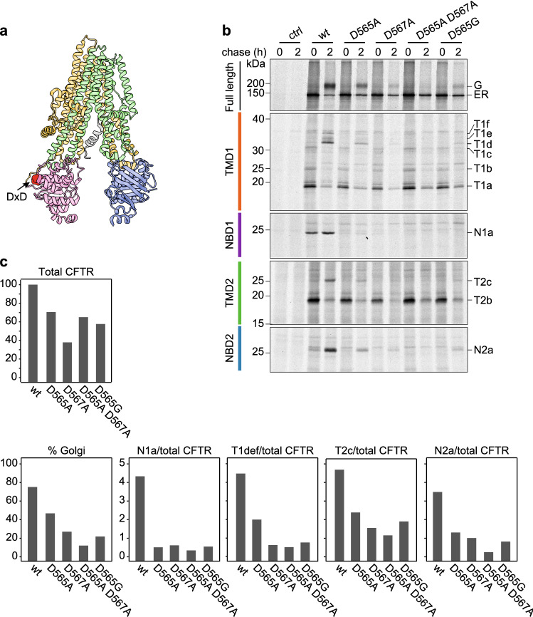
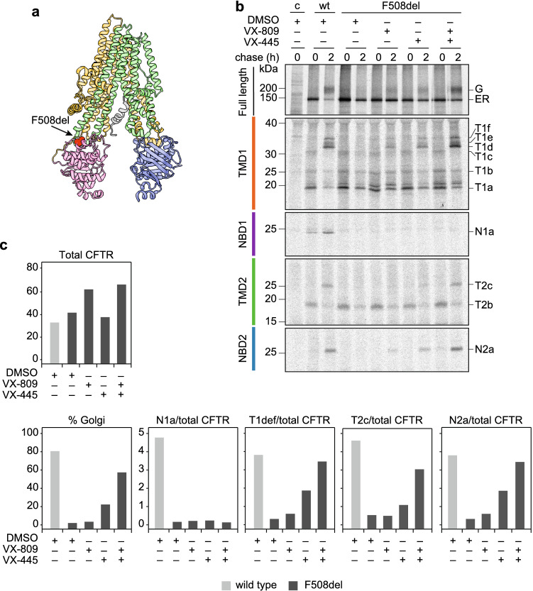
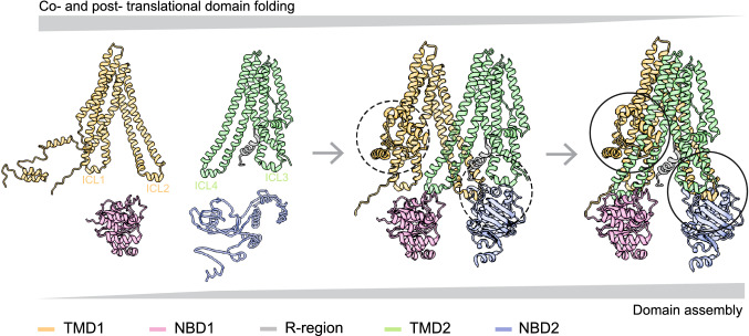
The question how proteins fold is especially pointed for large multi-domain, multi-spanning membrane proteins with complex topologies. We have uncovered the sequence of events that encompass proper folding of the ABC transporter CFTR in live cells by combining kinetic radiolabeling with protease-susceptibility assays. We found that CFTR folds in two clearly distinct stages. The first, co-translational, stage involves folding of the 2 transmembrane domains TMD1 and TMD2, plus one nucleotide-binding domain, NBD1. The second stage is a simultaneous, post-translational increase in protease resistance for both TMDs and NBD2, caused by assembly of these domains onto NBD1. Our assays probe every 2-3 residues (on average) in CFTR. This in-depth analysis at amino-acid level allows detailed analysis of domain folding and importantly also the next level: assembly of the domains into native, folded CFTR. Defects and changes brought about by medicines, chaperones, or mutations also are amenable to analysis. We here show that the well-known disease-causing mutation F508del, which established cystic fibrosis as protein-folding disease, caused co-translational misfolding of NBD1 but not TMD1 nor TMD2 in stage 1, leading to absence of stage-2 folding. Corrector drugs rescued stage 2 without rescuing NBD1. Likewise, the DxD motif in NBD1 that was identified to be required for export of CFTR from the ER we found to be required already upstream of export as CFTR mutated in this motif phenocopies F508del CFTR. The highly modular and stepwise folding process of such a large, complex protein explains the relatively high fidelity and correctability of its folding.

Keywords: ABC-transporter; COPII; Cystic fibrosis; Domain assembly; Protein folding; Secretory pathway.


Conflict of interest statement

The authors have no relevant financial or non-financial interests to disclose.

Figures

Fig. 1
Fig. 1
New antibodies against CFTR transmembrane domains recognize whole domains synthesized in vitro and in vivo. a Cartoon of CFTR in which red spheres represent the position of the epitopes to which antibodies were raised. Sites for N-linked glycosylation in TMD2 are shown. b TMD1 and TMD2 were translated in vitro and translocated in the presence of semi-intact cells as a source of ER membranes. Membrane fractions of in-vitro translated TMD1 and TMD2 were resolved by SDS-PAGE (left panel) or immunoprecipitated (IP) with E1-22 & TMD1C (middle), and I4N & TMD2C, respectively (right). Samples were resolved by 12% SDS-PAGE. c Schematic representation of constructs used in (b, d), with a table showing the used boundaries of CFTR domains. d HEK293T cells expressing single or multi-domain CFTR constructs were pulse labeled for 15 min and lysed immediately or after a chase of 2 h. CFTR was immunoprecipitated from detergent lysates with indicated antibodies and resolved by 10% SDS-PAGE
Fig. 2
Fig. 2
Antibodies against TMDs identify folding intermediates in vivo. a Workflow of radioactive pulse-chase-limited-proteolysis assay. b HEK293T cells expressing CFTR were pulse labeled for 15 min and chased for the indicated times. CFTR was immunoprecipitated using MrPink and immunoprecipitates were resolved by 7.5% SDS-PAGE. Remaining lysates were subjected to limited proteolysis (LP) with 25 µg/mL Proteinase K and protease-resistant fragments were immunoprecipitated with c E1-22, d TMD1C, e I4N, f TMD2C, g MrPink, and h 596 and resolved by 12% SDS-PAGE. wt, wild-type CFTR; T1a-T1f are TMD1-specific protease resistant fragments; T2a-c are TMD2-specific protease resistant fragments; N1a and N2a are protease resistant fragments specific for NBD1 and NBD2, respectively. i Quantification of the amount of CFTR transported to the Golgi complex and of the indicated fragments generated during limited proteolysis
Fig. 3
Fig. 3
Identification of TMD1 fragments. a HEK293T cells expressing CFTR were pulse labeled for 15 min and lysed immediately or after a chase of 2 h. Lysates were subjected to limited proteolysis with 25 µg/mL Proteinase K and protease-resistant fragments were immunoprecipitated with indicated antibodies. Position of the antigenic epitopes in TMD1 is marked with red spheres in the cartoon on the right. b HEK293T cells expressing N-terminally truncated versions ΔN48 to ΔN76 of CFTR were pulse-labeled for 15 min and lysed. Lysates were subjected to limited proteolysis with 25 µg/mL Proteinase K and immunoprecipitated with TMD1C. The downward shifts of the TMD1-derived fragments are marked in cyan boxes. c HEK293T cells expressing N-terminal CFTR truncations ΔN47 to ΔN51 were pulse labeled for 15 min and lysed immediately. Detergent lysates were subjected to limited proteolysis with 25 µg/mL Proteinase K and immunoprecipitated with E1-22. Lane intensity profiles (ImageQuant analysis) of the fragments of interest are shown below each panel. d Same as (c) but now with C-terminal truncations K381X to E395X. e Same as (b) but now for ΔN4 to ΔN45 CFTR and after a chase of 2 h. f Same as (c) with N-terminal truncations ΔN35 to ΔN45, but the 15-min pulse labeling was followed by a 1-h chase. g Same as (f) but now with N-terminal truncations ΔN2 to ΔN5. Experiments in panels (f, g) were done in the presence of VX-770 (3 µM) and/or VX-809 (3 µM) as indicated. The undigested samples corresponding to panels b–d and f–g are in Fig. S3a–e. All samples were resolved by 12% SDS-PAGE. wt wild-type CFTR
Fig. 4
Fig. 4
Summary map of TMD1 fragments. a TMD1 amino-acid sequence highlighting the Proteinase-K cleavage sites that result in TMD1 fragments T1a–T1f. All alpha helices are depicted as columns of 3 residues wide, with the exception of ICL1, which includes a coupling helix of only 5 residues (SRVLD). T1a extends from aa S50 to S256, T1c from S50 to L383, T1d from D36 to L383, T1e from F17 to L383, and T1f from M1 to L383. Key: light blue circles: Proteinase-K consensus cleavage residues; grey lines: antigenic epitopes; blue lines: N-terminal boundaries of fragments; red lines: C-terminal boundaries of fragments; dotted lines: possible cleavage area; Lh1: location of Lasso helix 1; Lh2, location of Lasso helix 2; elbow: location of the N-terminal elbow helix. b TMD1 proteolytic fragments in structure representation. The left panel shows fragments T1a and T1c with their fragment boundaries. The structure is based on CFTR models with the N-terminus in the cytoplasm [13, 15] because this better encapsulates the conformation of the protein during vectorial folding, before the domains have assembled; the cryo-EM structures represent the mature, domain-assembled form, which has not yet been reached. The right panel shows fragments T1d-f with their fragment boundaries. For this panel we used the cryo-EM structure (PDB: 5UAK) [14], as T1d-f represent domain-assembled, mature CFTR
Fig. 5
Fig. 5
Identification of TMD2 fragments. a HEK293T cells expressing CFTR were pulse labeled for 15 min and lysed immediately or after a chase of 2 h (left panel). Lysates were subjected to limited proteolysis with 25 µg/mL Proteinase K and protease-resistant fragments were immunoprecipitated with indicated antibodies. Position of the antigenic epitopes in TMD2 is marked with red spheres in the cartoon (middle panel). The right panel shows TMD2-fragment immunoprecipitates treated with PNGaseF. b HEK293T cells expressing CFTR mutants S1058K to L1065K were pulse labeled for 15 min and lysed immediately. c Same as (b) but with mutants M961K to L967K. d same as (b) with mutants T908K to S912K, but now after a chase of 2 h. e Same as (b) with C-terminal truncations N1184X to M1191X. Detergent cell lysates subjected to limited proteolysis with 25 µg/mL Proteinase K were immunoprecipitated with TMD2C. Samples were resolved by 12% SDS-PAGE. Lane intensity profiles (ImageQuant analysis) of the fragments of interest are shown below each panel. The undigested samples corresponding to panels (be) are in Fig. S4c–f. Wt wild-type CFTR, T2a, T2b, and T2c are TMD2-specific protease-resistant fragments. Red asterisks mark the wild-type peaks between which straight lines were drawn for reference
Fig. 6
Fig. 6
Summary map of TMD2 fragments. a Representation of the TMD2 amino-acid sequence highlighting the Proteinase-K cleavage sites that result in TMD2 fragments T2a-T2c. All alpha helices are depicted as columns of 3 residues wide. T2a extends from aa K1060 to M1191, T2b from N965 to M1191 and T2c from T910 to M1191. Key: light blue circles: Proteinase-K consensus cleavage residues; grey lines: antigenic epitopes; blue lines: N-terminal boundaries of fragments; red lines: C-terminal boundaries of fragments; dotted lines: possible cleavage area. b TMD2 proteolytic fragments in structure representation. The left panel shows fragments T2a and T2b with their fragment boundaries. As in Fig. 4b, the structure is based on CFTR models with the N-terminus in the cytoplasm [13, 15] because this better encapsulates the conformation of the protein during vectorial folding, before the domains have assembled; the cryo-EM structures represent the mature, domain-assembled form, which has not yet been reached. The right panel shows fragment T2c with its fragment boundaries. For this panel we did use the cryo-EM structure (PDB: 5UAK) [14], as T2c represents domain-assembled, mature CFTR
Fig. 7
Fig. 7
The N1a fragment represents NBD1 without RE and RI. a Secondary structure of NBD1 including Regulatory Insertion (RI) and C-terminal Regulatory Extension (RE), with antigenic epitopes indicated and the identity of the N1a fragment shown, resulting from cleavages in RI and RE, likely from residues L428 to F653. b NBD1 was translated in vitro at 30 °C for 30 min and proteolyzed on ice with different concentrations of Proteinase K; immunoprecipitations were done with the indicated antibodies. Full-length NBD1, 27-kDa and 25-kDa fragments are indicated. c Structure of NBD1 and interacting TMD elements ICL1 and ICL4 (PDB: 5UAK) [14] indicating the antigenic epitopes in red. N1a contains NBD1 without most of RI (as well as upstream sequence) and RE
Fig. 8
Fig. 8
The late NBD2 fragment contains almost all of NBD2. a Secondary structure of NBD2 with antigenic epitopes indicated and showing the identity of the N2a fragment, residues I1192-Q1439. b HEK293T cells expressing CFTR or not (control) were pulse labeled for 10 min and chased for 2 h. After digestion with 25 µg/mL Proteinase K, proteolytic fragments were immunoprecipitated in parallel with I4N, TMD2C, 596, 2–39.14, and 2-3.5 antibodies and resolved by 12% SDS-PAGE. c HEK293T cells expressing CFTR or not (control) were pulse labeled for 10 min and chased for 2 h. After digestion with 25 µg/mL Proteinase K, proteolytic fragments were immunoprecipitated in parallel with I4N, TMD2C, 596, 2–39.14, and 2-3.5 antibodies and resolved by 12% SDS-PAGE. d HEK293T cells expressing C-terminally truncated CFTR constructs F1437X to S1442X were pulse-labeled for 15 min and chased for 1 h and subsequently digested or not with 25 µg/mL Proteinase K for 15 min on ice. Non-digested lysates (upper panel) were immunoprecipitated with MrPink and proteolyzed samples with NBD2-specific antibody 596 (lower panel) and resolved by 7.5% or 12% SDS-PAGE, respectively. Lane intensity profiles (ImageQuant analysis) of N2a are shown below the panel. e Structure of NBD2 and interacting TMD elements ICL2 and ICL3 (PDB: 5UAK) [14] indicating the antigenic epitopes in red. Dotted lines denote unresolved residues in the structure
Fig. 9
Fig. 9
Folding analysis of CFTR mutated in an NBD1 di-acidic ER-export motif. a Structure of CFTR showing the location of the di-acidic ER exit motif (arrow & red-shaded area; PDB: 5UAK) [14]. b HEK293T cells expressing CFTR variants were pulse-labeled for 15 min and lysed immediately or after a chase of 2 h. CFTR was immunoprecipitated from non-proteolyzed lysate with MrPink and resolved by 7.5% SDS-PAGE. Proteolysis was done with 25 µg/mL Proteinase K and domain-specific fragments were immunoprecipitated with E1-22 (TMD1), MrPink (NBD1), TMD2C (TMD2), and 596 (NBD2) and analyzed by 12% SDS-PAGE. c Quantitation of total amounts of wild-type and mutant CFTR at 2-h chase time, of the % that had been transported to the Golgi complex and of the T1def (TMD1), T2c (TMD2), and N2a (NBD2) fragments, as measure of domain assembly
Fig. 10
Fig. 10
Correctors boost F508del CFTR domain assembly without correcting underlying defect in NBD1. a Structure of CFTR showing the location of F508del (arrow & red-shaded area; PDB: 5UAK) [14]. b HEK293T cells expressing wt CFTR and F508del CFTR were treated with corrector compounds VX-809, VX-445 or both (at 3 µM final concentration) during starvation, labeling and chase, and subsequently processed as in Fig. 9b. c Quantitation of wt CFTR and F508del CFTR transported to the Golgi complex and of domain assembly as the amount of fragment corrected for total amount of CFTR
Fig. 11
Fig. 11
Two-stage folding process of CFTR. The left structure illustrates stage 1 of folding: TMD1, NBD1, and TMD2 fold already co-translationally and acquire a structure as if expressed on their own [7, 31]. ICL1 docks onto NBD1 already during synthesis [31], whereas the N-terminus of TMD1 cannot have native structure yet and may hang off the ER membrane [13] and/or associate with NBD1 [15]. NBD1 reaches its native protease-resistant fold completely co-translationally, which does not change upon domain assembly. Structure is based on CFTR models with the N-terminus in the cytoplasm [13, 15]. The middle structure shows a putative domain-assembly intermediate, in which the domain interfaces form fast and cooperatively, yielding the right structure. The fully domain-assembled CFTR (right structure) has ICL1 of TMD1 and ICL4 of TMD2 docked onto NBD1. The TMDs assemble, such that ICL2 of TMD1 and ICL3 of TMD3 associate with and stabilize NBD2. The circles show the sites that gain significant protease resistance around the time CFTR leaves the ER, reaches the Golgi complex and obtains complex glycans. ICL2, ICL3, and NBD2 acquire increased protease resistance simultaneously (right circle), as does the N-terminus of TMD1, which becomes protease resistant by wrapping around TMD1 and TMD2 (left circle). Structure in middle and right panels are based on cryo-EM structure (PDB: 5UAK) [14]

References

    1. Uhlen M, Fagerberg L, Hallstrom BM, Lindskog C, Oksvold P, Mardinoglu A, et al. Proteomics. Tissue-based map of the human proteome. Science. 2015;347:1260419. doi: 10.1126/science.1260419. - DOI - PubMed
    1. Overington JP, Al-Lazikani B, Hopkins AL. How many drug targets are there? Nat Rev Drug Discov. 2006;5:993–996. doi: 10.1038/nrd2199. - DOI - PubMed
    1. Gulezian E, Crivello C, Bednenko J, Zafra C, Zhang Y, Colussi P, et al. Membrane protein production and formulation for drug discovery. Trends Pharmacol Sci. 2021;42:657–674. doi: 10.1016/j.tips.2021.05.006. - DOI - PubMed
    1. Skach WR. Cellular mechanisms of membrane protein folding. Nat Struct Mol Biol. 2009;16:606–612. doi: 10.1038/nsmb.1600. - DOI - PMC - PubMed
    1. Shiber A, Doring K, Friedrich U, Klann K, Merker D, Zedan M, et al. Cotranslational assembly of protein complexes in eukaryotes revealed by ribosome profiling. Nature. 2018;561:268–272. doi: 10.1038/s41586-018-0462-y. - DOI - PMC - PubMed

MeSH terms

Substances