Review
. 2023 Feb;65(2):343-370.
doi: 10.1111/jipb.13446.

Asymmetric cell division in plant development

Affiliations
Review

Asymmetric cell division in plant development

Yi Zhang et al. J Integr Plant Biol. 2023 Feb.

Abstract

Asymmetric cell division (ACD) is a fundamental process that generates new cell types during development in eukaryotic species. In plant development, post-embryonic organogenesis driven by ACD is universal and more important than in animals, in which organ pattern is preset during embryogenesis. Thus, plant development provides a powerful system to study molecular mechanisms underlying ACD. During the past decade, tremendous progress has been made in our understanding of the key components and mechanisms involved in this important process in plants. Here, we present an overview of how ACD is determined and regulated in multiple biological processes in plant development and compare their conservation and specificity among different model cell systems. We also summarize the molecular roles and mechanisms of the phytohormones in the regulation of plant ACD. Finally, we conclude with the overarching paradigms and principles that govern plant ACD and consider how new technologies can be exploited to fill the knowledge gaps and make new advances in the field.

Keywords: asymmetric cell division; peptide signaling; phytohormonal signaling; plant development; polarity proteins; transcription factors.


Conflict of interest statement

CONFLICTS OF INTEREST

The authors declare that they have no conflict of interest.

Figures

Figure 1.
Figure 1.. Plant asymmetric cell division (ACD) driven by asymmetric expression of transcription factors (TFs)
(A) ACD events during early embryogenesis generate 1) apical and basal lineages, 2) inner and outer layers in the proembryo, and 3) the lens-shaped quiescent center (QC) precursor (magenta). (B) The hypophyseal ACD is driven by the expression of TMO7 which is originally expressed in the upper provascular cells (light green) and diffused to the hypophysis (dark blue). The MP/ARF5 TF binds to the TMO7 promoter to drive its expression. (C) Schematic shows a longitudinal section of a developing root apical meristem (RAM) with distinct cell types (differently colored). Future division planes in ACD are marked by dashed lines. (D) Key transcription factors are required for the QC maintenance (middle), and they are differentially expressed to specify daughter-cell fates in the ACD of cortex/endodermal initial daughter (CEID) (left) and columella stem cell (CSC) (right), respectively. Middle: mobile transcription factors, SHR (grey ovals) and WOX5 (magenta ovals), have non-autonomous functions (arrows indicate protein movement) that are required for QC maintenance (middle). Left: the mobile TF SHR (gray ovals) diffuses from the vasculature tissue to the CEI and CEID, where it interacts with SCR to induce sequential ACDs via activating the expression of the cell-cycle gene CYCD6. Right: the CSC ACD is guided by the oscillating expression of FEZ that activates SMB to form a negative feedback loop to induce cell differentiation. The reappearance of FEZ in the apical daughter directs the next round of ACD.
Figure 2.
Figure 2.. Intrinsic factors in the regulation of plant ACD
(A) Schematics depict the process of zygotic ACD and differentially expressed TFs. Before zygotic ACD, WRKY2 works together with HDG11 and HDG12 to induce zygote polarization by directly upregulating WOX8 expression. Following the zygotic ACD, WOX2 and WOX8/9 are differentially inherited in apical and basal daughter cells, respectively. (B) Schematics of stomata development in Arabidopsis (dicot). Three bHLH TFs, SPCH, MUTE, and FAMA are consecutively required for stomatal initiation, transition, and differentiation through interacting with SCRM and SCRM2. (C) Schematics of stomatal ACDs for development and patterning in Arabidopsis. Amplifying ACDs occur in the meristemoid (M), and spacing ACDs occur in the stomatal lineage ground cell (SLGC). Stomatal ACD is guided by the polarity complex (green crescent) assembled by the scaffold proteins BASL, BRX members, and POLAR proteins. (D) Schematics of stomata development and subsidiary mother cell (SMC) ACD in monocots, such as maize or Brachypodium. The differentiation of the guard mother cell (GMC) is required for the specification of SMC (pink) and its subsequent ACD. Complementary polarized proteins, the PAN complex, and BdPOLAR, together with actin-mediated nuclear movement participate in the regulation of SMC ACD.
Figure 3.
Figure 3.. The dynamics of polarity proteins in regulating stomatal ACD in Arabidopsis and maize
(A) Diagrams show dynamically assembled polarity components in pre-mitotic (left) and post-mitotic (right) stomatal lineage cells expressing the polarity module (green crescent) in Arabidopsis. In the pre-mitotic cell (left), POLAR recruits the BIN2 kinase that suppresses the MAPKKK YDA’s inhibition on SPCH, so that SPCH abundantly accumulates in the ACD precursor cell to drive cell division. In the post-mitotic cell, BASL recruits the BSL1 phosphatase that dislodges BIN2 from the polarity module and activates the YDA kinase, leading to highly suppressed SPCH activity and lowered cell division potential. Thus, the dynamically assembled polarity components differentially determine the high cell-division potential and restricted cell division at different stages during ACD. (B) Model for ACD of subsidiary mother cell (SMC) in monocot stomatal development. Before the SMC ACD in maize, the SCAR/WAVE regulatory complex (WRC) (dark blue) is first assembled in the site of guard mother cell (GMC) contact, followed by sequential recruitment of PAN2, WPR proteins, PAN1 and ROP GTPases (light blue), that direct the actin patch formation and directional nuclear migration, ultimately leading to asymmetric placement of the division plate. An outer nuclear membrane protein MLKS2 (dashed green oval) is required for the process of polarity complex-guided nuclear migration in maize. In addition, transcription factor MUTE (yellow ovals), in both maize and Brachypodium, is originally expressed in the GMC, where it has an important role in inducing the GMC symmetric division, and then moves to the adjacent SMC, where it directly upregulates the expression of PAN1 and PAN2. In Brachypdium, BdPOLAR defines a polarity domain at the plasma membrane that is complementary to the PAN polarity and required for SMC ACD.
Figure 4.
Figure 4.. Extrinsic cue-mediated cell signaling regulates plant ACD
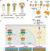
Schematics describe peptide ligand-mediated signal transduction that regulates zygotic and stomatal ACD, respectively. Zygotic ACD in Arabidopsis is triggered by the endosperm-derived ESF1 peptides that are likely perceived by the SERK receptor-like kinases at the cell surface. In the cytoplasm, membrane-associated BSK1/2 and SSP/BSK12 may directly activate the YDA-MKK4/5-MPK3/6 signaling pathway to stabilize WRKY2 protein that further directs the expression of WOX8 to specify the basal cell lineage after a zygotic ACD. The YDA-MAPK cascade is shared by stomatal ACD for suppression of stomatal initiation. Upstream of YDA, the extracellular EPF family peptides, including the positive peptides EPF1/2 and negative peptides STOMAGEN, are perceived by the membrane receptor complex comprised of the ERECTA, TMM, and SERK proteins. Downstream of the YDA-MAPK signaling, the master transcription factor SPCH initiates stomatal ACD by inducing thousands of gene expression, including the peptide ligand EPF2, and SPCH activity is directly suppressed by MPK3/6 activities for protein degradation. The YDA-MAPK signaling may also suppress later developmental processes driven by MUTE (dashed line) that activates the expression of EPF1 peptides in stomatal fate transition.
Figure 5.
Figure 5.. Auxin biosynthesis, transportation, and signaling regulate plant ACD
(A) The combined activity of local auxin biosynthesis (yellow cells) and directional transportation (magenta arrows) generates dynamic auxin maxima (orange cells) during early embryogenesis in Arabidopsis. The directional auxin transportation is mainly driven by the differentially polarized PIN1 and PIN7 as depicted. (B) An auxin depletion (light orange) presages stomatal differentiation. This process is coordinated by elevated expression of the efflux transporter PIN3 (green lines) in meristemoids (Ms) and guard mother cells (GMCs) that undergo terminal differentiation. (C) The canonical auxin signaling mediated by the TIR1/AFB receptors regulates multiple stages in lateral root (LR) development. The LR positioning is specified by the release of IAA28-mediated suppression of ARF5/6/7/18/19 that activate the expression of GATA23. The LR initiation ACD is promoted by the release of IAA14-mediated suppression of ARF7/19 that activates the expression of LBD16. The ACD patterning during the lateral root primordium (LRP) formation is regulated by the non-canonical auxin signaling mediated by the cell-surface receptors, ABP1, and homologs, that partner with the TMK1/4 receptor-like kinases. TMK1/4 directly activates MKK4/5 and MPK3/6 signaling to specify orientated cell divisions.
Figure 6.
Figure 6.. Phytohormonal signaling and crosstalk in the regulation of root apical meristem (RAM) and stomatal divisions
(A) The quiescent center (QC) division is fine-tuned by phytohormonal signaling. The WOX5 expression is enriched in the QC cells to restrict cell division. The expression of WOX5 can be both positively and negatively regulated by auxin-triggered activation of MP/ARF5 and ARF10/16, respectively. Cytokinin (CK) signaling suppresses the expression of WOX5 by activating the ARR1/12 transcription factors. Brassinosteroids (BR) trigger the activity of the BRI1 and BAK1 receptors that eventually promote the nuclear partition of the BES1 transcription factor in the QC cell, where BES1 interacts with the transcription co-repressor TPL to suppress the expression of BRAVO/MYB56 that functions to restrict QC cell division. Upon stresses or damages, BR signaling is elevated, so that the BES1-mediated suppression on BRAVO/MYB56 is strengthened to promote cell division for stem-cell replenishment. Upon wounding, jasmonic acid (JA) signaling induces the expression of MYC2 and ERF115 that release the inhibition of RBR on SCR so that elevated SCR functions together with SHR to promote cell division in the QC. (B) In Arabidopsis, the key transcription factor SPCH in stomatal development is heavily regulated in pre-division (left) and post-division (right) cells. In MMCs (pre-division cell, light pink), the SPCH protein is differentially phosphorylated by multiple kinases, including MPK6 and SnRK in the abscisic acid (ABA) signaling pathway, resulting in SPCH degradation. However, H2O2 signaling triggers the nuclear partition of the KIN10 kinase to phosphorylate SPCH for stabilization. Specific phosphorylation sites of SPCH are noted on each branch. JA signaling suppresses stomatal initiation through MYC2/3/4 that likely functions through SPCH. Ethylene signaling suppresses stomatal initiation by an unknown mechanism (likely through SPCH) which also influences the polarization events represented by BRXL2 (blue arrow-head shapes in the SLGC). In the SLGCs that undergo pavement differentiation or spacing division (light blue), SPCH is phosphorylated by BIN2 kinase in the BR signaling pathway for protein degradation. CK signaling promotes SPCH expression that induces the expression of CLE9/10 ligands and ARR-A TFs, both of which in turn suppress CK signaling. Thus, a locally reduced CK signaling is maintained in the SLGC.

Similar articles

Cited by

References

    1. Abrash EB, and Bergmann DC (2009). Asymmetric cell divisions: a view from plant development. Dev. Cell 16: 783–796. - PubMed
    1. Abrash EB, and Bergmann DC (2010). Regional specification of stomatal production by the putative ligand CHALLAH. Development 137: 447–455. - PubMed
    1. Adamowski M, and Friml J (2015). PIN-dependent auxin transport: Action, regulation, and evolution. Plant Cell 27: 20–32. - PMC - PubMed
    1. Adrian J, Chang J, Ballenger CE, Bargmann BO, Alassimone J, Davies KA, Lau OS, Matos JL, Hachez C, Lanctot A, Vatén A, Birnbaum KD, and Bergmann DC (2015). Transcriptome dynamics of the stomatal lineage: birth, amplification, and termination of a self-renewing population. Dev. Cell 33: 107–118. - PMC - PubMed
    1. Aichinger E, Kornet N, Friedrich T, and Laux T (2012). Plant stem cell niches. Ann. Rev. Plant Biol 63: 615–636. - PubMed

Substances

LinkOut - more resources