. 2023 Jan 25;51(2):935-951.
doi: 10.1093/nar/gkac1231.

Structural basis of Qng1-mediated salvage of the micronutrient queuine from queuosine-5'-monophosphate as the biological substrate

Affiliations

Structural basis of Qng1-mediated salvage of the micronutrient queuine from queuosine-5'-monophosphate as the biological substrate

Shr-Hau Hung et al. Nucleic Acids Res. .

Abstract

Eukaryotic life benefits from-and ofttimes critically relies upon-the de novo biosynthesis and supply of vitamins and micronutrients from bacteria. The micronutrient queuosine (Q), derived from diet and/or the gut microbiome, is used as a source of the nucleobase queuine, which once incorporated into the anticodon of tRNA contributes to translational efficiency and accuracy. Here, we report high-resolution, substrate-bound crystal structures of the Sphaerobacter thermophilus queuine salvage protein Qng1 (formerly DUF2419) and of its human ortholog QNG1 (C9orf64), which together with biochemical and genetic evidence demonstrate its function as the hydrolase releasing queuine from queuosine-5'-monophosphate as the biological substrate. We also show that QNG1 is highly expressed in the liver, with implications for Q salvage and recycling. The essential role of this family of hydrolases in supplying queuine in eukaryotes places it at the nexus of numerous (patho)physiological processes associated with queuine deficiency, including altered metabolism, proliferation, differentiation and cancer progression.


Figures

Graphical Abstract
Graphical Abstract
Queuosine is a microbiome-derived 7-deazaguanosine tRNA nucleoside important for the gut-brain axis. We report the structure and function of QNG1 as a queuosine-5′-monophosphate hydrolase essential for salvage of the nucleobase queuine from exogenous queuosine, following its phosphorylation by a cellular kinase.
Figure 1.
Figure 1.
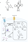
Queuine salvage by eukaryotic cells. (A) Queuosine structure and its location in bacterial and eukaryotic tRNAs carrying GUN anticodons (tyrosyl-, aspartyl-, asparaginyl- and histidyl-tRNA). (B) Current model for queuine uptake, incorporation in tRNA and salvage in the eukaryotic cell. Queuine (q) can be salvaged directly from the extracellular space, or alternatively from exogenous queuosine nucleoside (Q) via the recently discovered DUF2419 (C9orf64)-dependent pathway, incorporated into tRNA by the eTGT (comprised of QTRT1 and QTRT2 subunits) and further modified with mannose and galactose by unknown sugar transferases. Both q and Q enter the cell via unknown transporters, with early studies suggesting that imported queuosine may be converted to the 5′-nucleotide. Salvage of the queuine base from tRNA turnover could occur from Q, queuosine-5′-monophosphate (Q-5′MP) or queuosine-3′-monophosphate (Q-3′MP). The exact biochemical function of DUF2419 has remained unknown.
Figure 2.
Figure 2.
Qng1/QNG1-dependent q salvage from exogenous Q. (A, B) Qng1/QNG1 is required for salvage of q from exogenous Q, but not for salvage of exogenous q, by S. pombe and HeLa cells. (A) APB-Northern gel assay showing the queuosylation status of aspartyl tRNA extracted from S. pombe wild-type strain (top) and qng1Δ strain (bottom) grown in YES medium supplemented with 1% bactopeptone extract (BP), queuosine (Q) or queuine (q) in varying concentrations (100, 50 and 10 nM). (B) APB-Northern gel assay showing the queuosylation status of histidyl (top) and asparaginyl (bottom) tRNA extracted from wild-type and QNG1 (C9orf64)-knockout HeLa cells (CRISPR Clone 1 in Supplementary Figure S4b) grown in serum-free medium without any additions (–), or in the presence of queuine (q, 100 nM), queuosine (Q, 100 nM) or bactopeptone (BP, 1%). (C, D) UV absorbance traces at 260 nm from LC/MS analysis of in vitro time-course reactions showing release of q from synthetic Q (50 μM) in the presence of a high concentration (10 μM) of recombinant StQng1 (C) or HsQNG1 (D) at select time points (cyan and red lines). Active enzymes (after His6 tag removal) were used. UV traces of the control reactions (no-enzyme) at the highest time point (180 min) are also shown (blue line). Reaction conditions are identical except for temperature (50°C and 37°C for the bacterial and human enzyme, respectively, based on the living environment of the source organism). The identities of the substrate and product with corresponding retention times were verified by extracting the ion counts for the expected masses [M+H+] = m/z 278 for q and 410 for Q (Supplementary Figure S4c,d).
Figure 3.
Figure 3.
Qng1 structure and Q recognition. (A, B) Overall structure of Qng1. (A) Ribbon diagram of the StQng1 monomer. Secondary structure elements are shown in different colors and labeled. The C-terminus marks the general location of the active site. (B) Comparison of the crystal structures of StQng1 (left) and of human Ogg1 (right, PDB ID 1EBM (Bruner et al., 2000)) as an example of the HhH DNA glycosylase family, showing their common two-lobe architecture and HhH motifs (red). The two lobes are indicated with brackets. DNA bound to HsOgg1 (gold) marks the general DNA binding cleft in HhH DNA glycosylases, and the Qng1-specific surface insertions (green) differentiate it from DNA glycosylases. See full structure-based sequence alignment in Supplementary Figure S6. (C–F) Q recognition by Qng1. (C) Omit Fo-Fc map (resolution 2.35 Å, contour level 1.5 σ) in the active site region of StQng1 bound to queuosine. All residues of the cyclopentenediol binding pocket are shown in stick model. (D) View showing enzyme interactions with the 7-deazaguanine and ribose moieties of Q. Distances are in Ångstroms. (E) The terminal carboxylate is integral to the active site and catalysis. Superposition of the crystal structures of wild-type apo StQng1 (active, pink) and of the His6-tagged StQng1 bound to Q (inactive, cyan) allows visualizing substrate binding in the active site of the wild-type enzyme. The C-terminal peptide in the wild-type structure is bent toward the active site, which would position the terminal carboxylate within contact distance from the ribose of Q, participating in recognition together with K199. The invariant C-terminal Tyr323 side chain is anchored by hydrogen bonds and stacking against the ribose-pocket residue Y232. (F) View of the electrostatic surface potential in the active site region showing the positively charged pocket occupied by the malonate ion near the 5′-OH of bound Q, suggesting that Q-5′MP could be a substrate for Qng. In all panels, the Q molecule and malonate ion are shown in orange and pink stick model, respectively, and water molecules are shown as red spheres.
Figure 4.
Figure 4.
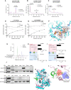
Q-5′-monophosphate is the biologically-relevant substrate of Qng1/QNG1. (A–C) LC/MS analysis of end-point StQng1 reactions containing various Q nucleotides as substrates. Active enzyme (after His6 tag removal) was used. UV absorbance traces at 260 nm from LC/MS analysis of StQng1 reactions containing 50 μM enzyme and 22.9 μM Q-3′MP (A), 17.9 μM Q-3′,5′DP (B) or 6.82 μM Q-5′MP (C) as substrates (red line), and of no-enzyme control reactions (blue line). Reaction times are 120, 90 and 60 min, respectively. Queuine release activity is robust when Q-5′MP is used as substrate (100% of Q consumed in a 60-min reaction), and absent or negligible when Q-3′MP or Q-3′,5′DP, respectively, are used as substrates (only 4.9% of Q-3′,5′MP consumed in a 90-min reaction and no Q-3′MP consumed in a 120-min reaction). Similar data for the human enzyme are shown in Supplementary Figure S8d-f and summarized in Supplementary Table S10. (D, E) Competition assays of StQng1 and HsQNG1 activities using Q and Q-5′MP as competing substrates. Substrate and product concentrations from LC/MS analysis of time-course reactions containing 2 μM StQng1 (D) or HsQNG1 (E) and 35 μM each Q and Q-5′MP, showing consumption of Q-5′MP, but not Q, and production of q. Time points are 5, 15, 30, 60, 120 and 180 min. Quantification was based on A260 peak areas in the LC chromatograms (Supplementary Figure S9a–d), normalized to the concentration of a Q standard in the no-enzyme control. Data represent triplicate reactions and errors are smaller than the displayed symbols (standard-error range ±0.05 to ±0.43 μM). (F) Omit Fo-Fc electron density map of StQng1 bound to Q-5′MP, in the active site region (resolution 2.4 Å, contour level 3 σ). Residues of the cyclopentendiol and phosphate binding pockets are shown. (G–I) HeLa cells harbouring a knockout of QNG1 accumulate intracellular Q-5′MP from exogenously supplied queuosine. (G) LC–MS/MS was used to quantify extracellular (Media) and intracellular (Cytosol) levels of Q, Q-5′MP and q in wild-type (WT, panel H) and QNG1-knockout HeLa cells (QNG1-KO, panel I) supplied with queuosine nucleoside (250 nM) for 24 h in the culture medium. Quantification was done using standard curves as described in Supplementary Methods (Supplementary Figure S10). (J) High-levels of QNG1 expression in mouse liver are consistent with a role in Q salvage from the gut. Western blot assessment of QNG1 (C9orf64) expression in HeLa cells and tissues from adult C57BL/6 mice. GAPDH serves as a loading control. MW, molecular weight markers; Int, small intestine. (K) Superposition of the crystal structures of wild-type HsQNG1 (pink) with bound q (orange stick model) onto His6-tagged (inactive) StQng1 (cyan) with bound Q-5′MP (ligand not shown). The extra helix in the human enzyme adjacent to the cyclopentenediol binding pocket is colored in green and indicated with an arrow. View is similar to panel F. (L) Close up view of the active site cavity in the human enzyme represented by its electrostatic surface potential, with bound q. The human-specific helix is colored in green, and bulky side chains W49 and L52 and the H53-D319 salt bridge abutting and constricting the cyclopentene binding pocket are shown in stick model. Water molecules are shown as red spheres.
Figure 5.
Figure 5.
Proposed mechanism of the Qng1 catalyzed reaction. (A) View of strictly conserved H-bonding network putatively leading to protonation of the purinic N7 atom. For a complete representation of the active site as it would be in the catalytically active state, the C-terminal Y323 is modeled based on its position in the crystal structure of wild-type (active) StQng1 (pink stick model). For clarity, the substrate phosphate moiety is shown in transparent stick model. (B) Proposed catalytic mechanism of Qng1. Our structural data argue strongly for bond cleavage and formation with retention of configuration, either via a covalent intermediate with D231 in a formal double-displacement mechanism, or via an SN1 mechanism in which D231 stabilizes the intermediate oxocarbenium ion electrostatically and water addition occurs from the same face as the departed base. These represent mechanistic extremes on a spectrum of potential reactivity, and to reflect this mechanistic ambiguity we show the reaction intermediate as possessing an extended, partial covalent bond between C1′ and D231, with partial oxocarbenium ion character. The interactions of Y323 and K199 are not shown after the first frame for clarity.

References

    1. Fergus C., Barnes D., Alqasem M.A., Kelly V.P.. The queuine micronutrient: charting a course from microbe to man. Nutrients. 2015; 7:2897–2929. - PMC - PubMed
    1. Kasai H., Ohashi Z., Harada F., Nishimura S., Oppenheimer N.J., Crain P.F., Liehr J.G., von Minden D.L., McCloskey J.A.. Structure of the modified nucleoside Q isolated from Escherichia coli transfer ribonucleic acid. 7-(4,5-cis-dihydroxy-1-cyclopenten-3-ylaminomethyl)-7-deazaguanosine. Biochemistry. 1975; 14:4198–4208. - PubMed
    1. Ohgi T., Kondo T., Goto T.. Total synthesis of optically pure nucleoside Q. Determination of absolute configuration of natural nucleoside Q. J. Am. Chem. Soc. 1979; 101:3629–3633.
    1. Hutinet G., Swairjo M.A., de Crécy-Lagard V.. Deazaguanine derivatives, examples of crosstalk between RNA and DNA modification pathways. RNA Biol. 2017; 14:1175–1184. - PMC - PubMed
    1. Randerath E., Agrawal H.P., Randerath K.. Specific lack of the hypermodified nucleoside, queuosine, in hepatoma mitochondrial aspartate transfer RNA and its possible biological significance. Cancer Res. 1984; 44:1167–1171. - PubMed

Publication types

Supplementary concepts