Review
. 2023 Mar;46(3):211-227.
doi: 10.1016/j.tins.2022.12.007. Epub 2023 Jan 5.

Persistent nociceptor hyperactivity as a painful evolutionary adaptation

Affiliations
Review

Persistent nociceptor hyperactivity as a painful evolutionary adaptation

Edgar T Walters et al. Trends Neurosci. 2023 Mar.

Abstract

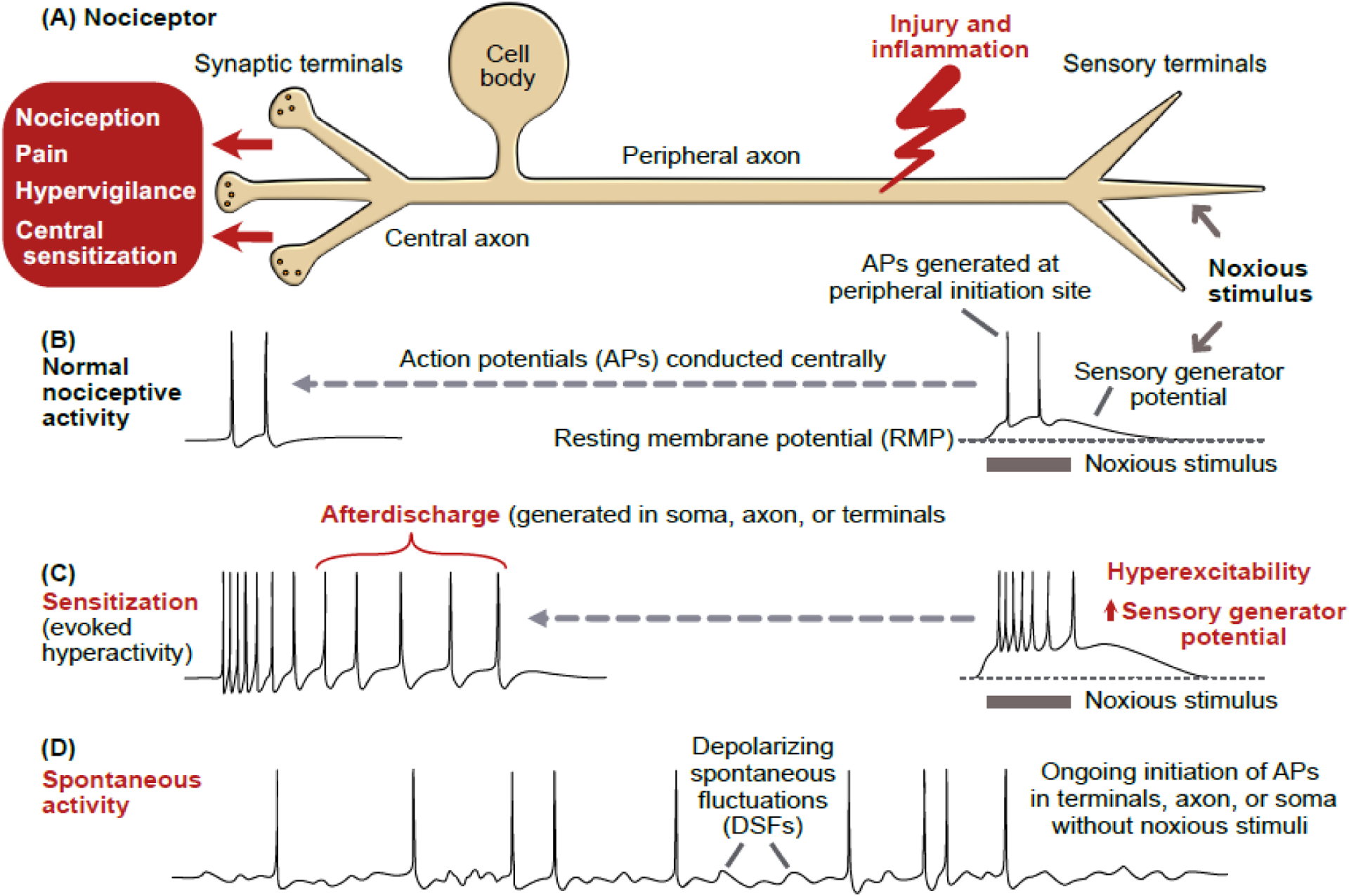
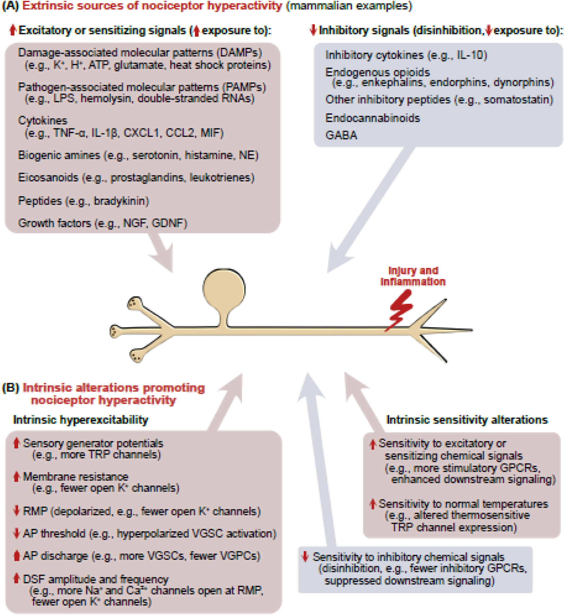
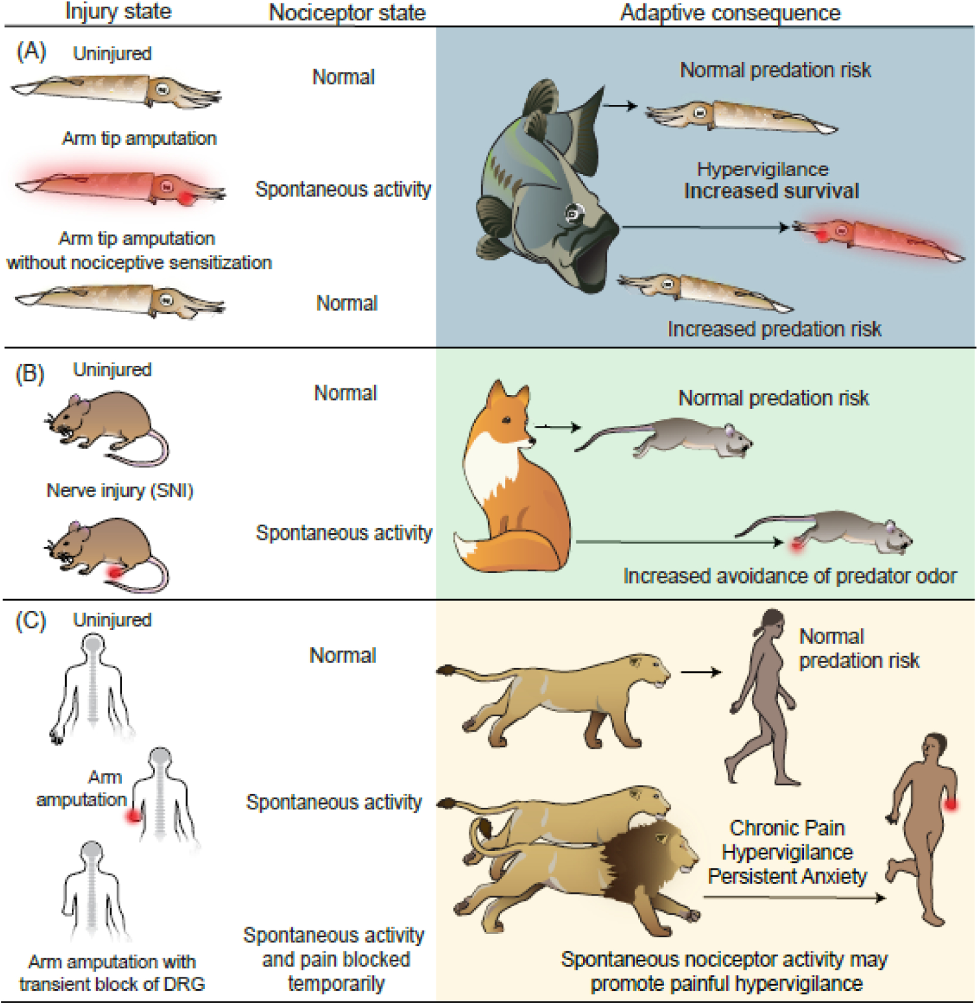
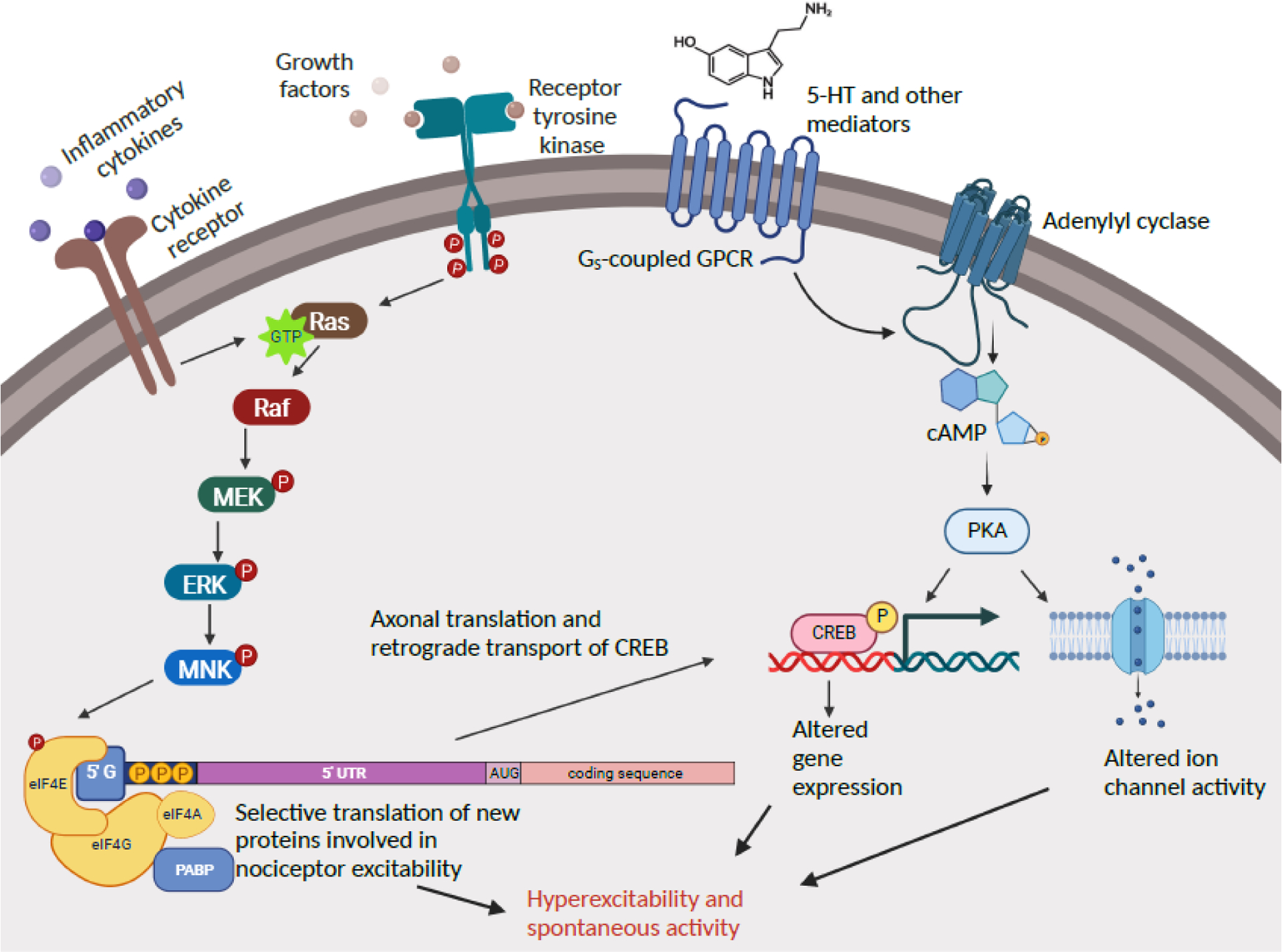
Chronic pain caused by injury or disease of the nervous system (neuropathic pain) has been linked to persistent electrical hyperactivity of the sensory neurons (nociceptors) specialized to detect damaging stimuli and/or inflammation. This pain and hyperactivity are considered maladaptive because both can persist long after injured tissues have healed and inflammation has resolved. While the assumption of maladaptiveness is appropriate in many diseases, accumulating evidence from diverse species, including humans, challenges the assumption that neuropathic pain and persistent nociceptor hyperactivity are always maladaptive. We review studies indicating that persistent nociceptor hyperactivity has undergone evolutionary selection in widespread, albeit selected, animal groups as a physiological response that can increase survival long after bodily injury, using both highly conserved and divergent underlying mechanisms.

Keywords: Aplysia; cephalopod; chronic pain; human; primary afferent neuron; spontaneous activity.


Conflict of interest statement

Declaration of interests T.J.P. is a co-founder of 4E Therapeutics, Doloromics, PARMedics, and NuvoNuro; he serves on the Board of Directors of 4E Therapeutics and Doloromics; he is an inventor on patents related to MNK inhibition for the treatment of pain. E.S.S. receives funding from AstraZeneca and GlaxoSmithKline. E.T.W., R.J.C., and G.G.N. declare no competing interests.

Figures

Figure 1.
Figure 1.. Nociceptor hyperactivity.
(A) Schematic of a primary nociceptor in vertebrates, gastropod molluscs, and leeches. The cell body is centrally located and distant from peripheral terminals where survivable injury and inflammation are most likely. In vertebrates, the cell body is in a ganglion near the central nervous system (CNS). In molluscs and leeches, nociceptor cell bodies are in ganglia within the CNS. (B) Normal nociceptive activity initiated by noxious stimulation of peripheral terminals produces a depolarizing sensory generator potential that may reach threshold for action potentials (APs). These APs are conducted to the CNS, sometimes evoking pain. (C) Illustration of sensitization of nociceptive activity evoked by the same noxious stimulus as in panel B, with the evoked hyperactivity following injury caused by a larger sensory generator potential, increased terminal excitability, and afterdischarge triggered by the evoked APs, which sometimes evoke pain, hypervigilance, and central sensitization. Experimentally, injection of pulses or ramps of depolarizing current (usually into the cell body) is often used to reveal hyperexcitability (see Box 1). (D) Spontaneous activity after injury, defined as ongoing activity generated in the absence of concurrent extrinsic stimulation of a neuron (the spontaneous activity is sometimes generated by depolarizing spontaneous fluctuations, DSFs) [100]. The term spontaneous activity is often used more loosely in referring to any nociceptor activity in the absence of evident ongoing noxious stimulation, including cases where intrinsic (cell autonomous) hyperexcitability and unobserved inflammatory signals combine to drive ongoing hyperactivity. Spontaneous activity and/or afterdischarge have been found in peripheral terminals, injured axons, and cell bodies of mammals and molluscs.
Figure 2.
Figure 2.. Sources of nociceptor hyperactivity following injury.
(A) Left: Some of the many extrinsic chemical signals implicated in mammals to excite and/or sensitize nociceptors. Many of these have multiple sources (e.g., glutamate and ATP are DAMPs released from ruptured cells but also are secreted exocytotically from various cell types including neurons). Right: Hyperactivity may also be promoted by reducing exposure to inhibitory chemical signals (disinhibition). (B) In principle, nociceptor hyperactivity can be promoted by intrinsic hyperexcitability via any or all the listed electrophysiological alterations [100,104]. Hyperactivity may also be promoted by increasing a nociceptor’s intrinsic sensitivity to excitatory or sensitizing sensory stimuli (including normal body temperature) or chemical signals, and by decreasing intrinsic sensitivity to inhibitory chemical signals. Abbreviations: AP, action potential; ATP, adenosine triphosphate, DSF, depolarizing spontaneous fluctuation; GABA, gamma-aminobutyric acid; GDNF, glial-derived neurotrophic factor; GPCR, G protein-coupled receptor; IL-1β, interleukin-1β; IL-10, interleukin 10; LPS, lipopolysaccharide; MIF, macrophage migration inhibitory factor; NGF, nerve growth factor; RMP, resting membrane potential; TNFα, tumor necrosis factor α; TRP, transient receptor potential; VGPC, voltage-gated potassium channel; VGSC, voltage-gated sodium channel.
Figure 3.
Figure 3.. Persistent nociceptor hyperactivity can be adaptive.
(A) Squid survival is enhanced by nociceptor activity after injury. Peripheral tissue injury causes long-lasting nociceptor hyperactivity expressed over much of the body surface [75] (red). In staged 30-minute encounters with fish predators [87], ~75% of uninjured subjects survived. Nearly half of squid previously receiving a minor arm amputation survived. Although selectively targeted by the fish, injured squid began their escape behavior farther from approaching fish (indicated by arrow length) than uninjured squid [87]. Squid that were injured without nociceptive sensitization and persistent nociceptor hyperactivity (prevented by transient block of nociceptor activity during amputation) waited longer to begin escape, with only ~20% surviving, indicating the survival benefit of persistent nociceptor hyperactivity after injury [86]. (B) Enhanced avoidance of a predator cue by mice with neuropathic pain. Mice with a spared nerve injury model exhibit nociceptor hyperactivity generated in the hindpaw (red) [138] and DRG [25,139] (not shown). When exposed to fox urine, injured mice chose a route that took them farther from the predator odor [88]. This suggests that injury-induced nociceptor hyperactivity promotes hypervigilance and increased risk aversion. (C) Speculative benefit of chronic nociceptor hyperactivity in ancestral humans at high risk for predation after injury (e.g., amputation injury in the illustration). The top two rows parallel the arguments for squid illustrated in panel A. The bottom row illustrates a situation of transient blockade of DRG APs in amputees that blocks ongoing pain [83]. If spontaneous activity in human nociceptors persistently drives hypervigilance as well as pain (as suggested by anxiety being a comorbidity of pain [90]), this hyperactivity should be protective for injured individuals who are more likely to be targeted by predators.
Figure 4.
Figure 4.. Shared signaling pathways driving nociceptor hyperactivity in Aplysia and rodents.
Two general cell signaling pathways have been implicated directly and indirectly in the maintenance and induction of persistent nociceptor hyperactivity both in a gastropod mollusc and in mammals: Ras-MNK signaling and cAMP-PKA signaling. These pathways are more complex than shown (e.g., cAMP has additional effectors implicated in nociceptor hyperactivity, and many protein kinases in addition to PKA can activate CREB), and each has additional interactions with the same and other cell signaling pathways. Only steps for which biochemical and/or pharmacological evidence is available are indicated. 5-HT, serotonin; 5’ UTR, 5’ untranslated region; cAMP, cyclic adenosine monophosphate; CREB, cAMP response element-binding protein; eIF4E, eukaryotic translation initiation factor 4E (correspondingly for eIF4G, eIF4A); ERK, extracellular-signal-regulated kinase; GPCR, G protein coupled receptor; Gs, stimulatory G protein; MEK, mitogen-activated protein kinase kinase; MNK, mitogen-activated protein kinase-interacting kinase; P, phosphorylation; PABP, poly-A-binding protein; PKA, protein kinase A; Raf, rapidly activated fibrosarcoma kinase; Ras, rat sarcoma GTPase. Created with BioRender.com.

References

    1. Williams GC and Nesse RM (1991) The dawn of Darwinian medicine. Q Rev Biol 66, 1–22 - PubMed
    1. Finnerup NB et al. (2021) Neuropathic Pain: From Mechanisms to Treatment. Physiol Rev 101, 259–301 - PubMed
    1. Sommer C et al. (2018) Inflammation in the pathophysiology of neuropathic pain. Pain 159, 595–602 - PubMed
    1. Walters ET (2021) Nociceptors and chronic pain. In Oxford Research Encyclopedia of Neuroscience (Murray S. Oxford University Press
    1. Roza C and Bernal L (2022) Electrophysiological characterization of ectopic spontaneous discharge in axotomized and intact fibers upon nerve transection: a role in spontaneous pain. Pflugers Arch 474, 387–396 - PubMed

Publication types