

Long Axial Field-of-View PET for Ultra-Low-Dose Imaging of Non-Hodgkin Lymphoma during Pregnancy
- PMID: 36611320
- PMCID: PMC9818305
- DOI: 10.3390/diagnostics13010028
Long Axial Field-of-View PET for Ultra-Low-Dose Imaging of Non-Hodgkin Lymphoma during Pregnancy
Abstract
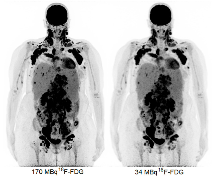
Generally, positron emission tomography imaging is not often performed in the case of pregnant patients. The careful weighing of the risks of radiation exposure to the fetus and benefits for cancer staging and the swift onset of treatment for the mother complicates decision making in clinical practice. In oncology, the most commonly used PET radiotracer is 2-deoxy-2-[fluorine-18] fluoro-D-glucose (18F-FDG), a glucose analog which has established roles in the daily routines for, among other applications, initial diagnosis, staging, (radiation) therapy planning, and response monitoring. The introduction of long axial Field-of-View (LAFOV) PET systems allows for PET imaging with a reduced level of injected 18F-FDG activity while maintaining the image quality. Here, we discuss the first reported case of a pregnant patient diagnosed with follicular lymphoma using LAFOV PET imaging for the staging and therapy selection. The acquired PET images show diagnostic quality images with clearly distinguishable areas of lymphadenopathy, even with only 34 MBq of injected 18F-FDG activity, leading to a considerable decrease in the level of radiation exposure to the fetus.
Keywords: 18F-FDG; LAFOV PET; follicular lymphoma; image quality; pregnant.
Conflict of interest statement
The authors declare no conflict of interest.
Figures


References
-
- Bastiaannet E., Groen B., Jager P.L., Cobben D.C.P., van der Graaf W.T.A., Vaalburg W., Hoekstra H.J. The value of FDG-PET in the detection, grading and response to therapy of soft tissue and bone sarcomas: A systematic review and meta-analysis. Cancer Treat. Rev. 2004;30:83–101. doi: 10.1016/j.ctrv.2003.07.004. - DOI - PubMed
Publication types
LinkOut - more resources
Full Text Sources
