Review
. 2022 Dec 31;13(1):139.
doi: 10.3390/diagnostics13010139.

Ischiofemoral Impingement Syndrome: Clinical and Imaging/Guidance Issues with Special Focus on Ultrasonography

Affiliations
Review

Ischiofemoral Impingement Syndrome: Clinical and Imaging/Guidance Issues with Special Focus on Ultrasonography

Wei-Ting Wu et al. Diagnostics (Basel). .

Abstract

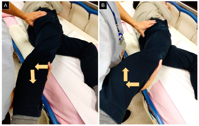
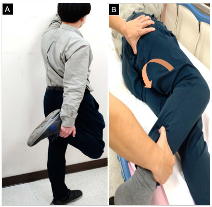
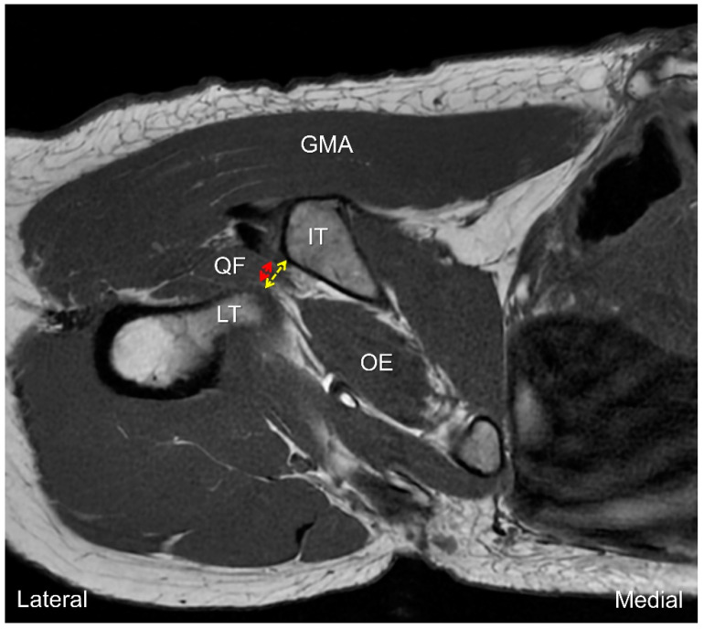
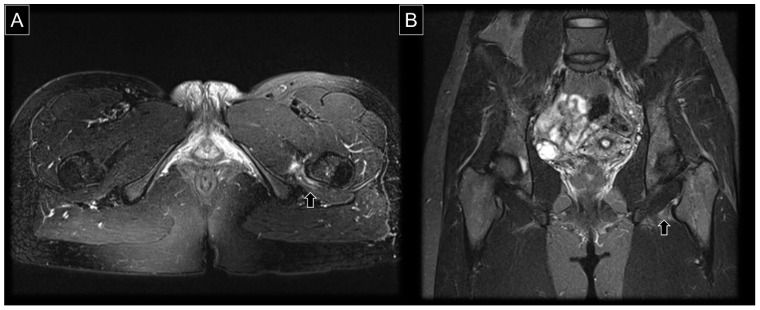
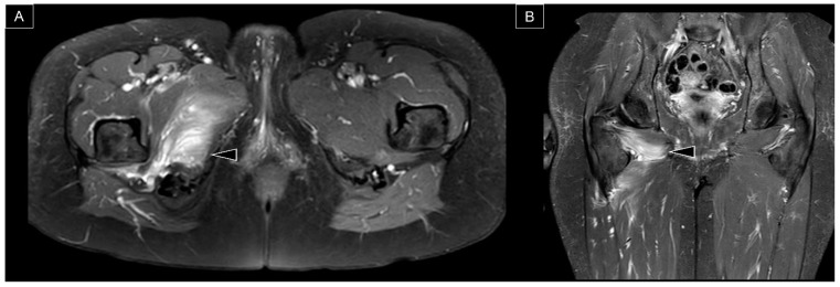
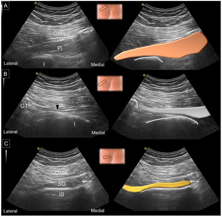
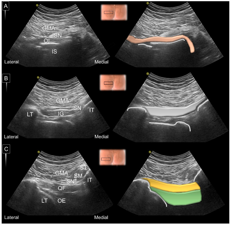
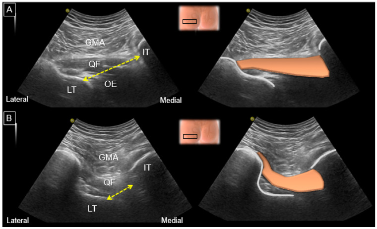
Ischiofemoral impingement syndrome is a neglected cause of posterior hip pain which is derived from narrowing of the space between the lateral aspect of the ischium and the medial aspect of the lesser trochanter. Its diagnosis is challenging and requires the combination of physical tests and imaging studies. In the present narrative review, we found that femoral anteversion predisposes patients to the narrowing of the ischiofemoral space and subsequent quadratus femoris muscle injury. Magnetic resonance imaging serves as the gold-standard diagnostic tool, which facilities the quantification of the ischiofemoral distance and the recognition of edema/fat infiltration/tearing of the quadratus femoris muscle. Ultrasound is useful for scrutinizing the integrity of deep gluteal muscles, and its capability to measure the ischiofemoral space is comparable to that of magnetic resonance. Various injection regimens can be applied to treat ischiofemoral impingement syndrome under ultrasound guidance and they appear to be safe and effective. Finally, more randomized controlled trials are needed to build solid bases of evidence on ultrasound-guided interventions in the management of ischiofemoral impingement syndrome.

Keywords: hip pain; ischiofemoral impingement; magnetic resonance imaging; quadratus femoris; ultrasound.


Conflict of interest statement

The authors declare no conflict of interest.

Figures

Figure 1
Figure 1
Cadaveric image of the posterior hip region, focusing on the ischiofemoral space. GMA: gluteus maximus muscle; GME: gluteus medius muscle; Pi: piriformis muscle; SG: superior gemellus muscle; OI: obturator internus muscle; IG: inferior gemellus muscle; QF: quadratus femoris muscle; LT: lesser trochanter; IT: ischial tuberosity; ST: semitendinosus; BF: biceps femoris; ADM: adductor magnus muscle; SN: sciatic nerve.
Figure 2
Figure 2
Cadaveric image providing an axial view of the ischiofemoral space. GMA: gluteus maximus muscle; ST: tendon of the semitendinosus; SM: tendon of the semimembranosus; QF: quadratus femoris muscle; OE: obturator externus muscle; LT: lesser trochanter; IT: ischial tuberosity; ADM: adductor magnus muscle; ADB: adductor brevis muscle; ADL: adductor longus muscle.
Figure 3
Figure 3
(A) Illustration of femoral anteversion (left) and compensatory toe-in posture (right); (B) illustration of femoral retroversion (left) and compensatory toe-out posture (right). * denotes the femoral anteversion angle.
Figure 4
Figure 4
Pain over the ischium can be reproduced by walking with a long stride (A) and relieved during short-stride walking (B).
Figure 5
Figure 5
(A) Tenderness over the ischium is elicited under passive hip extension and adduction, and it disappears during (B) hip extension and abduction. Arrows: direction of force applied by the examiner.
Figure 6
Figure 6
(A) Step 1: The volunteer’s hip is brought to extension, external rotation, and adduction to compress the ischiofemoral space in the upright position. (B) Step 2: The participant is in the lateral decubitus position with the knee being flexed and the hip being externally rotated (brown arrow). The examiner places the thumb over the ischium to check if the tenderness can be provoked.
Figure 7
Figure 7
Axial T1 weighted MRI for measurement of the ischiofemoral (yellow dashed line) and quadratus femoris (red dashed line) spaces. GMA: gluteus maximus muscle; QF: quadratus femoris muscle; OE: obturator externus muscle; LT: lesser trochanter; IT: ischial tuberosity.
Figure 8
Figure 8
(A) Axial and (B) coronal views of T2 weighted MRI in a case with IIS. The arrows indicate swelling and perifocal edema of the quadratus femoris muscle.
Figure 9
Figure 9
(A) Axial and (B) coronal views of T2 weighted MRI in another case with post-traumatic IIS, with edema and hematoma inside the quadratus femoris and obturator externus muscles (arrowheads).
Figure 10
Figure 10
US images and schematic drawings of (A) the piriformis muscle and (B) its tendon (arrowhead, gray color) and (C) the superior gemellus muscle. GMA: gluteus maximus muscle; Pi: piriformis muscle, brown color; SG: superior gemellus muscle, yellow color; IS: ischial spine; I: ischium; GT: greater trochanter. Black hollow square: transducer position.
Figure 11
Figure 11
US images and schematic drawings of (A) the obturator internus, (B) the inferior gemellus and (C) the quadratus femoris and obturator externus muscles. GMA: gluteus maximus muscle; IS: ischial spine; OI: obturator internus muscle, light brown color; SN: sciatic nerve; IG: inferior gemellus muscle, gray color; QF: quadratus femoris muscle, yellow color; OE: obturator externus muscle, green shade; LT: lesser trochanter; IT: ischial tuberosity; ST: semitendinosus; SM, semimembranosus muscle.
Figure 12
Figure 12
US images and schematic drawings for measuring of the ischiofemoral distance (yellow dashed line) during (A) internal and (B) external femoral rotations. GMA: gluteus maximus muscle; QF: quadratus femoris muscle, brown color; OE: obturator externus muscle; LT: lesser trochanter; IT: ischial tuberosity.
Figure 13
Figure 13
US images and schematic drawings of the sciatic nerve (SN, yellow color) in its long axis in the (A) neutral position and (B) with hip internal rotation. GMA: gluteus maximus muscle; SG: superior gemellus muscle; IS: ischial spine; OI: obturator internus muscle; IG: inferior gemellus muscle; QF: quadratus femoris muscle; arrowhead, kinking of the sciatic nerve.
Figure 14
Figure 14
US images and guided injection of the quadratus femoris muscle performed using (A) the in-plane lateral-to-medial approach. (B) Note the intra-muscular distribution of the injectate. Black arrows: needle trajectory. GMA: gluteus maximus muscle; QF: quadratus femoris muscle, brown shade; LT: lesser trochanter; SN: sciatic nerve.

References

    1. Sundén-Lundius A., Johnsson B., Lohmander S., Ekdahl C. Prevalence of self-reported hip disorders, relations to age, gender, pain, stiffness, weakness and other joint disorders. Adv. Physiother. 2005;7:108–113. doi: 10.1080/14038190510010395. - DOI
    1. Stafford G.H., Villar R.N. Ischiofemoral impingement. J. Bone Jt. Surg. Br. 2011;93:1300–1302. doi: 10.1302/0301-620X.93B10.26714. - DOI - PubMed
    1. Lee S., Kim I., Lee S.M., Lee J. Ischiofemoral impingement syndrome. Ann. Rehabil. Med. 2013;37:143–146. doi: 10.5535/arm.2013.37.1.143. - DOI - PMC - PubMed
    1. Wu W.T., Lin C.Y., Shu Y.C., Chen L.R., Ozcakar L., Chang K.V. Subacromial Motion Metrics in Painful Shoulder Impingement: A Dynamic Quantitative Ultrasonography Analysis. Arch. Phys. Med. Rehabil. 2022 doi: 10.1016/j.apmr.2022.08.010. - DOI - PubMed
    1. Chang K.V., Wu W.T., Lew H.L., Ozcakar L. Ultrasound Imaging and Guided Injection for the Lateral and Posterior Hip. Am. J. Phys. Med. Rehabil. 2018;97:285–291. doi: 10.1097/PHM.0000000000000895. - DOI - PubMed

LinkOut - more resources