. 2022 Dec 28;12(1):125.
doi: 10.3390/cells12010125.

iNOS Deletion in Alveolar Epithelium Cannot Reverse the Elastase-Induced Emphysema in Mice

Affiliations

iNOS Deletion in Alveolar Epithelium Cannot Reverse the Elastase-Induced Emphysema in Mice

Marija Gredic et al. Cells. .

Abstract

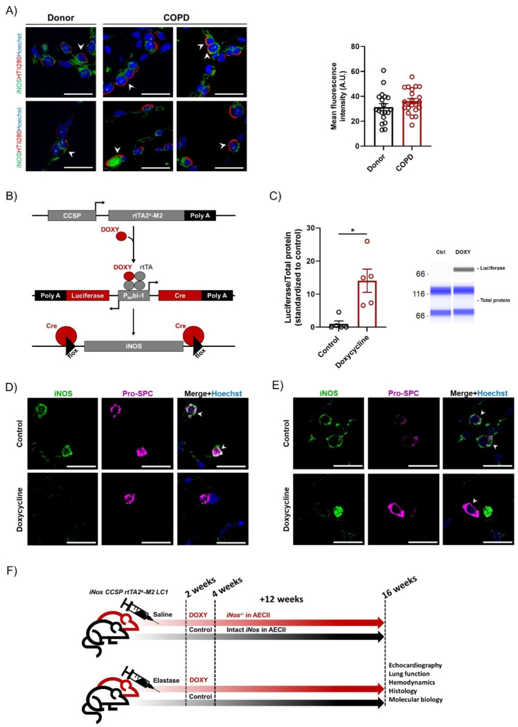
Background: Chronic obstructive pulmonary disease (COPD) is the third leading cause of death worldwide. In addition to chronic bronchitis and emphysema, patients often develop at least mild pulmonary hypertension (PH). We previously demonstrated that inhibition of inducible nitric oxide synthase (iNOS) prevents and reverses emphysema and PH in mice. Interestingly, strong iNOS upregulation was found in alveolar epithelial type II cells (AECII) in emphysematous murine lungs, and peroxynitrite, which can be formed from iNOS-derived NO, was shown to induce AECII apoptosis in vitro. However, the specific cell type(s) that drive(s) iNOS-dependent lung regeneration in emphysema/PH has (have) not been identified yet.

Aim: we tested whether iNOS knockout in AECII affects established elastase-induced emphysema in mice.

Methods: four weeks after a single intratracheal instillation of porcine pancreatic elastase for the induction of emphysema and PH, we induced iNOS knockout in AECII in mice, and gave an additional twelve weeks for the potential recovery.

Results: iNOS knockout in AECII did not reduce elastase-induced functional and structural lung changes such as increased lung compliance, decreased mean linear intercept and increased airspace, decreased right ventricular function, increased right ventricular systolic pressure and increased pulmonary vascular muscularization. In vitro, iNOS inhibition did not reduce apoptosis of AECII following exposure to a noxious stimulus.

Conclusion: taken together, our data demonstrate that iNOS deletion in AECII is not sufficient for the regeneration of emphysematous murine lungs, and suggest that iNOS expression in pulmonary vascular or stromal cells might be critically important in this regard.

Keywords: AECII; COPD; emphysema; iNOS; lung epithelium.


Conflict of interest statement

The authors declare no conflict of interest; portions of the doctoral thesis of Vinita Sharma are incorporated into this report.

Figures

Figure 1
Figure 1
iNOS is successfully deleted in AECII using iNos CCSP rtTA2s-M2 LC1 mouse line. (A) Representative images of human tissue sections from donor and COPD lungs, co-stained against iNOS (green) and AECII apical membrane protein HTII280 (red), and counter-stained with Hoechst. Fluorescence intensity of the iNOS staining in AECII (HTII280-positive cells) was quantified. Arrows indicate double-positive cells. Scale bar = 20 µm. For the signal intensity quantification, every point on the graph represents quantification of the mean fluorescence intensity in one AECII. No fewer than 4 images per group were quantified. (B) Schematic representation of the genetic constructs allowing for club-cell secretory protein (CCSP)-reverse tetracycline-dependent transactivator (rtTA2s-M2)–mediated, doxycycline-controlled luciferase (Luc) and Cre expression and iNOS deletion in mice. (C) Automated Western-blot analysis (n = 5) of luciferase expression in AECII isolated from mice fed with either normal (control, ctrl) or doxycycline-containing food. (D,E) Representative images of lung sections from control and AECII-specific iNOS knockout mice, immediately (D) and 12 weeks (E) after the termination of doxycycline feeding for cell type-specific iNOS knockout induction. The samples were co-stained against pro-SPC (magenta) and iNOS (green) and counterstained with Hoechst (blue). Scale bar = 20µm. Arrow: AECII (pro-SPC-positive) cells. (F) Schematic representation of experimental design used to assess the effect of iNOS expression in AECII on the regeneration of alveolar epithelium and the reversal of pulmonary hypertension in an elastase mouse model. DOXY: Doxycycline. A.U.: Arbitrary units. Graphs show mean ± SEM. * p < 0.05. An independent t-test was used.
Figure 2
Figure 2
iNOS knockout in AECII cells does not promote reverse remodeling of the pulmonary vasculature upon elastase instillation. Two weeks after intratracheal instillation of saline or elastase, iNos CCSP rtTA2s-M2 LC1 mice were fed with either standard (control) or doxycycline-containing (Doxycycline, DOXY) chow for additional two weeks. Twelve weeks later, right heart function and structure as well as the vascular structure were investigated. (A,B) Right ventricular systolic pressure (RVSP) (n = 6–7 per group). (CE) Echocardiographic assessment of right ventricular function and dimensions (n = 6 per group), depicted as (C) the ratio (PAT/PET) of pulmonary acceleration time (PAT) and pulmonary ejection time (PET), (D) tricuspid annular plane systolic excursion (TAPSE), and (E) right ventricular wall thickness (RVWT). (F) Changes in right ventricular weight shown as the ratio of the right ventricular (RV) and the left ventricular plus septum (LV + septum) mass (n = 7–8). LV mass was not significantly altered. Graphs show mean ± SEM. * p < 0.05. Two-way ANOVA (with Tukey’s multiple comparison post-hoc test) was used. (G) Representative images of pulmonary vessels in lung sections from control and doxycycline-treated mice instilled either with saline or elastase, co-stained against α-smooth muscle actin (α-SMA, magenta), von Willebrand factor (vWF, green), and counterstained with Hoechst (blue). Scale bar = 50 µm.
Figure 3
Figure 3
Deletion of iNOS in AECII cannot ameliorate elastase-induced changes in lung function. Two weeks after intratracheal instillation of saline or elastase, iNos CCSP rtTA2s-M2 LC1 mice were fed with either standard (control) or doxycycline-containing (Doxycycline) chow for additional two weeks. Twelve weeks later, micro-computed tomography (µCT) and lung function measurements were performed. (A,B) µCT-based assessment (n = 3–7) of (A) functional residual capacity (FRC) and (B) lung density. HU: Hounsfield units. (CF) Lung function (n = 7–8) presented as (C) static compliance, (D) respiratory pressure-volume (P-V) loops, (E) tissue damping, (F) area enclosed by the P-V loop. Graphs show mean ± SEM. * p < 0.05. Two-way ANOVA (with Tukey’s multiple comparison post-hoc test) was used.
Figure 4
Figure 4
iNOS knockout in AECII cells does not influence regeneration of alveolar epithelium in mice with severe emphysema. (AC) Alveolar morphometry (n = 7–8) showing (A) the percentage of airspace, (B) septal wall thickness and (C) mean linear intercept in saline- or elastase-treated iNos CCSP rtTA2s-M2 LC1 mice fed either with normal (control) or doxycycline-containing (Doxycycline) chow, after the 12-week follow-up observation period. Graphs show mean ± SEM. * p < 0.05. Two-way ANOVA (with Tukey´s multiple comparison post-hoc test) was used. (D) Representative images of lung sections from control and doxycycline-fed mice, 12 weeks after elastase instillation, stained with hematoxylin-eosin (H&E). Scale bar = 200 µm.
Figure 5
Figure 5
iNOS knockout in AECII cells influences Cytochrome c expression in elastase-treated mice. Western-blot analysis (n = 3–4) of (A) Cyclin D1, (B) Cleaved caspase 3, (C) Cytochrome c, (D) Bcl-2 (E) Bax and (F) MMP-8 in lung homogenates of saline- or elastase-treated iNos CCSP rtTA2s-M2 LC1 mice fed either with normal (control, Ctrl) or doxycycline (Doxycycline, DOXY)-containing chow, after the 12-week follow-up period. Data are given as the ratio between the protein of interest and β-actin, standardized to saline control. Graphs show mean ± SEM. * p < 0.05. Two-way ANOVA (with Tukey’s multiple comparisons post-hoc test) was used.
Figure 6
Figure 6
Bronchoalveolar lavage fluid from elastase-treated mice decreases apoptosis of mouse lung epithelial (MLE-12) cell-line in vitro. MLE-12 cells were treated with cigarette smoke extract (CSE) for 6 h, and then left for an additional 16 h in the presence or absence of L-NIL. (A) Proliferation of MLE-12 cells during the 16 h observation period assessed using the BrdU incorporation assay and standardized to control (n = 8). (B) Metabolic activity of MLE-12 cells assessed after the 16 h observation period using the alamarBlue assay (n = 8). (C) Apoptosis of MLE-12 cells after the 16 h observation period given as the confluence of cells labeled with Annexin XII-based polarity-sensitive probe, standardized to total cell confluence (n = 8). (D) Metabolic activity of primary murine alveolar epithelial type 2 cells (AECII) assessed using alamarBlue assay during the 16 h observation period (n = 8). (EH) MLE-12 cells were treated with 10% of either PBS (control) or bronchoalveolar lavage fluid (BALF) from saline- or elastase-treated iNos CCSP rtTA2s-M2 LC1 mice, fed either with normal (control) or doxycycline-containing chow as above, taken after the 12-week observation period. (E) Proliferation of MLE-12 cells during 24 h of treatment, assessed using the BrdU incorporation assay and standardized to control (n = 12). (F) Metabolic activity of MLE-12 cells after 24 h of treatment, measured using the alamarBlue assay and standardized to control (n = 18). Graphs show mean ± SEM. * p < 0.05. One-way ANOVA (with Tukey’s multiple comparison post-hoc test) was used for statistical analysis. (G) Apoptosis of MLE-12 cells during a 48 h long treatment given as the confluence of cells labeled with Annexin XII-based polarity-sensitive probe, standardized to total cell confluence (n = 18). Graphs show mean ± SEM. * p < 0.05. Two-way ANOVA (with Tukey’s multiple comparison post-hoc test) was used for statistical analysis. (H) Representative photos from apoptosis-kinetic assay. Green: Annexin XII-based polarity-sensitive probe. Scale bar = 300 µm.

Similar articles

Cited by

References

    1. Quaderi S.A., Hurst J.R. The unmet global burden of COPD. Glob. Health Epidemiol. Genom. 2018;3:e4. doi: 10.1017/gheg.2018.1. - DOI - PMC - PubMed
    1. Lindberg A., Lindberg L., Sawalha S., Nilsson U., Stridsman C., Lundback B., Backman H. Large underreporting of COPD as cause of death-results from a population-based cohort study. Respir. Med. 2021;186:106518. doi: 10.1016/j.rmed.2021.106518. - DOI - PubMed
    1. Christenson S.A., Smith B.M., Bafadhel M., Putcha N. Chronic obstructive pulmonary disease. Lancet. 2022;399:2227–2242. doi: 10.1016/S0140-6736(22)00470-6. - DOI - PubMed
    1. Celli B., Fabbri L., Criner G., Martinez F.J., Mannino D., Vogelmeier C., Montes de Oca M., Papi A., Sin D.D., Han M.K., et al. Definition and Nomenclature of Chronic Obstructive Pulmonary Disease: Time for its Revision. Am. J. Respir. Crit. Care Med. 2022;206:1317–1325. doi: 10.1164/rccm.202204-0671PP. - DOI - PMC - PubMed
    1. Confalonieri M., Braga L., Salton F., Ruaro B., Confalonieri P. COPD Definition: Is It Time to Incorporate Also the Concept of Lung Regeneration’s Failure? Am. J. Respir. Crit. Care Med. 2022 doi: 10.1164/rccm.202208-1508LE. - DOI - PMC - PubMed

Publication types

Substances

LinkOut - more resources