. 2022 Dec 29;12(1):136.
doi: 10.3390/cells12010136.

Human Neutrophils Generate Extracellular Vesicles That Modulate Their Functional Responses

Affiliations

Human Neutrophils Generate Extracellular Vesicles That Modulate Their Functional Responses

María José Hurtado Gutiérrez et al. Cells. .

Abstract

Neutrophils influence innate and adaptive immunity by releasing various cytokines and chemokines, by generating neutrophil extracellular traps (NETs), and by modulating their own survival. Neutrophils also produce extracellular vesicles (EVs) termed ectosomes, which influence the function of other immune cells. Here, we studied neutrophil-derived ectosomes (NDEs) and whether they can modulate autologous neutrophil responses. We first characterized EV production by neutrophils, following MISEV 2018 guidelines to facilitate comparisons with other studies. We found that such EVs are principally NDEs, that they are rapidly released in response to several (but not all) physiological stimuli, and that a number of signaling pathways are involved in the induction of this response. When co-incubated with autologous neutrophils, NDE constituents were rapidly incorporated into recipient cells and this triggered and/or modulated neutrophil responses. The pro-survival effect of GM-CSF, G-CSF, IFNγ, and dexamethasone was reversed; CXCL8 and NET formation were induced in otherwise unstimulated neutrophils; the induction of inflammatory chemokines by TNFα was modulated depending on the activation state of the NDEs' parent cells; and inducible NET generation was attenuated. Our data show that NDE generation modulates neutrophil responses in an autocrine and paracrine manner, and indicate that this probably represents an important aspect of how neutrophils shape their environment and cellular interactions.

Keywords: NETs; apoptosis; autocrine; chemokines; extracellular vesicles; neutrophils.


Conflict of interest statement

The authors declare no conflict of interest. PPMcD is also employed as Executive Director, Research at Insmed Inc. Insmed was, however, not involved in this work; did not fund it; and does not endorse it, implicitly or otherwise.

Figures
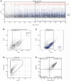
Figure 1
Figure 1
Characterization of neutrophil EVs by high resolution flow cytometry. Neutrophils were cultured in the presence of 100 nM fMLP for 30 min; culture supernatants were collected, centrifuged (1000× g, 10 min, 4 °C) to pellet intact cells, and the resulting supernatants were spun again (18,000× g, 15 min, 4 °C). The EV pellets were resuspended in 0.2 µ filtered HBSS and stained with Calcein Blue AM, prior to flow cytometry analyses. (A) Calcein-stained EV suspensions (blue) spiked with intact neutrophils (red) were monitored over time and data acquisitions were started after 1 min, i.e., when fluctuations in the cell and event numbers had stabilized. (B) Analysis of EV suspensions spiked with cytometry calibration beads (measuring 200, 500 and 760 nm) identified a region in which ectosomes are expected to localize and a size gate was established between 100 and 760 nm. (C) Distribution of calcein-stained EV suspensions (blue) from cultured neutrophil supernatants within this size gate. (D) Analysis of EVs for calcein fluorescence was used to set a gate for calcein-positive events. (E) Analysis of calcein-positive EVs for CD66b and Annexin-V fluorescence. Triple-positive EVs identified thusly are considered NDEs. The experiment shown in this figure is representative of NDE counts throughout the article.
Figure 2
Figure 2
Isolation and properties of NDEs released by cultured neutrophils. (A) Neutrophils were cultured (15 min, 37 °C) in the presence of 20 µM GW4869 (or its diluent, 0.1% DMSO) prior to further incubation in the absence (“ctrl”) or presence of 100 nM fMLP for 120 min. Culture supernatants were collected, and NDEs were isolated and counted as described in Methods. Mean ± s.e.m. from 3 independent experiments. §, p < 0.01 vs fMLP-stimulated cells. (B) Detail of the flow cytometry analysis of the NDEs described in panel A, following the steps described in Figure 1A–C. Calcein Blue-positive events are shown. The experiment depicted is representative of three. (C) Transmission electron micrograph of isolated NDEs from fMLP-stimulated cells. Red arrows point to intact vesicles. The experiment depicted is representative of three. (D) Culture supernatants from neutrophils stimulated for 60 min with 100 nM fMLP were centrifuged at 1000 g; the resulting supernatants were centrifuged at 18,000 g and the pellets (“18K”) were processed for flow cytometry analysis of calcein-positive EVs as described in Methods. The 18,000 supernatants were further centrifuged at 100,000× g (2 h, 4 °C) and the resulting pellets (“100K”) processed for flow cytometry analysis of calcein-positive EVs. The experiment depicted is representative of three. (E) Samples depicted in panel D were also analyzed for total protein determination in Bradford assays.
Figure 3
Figure 3
Induction of NDE generation and upstream signaling pathways. (A) Neutrophils were cultured for 30 min at 37 °C in the absence (“–”) or presence of 100 nM fMLP, 100 U/mL TNFα, 100 ng/mL LPS, or 1 nM GM-CSF (upper panel); or 1000 U/mL G-CSF, 100 nM dexamethasone, or 100 U/mL IFNγ (lower panel). Culture supernatants were collected, and NDEs were isolated and counted as described in Methods. Mean ± s.e.m. from at least 3 independent experiments. ** p < 0.01 vs. unstimulated cells. (B) Neutrophils were cultured in the absence (“unstim”) or presence of 100 nM fMLP or 100 U/mL TNFα for the indicated times. Culture supernatants were collected, and NDEs were isolated and counted as described in Methods. The experiment depicted is representative of three. (C) Neutrophils were cultured (15 min, 37 °C) in the presence of the following signaling pathway inhibitors (or their diluent, 0.1% DMSO): piceatannol (10 µM) for Syk; Src-I1 (10 µM) for Src; 5Z-7-oxozeaenol (1 µM) for TAK1; SB203580 (1 µM) for p38 MAPK; UO126 (10 µM) for MEK; SP600125 (20 µM) for JNK; and LY294002 (10 µM) for PI3K. Cells were then further incubated in the absence (“–”) or presence of 100 nM fMLP for 120 min. Culture supernatants were collected, and NDEs were isolated and counted as described in Methods. Mean ± s.e.m. from at least 3 independent experiments. * p < 0.05; ** p < 0.01; *** p < 0.001; vs. stimulus alone. §, p < 0.01 vs. unstimulated cells.
Figure 4
Figure 4
Isolation and properties of NDEs released by cavitated neutrophils. (A) Cavitates from neutrophils stimulated for 60 min with 100 nM fMLP were centrifuged at 1000× g; the resulting supernatants were centrifuged at 4000× g and the pellets (“4K”) were processed for flow cytometry analysis of NDEs as described in Methods. The post 4000 supernatants were centrifuged at 18,000× g and the pellets (“18K”) were processed for flow cytometry analysis of MNDEs, while the 18,000 supernatants were further centrifuged at 100,000× g (2 h, 4 °C) and the resulting pellets (“100K”) were processed for flow cytometry analysis of NDEs. The experiment depicted is representative of three. (B) Samples depicted in panel A were also analyzed for total protein determination in Bradford assays. (C) Neutrophils were cultured in the absence or presence of 100 nM fMLP for 60 min and NDEs were isolated from either culture supernatants or cavitates, and processed for flow cytometry analysis of NDEs as described in Methods. Mean ± s.e.m. from 3 independent experiments. (D) Transmission electron micrograph of isolated NDEs from the 4000× g pellets of cavitates from fMLP-stimulated cells. The micrograph is representative of three independent experiments.
Figure 5
Figure 5
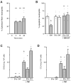
Incorporation of NDEs into autologous neutrophils and its consequence on their apoptotic rate and chemokine production. (A) NDEs from unstimulated neutrophils disrupted by nitrogen cavitation were stained with Cytotrack Red, washed, and co-cultured with autologous neutrophils at a ratio of 5 NDEs per recipient cell (NDEs were quantified as described in Figure 1 for triple-positive EVs). Reactions were stopped at the indicated times, and the cells were collected and analyzed by flow cytometry for Cytotrack Red fluorescence. Mean ± s.e.m. from at least 3 independent experiments. ***, p < 0.001; vs. unstimulated cells. (B) Neutrophils were cultured for 18 h in the absence of any stimulus (“–”) or in the presence of 1 nM GM-CSF, NDEs from the cavitates of unstimulated neutrophils (“EVu”, at a 5:1 NDE:cell ratio), NDEs from the cavitates of fMLP-stimulated neutrophils (“EVu”, at a 5:1 NDE:cell ratio), or a combination thereof. Cells were then processed for flow cytometry analysis of apoptotic cells (AnnexinV+, PI). Mean ± s.e.m. from at least 5 independent experiments. *, p < 0.05; **, p < 0.01; vs. GM-CSF alone. §, p < 0.001 vs. unstimulated cells. (C) Neutrophils were cultured for 6 h in the absence of any stimulus (“–”) or in the presence of 100 U/mL TNFα, NDEs from the cavitates of unstimulated neutrophils (“EVu”, at a 5:1 NDE:cell ratio), NDEs from the cavitates of fMLP-stimulated neutrophils (“EVf”, at a 5:1 NDE:cell ratio), or a combination thereof. Culture supernatants were analyzed by ELISA for their CXCL8 content. Mean ± s.e.m. from at least 4 independent experiments, each performed in duplicate. §, p < 0.001 vs. unstimulated cells; *, p < 0.05; **, p < 0.01; vs. matched condition without NDEs. §, p < 0.001 vs. unstimulated cells. (D) The same samples described in panel C were analyzed by ELISA for their CCL4 content. Mean ± s.e.m. from at least 3 independent experiments, each performed in duplicate. §, p < 0.001 vs. unstimulated cells; *, p < 0.05 vs. matched condition without NDEs.
Figure 6
Figure 6
Effect of NDEs on NET production by autologous neutrophils. Cells were cultured for 4 h in the absence of any stimulus (“–”) or in the presence of 100 nM fMLP, NDEs from the cavitates of unstimulated neutrophils (“EVu”, at a 5:1 NDE:cell ratio), NDEs from the cavitates of fMLP-activated neutrophils (“EVf”, at a 5:1 NDE:cell ratio), or a combination thereof. NDEs were quantified as described in Figure 1 for triple-positive EVs. Samples were then further incubated in the presence or absence of 10 U/mL DNAse I for another 30 min to digest extracellular DNA. NET formation was assessed using MPO detection, as described in Methods. (A) A representative experiment is shown (10× magnification). (B) For each experimental condition, MPO fluorescence from DNAse-treated samples was subtracted from that of untreated samples so as to only quantify the NET-specific signal; these values were then standardized for the total number of nuclei. Mean ± s.e.m. of the standardized NET index from 3 independent experiments. §, p < 0.001 vs. unstimulated cells; *, p < 0.05 vs. matched condition without NDEs.

References

    1. Metschnikoff E. Immunität bei Infektionskrankheiten. Verlag Gustav Fischer; Jena, Germany: 1902.
    1. Brinkmann V., Reichard U., Goosmann C., Fauler B., Uhlemann Y., Weiss D.S., Weinrauch Y., Zychlinsky A. Neutrophil extracellular traps kill bacteria. Science. 2004;303:1532–1535. doi: 10.1126/science.1092385. - DOI - PubMed
    1. Khoyratty T.E., Ai Z., Ballesteros I., Eames H.L., Mathie S., Martín-Salamanca S., Wang L., Hemmings A., Willemsen N., von Werz V., et al. Distinct transcription factor networks control neutrophil-driven inflammation. Nat. Immunol. 2021;22:1093–1106. doi: 10.1038/s41590-021-00968-4. - DOI - PMC - PubMed
    1. Veglia F., Hashimoto A., Dweep H., Sanseviero E., De Leo A., Tcyganov E., Kossenkov A., Mulligan C., Nam B., Masters G., et al. Analysis of classical neutrophils and polymorphonuclear myeloid-derived suppressor cells in cancer patients and tumor-bearing mice. J. Exp. Med. 2021;218:e20201803. doi: 10.1084/jem.20201803. - DOI - PMC - PubMed
    1. Tamassia N., Bianchetto-Aguilera F., Arruda-Silva F.A., Gardiman E., Gasperini S., Calzetti F., Cassatella M.A. Cytokine production by human neutrophils: Revisiting the “dark side of the moon”. Eur. J. Clin. Investig. 2018;48:e12952. doi: 10.1111/eci.12952. - DOI - PubMed

Publication types

Grants and funding

LinkOut - more resources