

Improved Diagnosis of Iron Deficiency Anemia in the Critically Ill via Fluorescence Flowcytometric Hemoglobin Biomarkers
- PMID: 36611936
- PMCID: PMC9818818
- DOI: 10.3390/cells12010140
Improved Diagnosis of Iron Deficiency Anemia in the Critically Ill via Fluorescence Flowcytometric Hemoglobin Biomarkers
Abstract
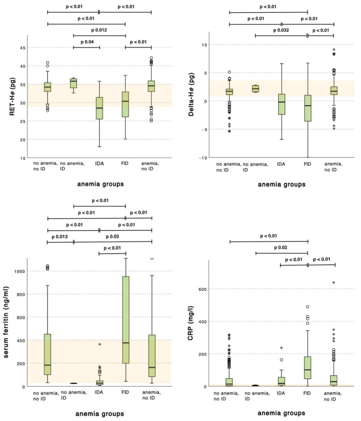
Background: Iron deficiency anemia (IDA) is common in critically ill patients treated in the intensive care unit (ICU), and it can lead to severe consequences. Precise and immediate diagnostics are not available, but they are inevitably needed to administer adequate therapy. Serological parameters such as serum ferritin and transferrin saturation (TSAT) are heavily influenced by simultaneous inflammation reactions, resulting in the need for more suitable parameters. Reticulocyte biomarkers such as reticulocyte hemoglobin content (RET-He) and Delta-hemoglobin equivalent (Delta-He) determined by fluorescence flowcytometry are more specific for the diagnosis of IDA-based anemia and should be investigated for this purpose.
Methods: In a prospective cohort single-center study, serum ferritin and transferrin saturation (TSAT) were collected and compared to RET-He and Delta-He by performing a receiver operating curve (ROC) analysis. The sensitivity and specificity of a single variable or the combination of two variables, as well as cutoff values, for the diagnosis of IDA were calculated. A group comparison for IDA patients without IDA was performed for a control group.
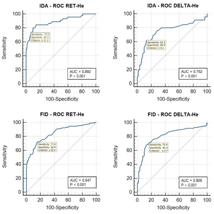
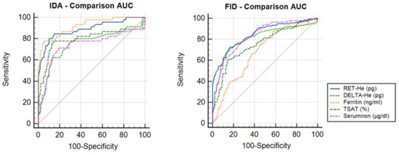
Results: A total of 314 patients were enrolled from an interdisciplinary ICU. RET-He (area under the curve (AUC) 0.847) and Delta-He (AUC 0.807) did indicate iron-deficient anemia that was more specific and sensitive in comparison to serum ferritin (AUC 0.678) and TSAT (AUC 0.754). The detection of functional iron deficiency (FID) occurred in 28.3% of cases with anemia.
Conclusions: Determination of RET-He and Delta-He allows for the increased precision and sensitivity of iron-deficient anemia in the ICU.
Keywords: anemia; functional iron deficiency; intensive care; iron deficiency; reticulocyte hemoglobin content (RET-He); reticulocyte parameters.
Conflict of interest statement
C.H. and M.Z. (Mathias Zimmermann) received fees (honoraria, consulting fees, lecture fees) from Sysmex Germany GmbH, Norderstedt, Germany. C.H. was funded by the “Patient Blood Management Academy, Germany (grants 2019/2021) and received consulting and lecture fees from Dräger GmbH & Co.KG Lübeck, Germany, Sedana Medical GmbH, Germany; and Vifor Pharma GmbH, München, Germany. A.Z. received consultant/lecture fees: Astute Medical, BioMerieux, Baxter, Fresenius, Braun, AM Pharma, Guard Therapeutics, Novartis, Bayer, Amomed, Ratiopharm, Astellas. Grants: Baxter: Fresenius, Astute Medical, Astellas, BioMerieux, DFG, BMBF, and GIF. M.L.R. and M.Z. declare no conflict of interest.
Figures
Similar articles
-
The role of reticulocyte hemoglobin equivalent on the evaluation of iron deficiency and iron deficiency anemia in pediatric cyanotic heart disease: a diagnostic study in Indonesia.BMC Pediatr. 2024 Aug 23;24(1):541. doi: 10.1186/s12887-024-05000-w. BMC Pediatr. 2024. PMID: 39174917 Free PMC article.
-
Reticulocyte hemoglobin equivalent as a marker to assess iron deficiency: A large pediatric tertiary care hospital study.Int J Lab Hematol. 2024 Feb;46(1):148-155. doi: 10.1111/ijlh.14188. Epub 2023 Oct 18. Int J Lab Hematol. 2024. PMID: 37850393
-
Reticulocyte Hemoglobin Equivalent has Comparable Predictive Accuracy as Conventional Serum Iron Indices for Predicting Iron Deficiency and Anemia in a Nonhuman Primate model of Infantile Iron Deficiency.J Nutr. 2023 Jan;153(1):148-157. doi: 10.1016/j.tjnut.2022.11.002. Epub 2022 Dec 20. J Nutr. 2023. PMID: 36913448 Free PMC article.
-
The Role of Reticulocyte Hemoglobin Content for Diagnosis of Iron Deficiency and Iron Deficiency Anemia, and Monitoring of Iron Therapy: a Literature Review.Clin Lab. 2019 Dec 1;65(12). doi: 10.7754/Clin.Lab.2019.190315. Clin Lab. 2019. PMID: 31850722 Review.
-
Implementing Reticulocyte Hemoglobin Into Current Hematology Algorithms.Am J Clin Pathol. 2022 Nov 3;158(5):574-582. doi: 10.1093/ajcp/aqac103. Am J Clin Pathol. 2022. PMID: 36048898
Cited by
-
The incidence and factors associated with anemia in elective surgical patients admitted to a surgical intensive care unit: a retrospective cohort study.Eur J Med Res. 2024 May 19;29(1):290. doi: 10.1186/s40001-024-01887-4. Eur J Med Res. 2024. PMID: 38764061 Free PMC article.
-
Utility of reticulocyte hemoglobin as a new predictor of anemia in intensive care unit patients.Front Med (Lausanne). 2025 Apr 9;12:1577047. doi: 10.3389/fmed.2025.1577047. eCollection 2025. Front Med (Lausanne). 2025. PMID: 40270491 Free PMC article.
-
Comparison of Standard and New Iron Status Biomarkers: A Prospective Cohort Study in Sepsis Patients.Healthcare (Basel). 2023 Mar 30;11(7):995. doi: 10.3390/healthcare11070995. Healthcare (Basel). 2023. PMID: 37046922 Free PMC article.
-
Iron deficiency in sepsis patients managed with divided doses of iron dextran: a prospective cohort study.Sci Rep. 2023 Mar 31;13(1):5264. doi: 10.1038/s41598-023-32002-y. Sci Rep. 2023. PMID: 37002279 Free PMC article.
-
Impact of neutrophil percentage-to-albumin ratio on mortality in iron-deficiency anemia patients: a retrospective study using MIMIC-IV database.Eur J Med Res. 2025 Jan 4;30(1):4. doi: 10.1186/s40001-024-02268-7. Eur J Med Res. 2025. PMID: 39754182 Free PMC article.
References
-
- Manal M., Naglaa M., Kareem M.F., Wael S.A.E. Anemia in Critically Ill Patients; Prevalence and Prognostic Implications. Med. J. Cairo Univ. 2020;88:2121–2129. doi: 10.21608/mjcu.2020.125162. - DOI
Publication types
MeSH terms
Substances
LinkOut - more resources
Full Text Sources
Medical
