. 2022 Dec 31;12(1):177.
doi: 10.3390/cells12010177.

SESN2 Knockdown Increases Betulinic Acid-Induced Radiosensitivity of Hypoxic Breast Cancer Cells

Affiliations

SESN2 Knockdown Increases Betulinic Acid-Induced Radiosensitivity of Hypoxic Breast Cancer Cells

Antje Güttler et al. Cells. .

Abstract

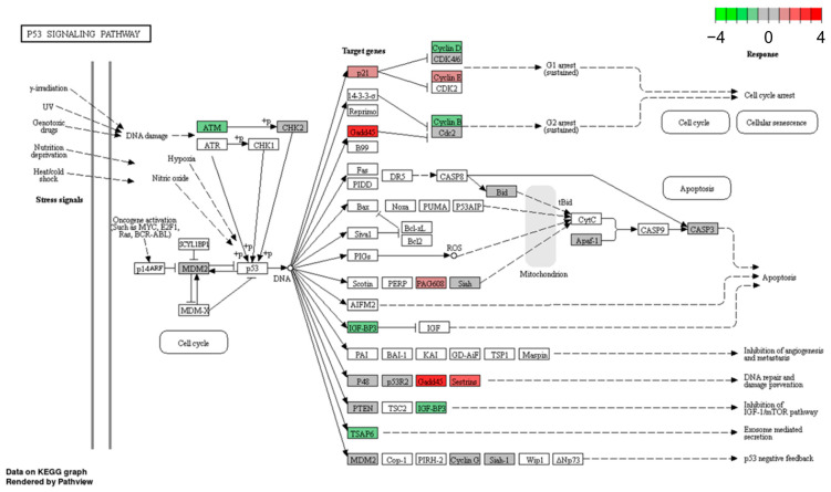
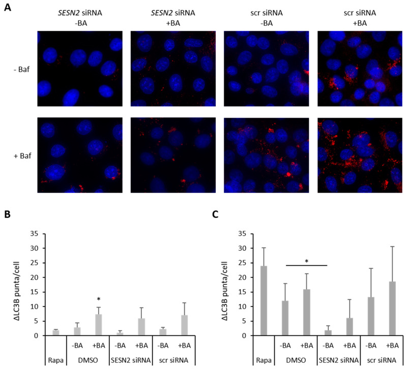
Betulinic acid (BA) is a natural compound well known for its anti-inflammatory, anti-viral, anti-bacterial, anti-malarial effects and anti-tumor properties. Its enhanced cytotoxicity in tumor cells and induction of cell death in various cancer entities qualifies BA as an interesting candidate for novel treatment concepts. Our analyses showed enhanced cytotoxicity and radiosensitization under hypoxic conditions in human breast cancer cells. So far, the underlying mechanisms are unknown. Therefore, we investigated the BA-treated human breast cancer cell lines MDA-MB-231 and MCF-7 under normoxic and hypoxic conditions based on microarray technology. Hypoxia and BA regulated a variety of genes in both breast cancer cell lines. KEGG pathway analysis identified an enrichment of the p53 pathway in MCF-7 cells (wtp53) under hypoxia. In MDA-MB-231 cells (mtp53) an additional BA incubation was required to activate the p53 signaling pathway. Fourteen down-regulated and up-regulated genes of the p53 pathway were selected for further validation via qRT-PCR in a panel of five breast cancer cell lines. The stress-induced gene Sestrin-2 (SESN2) was identified as one of the most strongly up-regulated genes after BA treatment. Knockdown of SESN2 enhanced BA-induced ROS production, DNA damage, radiosensitivity and reduced autophagy in breast cancer cells. Our results identified SESN2 as an important target to enhance the radiobiological and anti-tumor effects of BA on breast cancer cells.

Keywords: autophagy; betulinic acid; breast cancer; knockdown; radiosensitivity; sestrin-2.


Conflict of interest statement

The authors declare no conflict of interest.

Figures

Figure 1
Figure 1
Heat map and Venn diagram of 10 µM BA-treated cells. Transcriptomic profiles of breast cancer cell lines under normoxia and hypoxia used cDNA microarray are presented. MDA-MB-231 and MCF-7 cells were incubated with 10 µM BA under normoxia (21% O2) and hypoxia (0.1% O2) for 48 h. (A) Heat map displays genes up-regulated relative to DMSO treated control cells in red and down-regulated genes in blue. (B) Differential gene expression was calculated as log2-fold-change > 0.5 or <−0.5 of DMSO control cells in at least three independent experiments (p < 0.05).
Figure 2
Figure 2
Hypoxia induced p53 KEGG pathway in MCF-7 cells (wtp53) (Benjamini-Hochberg adjusted p < 0.05). Apaf-1, apoptotic peptidase activating factor 1; ATM, ataxia telangiectasia mutated; CDK2/4/6, cyclin-dependent kinase 2/4/6; CHK1, cell cycle checkpoint kinase; CytC, cytochrome C; DR5, death receptor 5; GADD45, growth arrest and DNA damage-inducible protein 45; IGFBP3, insulin-like growth factor-binding protein 3; MDM2, mouse double minute 2 homolog; PIGs, p53-induced gene; PTEN, phosphatase and tensin homolog; p21 (=CDKN1A), cyclin-dependent kinase inhibitor 1A; p48 (=PTF1A), pancreas associated transcription factor 1a; p53R2 (=RRM2B), ribonucleotide reductase regulatory TP53 inducible subunit M2B); SESN2, sestrin-2; Siah-1, seven in absentia homolog 1; TSAP6 (=STEAP3), tumor suppressor-activated pathway protein 6; 14-3-3σ, Stratifin.
Figure 3
Figure 3
BA-induced p53 KEGG pathway in MDA-MB-231 cells (mtp53) under hypoxia (Benjamini-Hochberg adjusted p = 0.01). Apaf-1, apoptotic peptidase activating factor 1; ATM, ataxia telangiectasia mutated; Bid, BH3 interacting domain death agonist; CASP3, Caspase 3; CDK4/6, cyclin-dependent kinase 4/6; Cdc2 (=CDK1), cell division cycle 2; CHK2, cell cycle checkpoint kinase; GADD45, growth arrest and DNA damage-inducible protein 45; IGFBP3, insulin-like growth factor-binding protein 3; MDM2, mouse double minute 2 homolog; PAG608 (ZMAT3), p53-activated gene 608 protein; PTEN, phosphatase and tensin homolog; p21 (=CDKN1A), cyclin-dependent kinase inhibitor 1A; p48 (=PTF1A), pancreas associated transcription factor 1a; p53R2 (=RRM2B), ribonucleotide reductase regulatory TP53 inducible subunit M2B); SESN2, sestrin-2; Siah-1, seven in absentia homolog 1; TSAP6 (=STEAP3), tumor suppressor-activated pathway protein 6.
Figure 4
Figure 4
Western blot of SESN2 protein expression. Five breast cancer cell lines with different p53 status were analyzed for SESN2 (54 kDa) protein level after treatment with 10 µM and 20 µM BA under normoxic (21% O2) and hypoxic (0.1% O2) conditions for 48 h. A representative blot of at least three independent experiments is shown for each cell line. Actin (42 kDa) was used as a loading control. UT—untreated; D—DMSO.
Figure 5
Figure 5
Knockdown of SESN2 expression in human breast cancer cells. Breast cancer cells were transfected with siRNA against SESN2 and additionally treated with 20 µM BA for another 24 h under normoxic and hypoxic conditions. A-C. qRT-PCR: Effective SESN2 knockdown is shown without and after BA treatment in MDA-MB-231 (A), HS578T (B) and MCF-7 (C) cell line. Data represent mean values (+SD) of at least three independent experiments. Significant p values are highlighted with asterisks (* p ≤ 0.05). D-F. Western blot of MDA-MB-231 (D), HS578T (E) and MCF-7 (F) cells: BA treatment induced SESN2 (55 kDa) protein level, which is prevented by SESN2 knockdown. A representative blot is shown and Actin (42 kDa) is used as a loading control. src siRNA – scrambled siRNA.
Figure 6
Figure 6
Bafilomycin-Clamp-Assay. MDA-MB-231 cells were transfected with siRNA against SESN2 and treated with BA under hypoxic conditions (0.1% O2) with and without BafA1. LC3B puncta per cell were counted out of at least 100 cells. (A) Representative immunofluorescence staining of LC3B (red) and cell nuclei (blue: DAPI). (B) LC3B puncta per cell after siRNA and BA treatment without BAf1A. (C) Autophagic flux (ΔLC3B puncta) of MDA-MB-231 cells under hypoxic conditions. Data represent mean values (+SD) of three independent experiments. All data were referred to scr siRNA without BA treatment. Significant p values are highlighted with asterisks (* p ≤ 0.05).
Figure 7
Figure 7
SESN2 silencing enhances ROS production of BA-treated breast cancer cells. Breast cancer cell lines MDA-MB-231 (A), HS578T (B) and MCF-7 (C) were transfected with siRNA against SESN2 and treated with BA under hypoxic conditions (0.1% O2). Intracellular ROS level was measured with CM-H2DCFDA staining. Data represent mean values (+SD) of at least three independent experiments. All data were referred to scr siRNA without BA treatment (=100%). Significant p values are highlighted with asterisks (* p ≤ 0.05; ** p ≤ 0.01).
Figure 8
Figure 8
SESN2 silencing enhances DNA damage of BA-treated breast cancer cells. (A) Representative immunofluorescence staining of γH2AX foci (green) and cell nuclei (DAPI: blue) of non-irradiated MDA-MB-231 cells. Breast cancer cell lines MDA-MB-231 (B) and HS578T (C) were transfected with siRNA against SESN2, treated with BA and irradiated with 4 Gy under hypoxic conditions (0.1% O2). γH2AX foci per cell were counted out of 100 cells. Data represent mean values (+SD) of at least three independent experiments. All data were referred to scr siRNA without BA treatment of non-irradiated (0 Gy) or irradiated cells (4 Gy). Significant p values are highlighted with asterisks (* p ≤ 0.05; ** p ≤ 0.01; *** p = 0.001).
Figure 9
Figure 9
Radiosensitivity of breast cancer cells. Breast cancer cells ((A) MDA-MB-231, (B) HS578T, (C) MCF-7) were transfected with siRNA against SESN2 and treated with 20 µM BA under hypoxic conditions (0.1% O2). 24 h after BA treatment cells were irradiated under hypoxic conditions with different doses depending on cell line. Afterwards, 150 to 10,000 single cells were plated depending on cell line and radiation dose. After approximately 12 days, colonies were stained and counted. Data represent mean values (+SD) of at least three independent experiments.

References

    1. Sung H., Ferlay J., Siegel R.L., Laversanne M., Soerjomataram I., Jemal A., Bray F. Global Cancer Statistics 2020: GLOBOCAN Estimates of Incidence and Mortality Worldwide for 36 Cancers in 185 Countries. CA A Cancer J. Clin. 2021;71:209–249. doi: 10.3322/caac.21660. - DOI - PubMed
    1. Lin N.U., Vanderplas A., Hughes M.E., Theriault R.L., Edge S.B., Wong Y.-N., Blayney D.W., Niland J.C., Winer E.P., Weeks J.C. Clinicopathologic features, patterns of recurrence, and survival among women with triple-negative breast cancer in the National Comprehensive Cancer Network. Cancer. 2012;118:5463–5472. doi: 10.1002/cncr.27581. - DOI - PMC - PubMed
    1. Zhang Y., Zhang H., Wang M., Schmid T., Xin Z., Kozhuharova L., Yu W.-K., Huang Y., Cai F., Biskup E. Hypoxia in Breast Cancer-Scientific Translation to Therapeutic and Diagnostic Clinical Applications. Front. Oncol. 2021;11:652266. doi: 10.3389/fonc.2021.652266. - DOI - PMC - PubMed
    1. Semenza G.L. The hypoxic tumor microenvironment: A driving force for breast cancer progression. Biochim. Biophys. Acta. 2016;1863:382–391. doi: 10.1016/j.bbamcr.2015.05.036. - DOI - PMC - PubMed
    1. Jiang W., Li X., Dong S., Zhou W. Betulinic acid in the treatment of tumour diseases: Application and research progress. Biomed. Pharmacother. 2021;142:111990. doi: 10.1016/j.biopha.2021.111990. - DOI - PubMed

Publication types