

Azilsartan Modulates HMGB1/NF-κB/p38/ERK1/2/JNK and Apoptosis Pathways during Renal Ischemia Reperfusion Injury
- PMID: 36611978
- PMCID: PMC9818604
- DOI: 10.3390/cells12010185
Azilsartan Modulates HMGB1/NF-κB/p38/ERK1/2/JNK and Apoptosis Pathways during Renal Ischemia Reperfusion Injury
Abstract
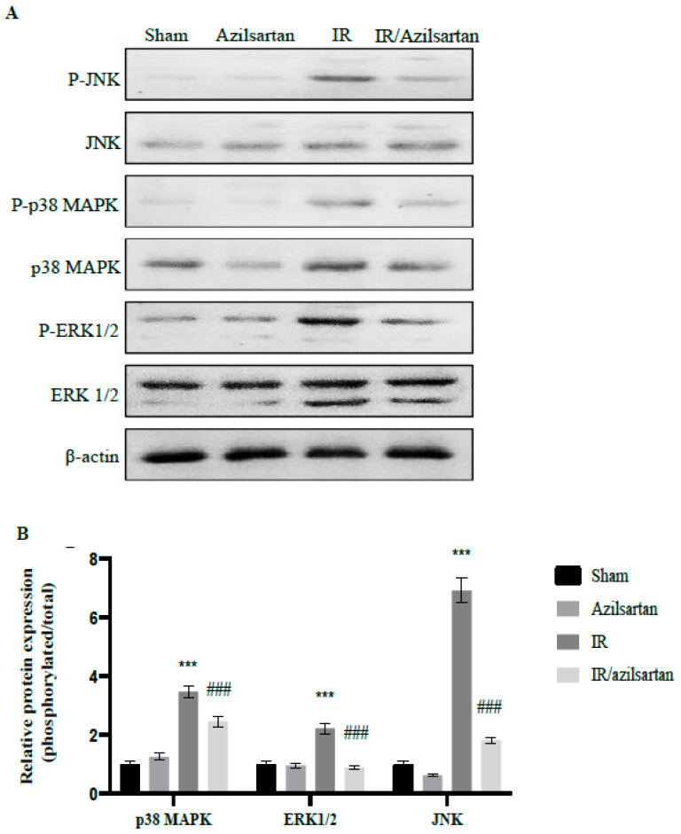
Renal ischemia/reperfusion (IR) injury is characterized by an unexpected impairment of blood flow to the kidney. Azilsartan is an angiotensin receptor blocker that is approved for the management of hypertension. The present study aimed to investigate, on molecular basics, the nephroprotective activity of azilsartan on renal IR injury in rats. Rats were assigned into four groups: (1) Sham group, (2) Azilsartan group, (3) IR group, and (4) IR/Azilsartan-treated group. Histological examination and renal function were evaluated. Levels of KIM-1, HMGB1, caspase 3, GPX, SOD, NF-κB, and p53 proteins were investigated using ELISA. mRNA levels of IL-1β, IL6, IL10, TNF-α, NF-κB, p53, and bax were assessed by qRT-PCR. Expression of p38, JNK, and ERK1/2 proteins was investigated by Western blotting. IR injury resulted in tissue damage, elevation of creatinine, BUN, KIM-1, HMGB1, caspase 3, NF-κB, and p53 levels, decreasing GPX and SOD activities, and up-regulation of NF-κB, IL-1β, IL6, TNF-α, p53, and bax genes. Furthermore, it up-regulated the expression of phosphorylated/total ratio of p38, ERK1/2, and JNK proteins. Interestingly, treatment of the injured rats with azilsartan significantly alleviated IR injury-induced histopathological and biochemical changes. It reduced the creatinine, BUN, KIM-1, HMGB1, caspase-3, NF-κB, and p53 levels, elevated GPX and SOD activities, down-regulated the expression of NF-κB, IL-1β, IL6, TNF-α, p53, and bax genes, and up-regulated IL10 gene expression. Furthermore, it decreased the phosphorylated/total ratio of p38, ERK1/2, and JNK proteins. Azilsartan exhibited nephroprotective activity in IR-injured rats via its antioxidant effect, suppression of inflammation, attenuation of apoptosis, and inhibition of HMGB1/NF-κB/p38/ERK1/2/JNK signaling pathway.
Keywords: ERK1/2; JNK; NF-κB; apoptosis; azilsartan; renal ischemia/reperfusion injury.
Conflict of interest statement
The authors declare no conflict of interest.
Figures


Similar articles
-
Pantoprazole Attenuates MAPK (ERK1/2, JNK, p38)-NF-κB and Apoptosis Signaling Pathways after Renal Ischemia/Reperfusion Injury in Rats.Int J Mol Sci. 2021 Oct 1;22(19):10669. doi: 10.3390/ijms221910669. Int J Mol Sci. 2021. PMID: 34639009 Free PMC article.
-
[miR-155-5p alleviates lipopolysaccharide-induced inflammatory damage of human SH-SY5Y neuroblastoma cells by down-regulating SOCS1].Xi Bao Yu Fen Zi Mian Yi Xue Za Zhi. 2023 Mar;39(3):220-229. Xi Bao Yu Fen Zi Mian Yi Xue Za Zhi. 2023. PMID: 36946346 Chinese.
-
[Acacetin protects rats from cerebral ischemia-reperfusion injury by regulating TLR4/NLRP3 signaling pathway].Zhongguo Zhong Yao Za Zhi. 2023 Nov;48(22):6107-6114. doi: 10.19540/j.cnki.cjcmm.20230719.401. Zhongguo Zhong Yao Za Zhi. 2023. PMID: 38114218 Chinese.
-
Neuropharmacological effects of calycosin: a translational review of molecular mechanisms and therapeutic applications.Naunyn Schmiedebergs Arch Pharmacol. 2025 Apr 16. doi: 10.1007/s00210-025-04154-3. Online ahead of print. Naunyn Schmiedebergs Arch Pharmacol. 2025. PMID: 40237798 Review.
-
Pasteurella multocida causes liver injury in ducks by mediating inflammatory, apoptotic and autophagic pathways.Microb Pathog. 2023 Nov;184:106336. doi: 10.1016/j.micpath.2023.106336. Epub 2023 Sep 7. Microb Pathog. 2023. PMID: 37683832 Review.
Cited by
-
Modulating Nrf-2/HO-1, apoptosis and oxidative stress signaling pathways by gabapentin ameliorates sepsis-induced acute kidney injury.Naunyn Schmiedebergs Arch Pharmacol. 2024 Feb;397(2):947-958. doi: 10.1007/s00210-023-02650-y. Epub 2023 Aug 7. Naunyn Schmiedebergs Arch Pharmacol. 2024. PMID: 37548662 Free PMC article.
-
Protective effect of irbesartan against hepatic ischemia-reperfusion injury in rats: role of ERK, STAT3, and PPAR-γ inflammatory pathways in rats.Naunyn Schmiedebergs Arch Pharmacol. 2025 Feb;398(2):1681-1693. doi: 10.1007/s00210-024-03301-6. Epub 2024 Aug 21. Naunyn Schmiedebergs Arch Pharmacol. 2025. PMID: 39167169 Free PMC article.
-
Nebivolol ameliorates sepsis-evoked kidney dysfunction by targeting oxidative stress and TGF-β/Smad/p53 pathway.Sci Rep. 2024 Jun 26;14(1):14735. doi: 10.1038/s41598-024-64577-5. Sci Rep. 2024. PMID: 38926458 Free PMC article.
-
Vincamine Ameliorates Epithelial-Mesenchymal Transition in Bleomycin-Induced Pulmonary Fibrosis in Rats; Targeting TGF-β/MAPK/Snai1 Pathway.Molecules. 2023 Jun 9;28(12):4665. doi: 10.3390/molecules28124665. Molecules. 2023. PMID: 37375218 Free PMC article.
-
Lycium barbarum glycopeptide mitigates retinal ischemia-reperfusion injury through its anti-inflammatory, anti- senescence, and anti-apoptosis properties.Sci Rep. 2025 Jul 30;15(1):27806. doi: 10.1038/s41598-025-10763-y. Sci Rep. 2025. PMID: 40739315 Free PMC article.
References
MeSH terms
Substances
LinkOut - more resources
Full Text Sources
Research Materials
Miscellaneous
