Review
. 2023 Jan 2;15(1):308.
doi: 10.3390/cancers15010308.

Emerging Indications for Interventional Oncology: Expert Discussion on New Locoregional Treatments

Affiliations
Review

Emerging Indications for Interventional Oncology: Expert Discussion on New Locoregional Treatments

Roberto Iezzi et al. Cancers (Basel). .

Abstract

Interventional oncology (IO) employs image-guided techniques to perform minimally invasive procedures, providing lower-risk alternatives to many traditional medical and surgical therapies for cancer patients. Since its advent, due to rapidly evolving research development, its role has expanded to encompass the diagnosis and treatment of diseases across multiple body systems. In detail, interventional oncology is expanding its role across a wide spectrum of disease sites, offering a potential cure, control, or palliative care for many types of cancer patients. Due to its widespread use, a comprehensive review of the new indications for locoregional procedures is mandatory. This article summarizes the expert discussion and report from the "MIOLive Meet SIO" (Society of Interventional Oncology) session during the last MIOLive 2022 (Mediterranean Interventional Oncology Live) congress held in Rome, Italy, integrating evidence-reported literature and experience-based perceptions. The aim of this paper is to provide an updated review of the new techniques and devices available for innovative indications not only to residents and fellows but also to colleagues approaching locoregional treatments.

Keywords: ablation; bone metastases; breast cancer; cementoplasty; cholangiocellular carcinoma; head and neck cancer; interventional oncology; lymphatics; radioembolization; spinal tumors.


Conflict of interest statement

The authors declare no conflict of interest.

Figures

Figure 1
Figure 1
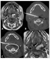
56-year-old male with neuropathic pain due to right oropharyngeal squamocellular carcinoma recurrence after tracheostomy, neck lymph node dissection, mandibulotomy, right oropharyngeal resection with free anterolateral thigh flap. (A) T1-weighted postcontrast axial MRI scan showing malignant involvement of trigeminal nerve (circle). (B) Axial CT scan showing cryoablation needle placement. (C) Intraprocedural axial CT scan showing ice ball formation around the needle (circle).
Figure 2
Figure 2
65-year-old male with trismus, gagging, airway compromise, and bleeding episodes due to recurrent nasopharyngeal carcinoma of the left retropharyngeal/paravertebral space with circumferential carotid artery involvement, after previous radiation therapy and left neck dissection. (A) T1-weighted postcontrast axial MRI scan showing malignant involvement of the left retropharyngeal space (circle). Notice the tongue (*) which can flop backward during the general anesthesia and could contact the ablation site; care must be taken in blocking the tongue prior to ablation. (B) Axial CT scan showing cryoablation needle placement through the retromolar trigone. (C) Axial CT scan showing the cryoablation needle near the previously embolized left carotid artery (arrow), which can lead to diving reflex.
Figure 3
Figure 3
63-year-old male with recurrence of supraglottic squamocellular carcinoma which underwent previous total pharyngolaryngectomy and bilateral neck dissection. (A) T1-weighted unenhanced axial MRI scan showing the tumor recurrence (X), the mucosa of the neopharynx (arrow), the right submandibular gland (black circle), and the right carotid space (white circle). (B) Axial CT scan showing the cryoprobe positioned in the lesion through a trans-submandibular gland approach. (C) Axial CT scan showing parallel needle placement for hydrodissection of peritumoral tissue. (D) One-month T1-weighted unenhanced axial MRI scan showing the postablation tumor shrinkage (arrow).
Figure 4
Figure 4
70-year-old male with airway impairment and left postauricular discharge. Previous nasopharyngeal carcinoma and radiation-induced squamous cell carcinoma of maxilla which underwent maxillectomy, mandibulectomy, parotidectomy, and myocutaneous flap reconstruction. (A) T1-weighted postcontrast axial MRI scan showing extensive tumor recurrence (white circle), posterior to the tongue (*), threatening the airways and invading the left external auditory canal. (B) Axial CT scan showing cryoablation needle placement through the myocutaneous flap. (C) Coronal-oblique multiplanar reconstruction CT scan showing the ice ball formed by the three cryoprobes.
Figure 5
Figure 5
64-year-old female patient with left invasive T1 breast cancer of 11 mm with a molecular subtype Luminal-A. Contrast-enhanced T1-weighted MRI scan showing (A) baseline neoplastic lesion and (B) 5-month follow-up after microwave ablation with good ablation margins and central necrosis.
Figure 6
Figure 6
85-year-old female patient with invasive right breast cancer with axillary metastases. Diffusion-weighted imaging MRI scan showing (A) baseline large neoplastic lesion infiltrating the skin and soft tissues (arrows) and (B) 4-month follow-up after microwave ablation with absent signal restriction (arrows).
Figure 7
Figure 7
Ultrasound scan with linear probe showing the tip of the needle used for hydrodissection (arrow) to inject saline between the tumor and the skin or ductal structures.
Figure 8
Figure 8
79-year-old male patient presenting with pain in the right upper abdominal quadrant due to intrahepatic cholangiocellular carcinoma. Performance Status One, no surgical indication; the patient underwent eight cycles of chemotherapy. (A) Contrast-enhanced axial CT scan showing a 3.5 cm lesion of the I-IV segment adjacent to the inferior vena cava and to the right hepatic vein. (B) Contrast-enhanced coronal CT scan showing how the lesion is adjacent to the portal vein. (C) Digital subtraction angiography runs showing super selective catheterization of the target vessel with intra-arterial administration of marked albumin macroaggregates, which serve as the diagnostic phase of radioembolization. (D) Single-photon emission computed tomography oblique reconstruction scan showing focal and selective uptake of marked albumin macroaggregates by the target lesion, confirming the catheter position for the therapeutic radioembolization.
Figure 9
Figure 9
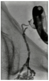
Digital subtraction lymphangiography of a 54-year-old male with lymphocele after prostatectomy. A percutaneous drainage catheter was placed in the lymphocele; then a right obturatory lymphangiography and lymphatic embolization with cyanoacrylate were performed. The percutaneous drainage catheter was removed 2 days later.
Figure 10
Figure 10
(A) Pelvic X-ray showing osteolytic lesion of left periacetabular region (circle). (B) Axial CT scan showing cryoablation needle inserted in the osteolytic lesion. (C) Axial CT scan showing postcryoablation cementoplasty performed to augment the ablative results and to prevent bone fractures.

References

    1. Helmberger T. The evolution of interventional oncology in the 21st century. Br. J. Radiol. 2020;93:20200112. doi: 10.1259/bjr.20200112. - DOI - PMC - PubMed
    1. Wilseck Z.M., Lin L., Gemmete J.J., Pandey A.S., Srinivasan A., Chaudhary N. Skull Base Neurointerventional Techniques. Neuroimaging Clin. 2021;31:649–664. doi: 10.1016/j.nic.2021.06.007. - DOI - PubMed
    1. Sagoo N.S., Haider A.S., Chen A.L., Vannabouathong C., Larsen K., Sharma R., Palmisciano P., Alamer O.B., Igbinigie M., Wells D.B., et al. Radiofrequency ablation for spinal osteoid osteoma: A systematic review of safety and treatment outcomes. Surg. Oncol. 2022;41:101747. doi: 10.1016/j.suronc.2022.101747. - DOI - PubMed
    1. Koch G., Cazzato R.L., Gilkison A., Caudrelier J., Garnon J., Gangi A. Percutaneous Treatments of Benign Bone Tumors. Semin. Interv. Radiol. 2018;35:324–332. doi: 10.1055/s-0038-1673640. - DOI - PMC - PubMed
    1. Auloge P., Garnon J., Robinson J.M., Thenint M.A., Koch G., Caudrelier J., Weiss J., Cazzato R.L., Kurtz J.E., Gangi A. Percutaneous cryoablation for advanced and refractory extra-abdominal desmoid tumors. Int. J. Clin. Oncol. 2021;26:1147–1158. doi: 10.1007/s10147-021-01887-y. - DOI - PubMed

LinkOut - more resources