. 2022 Dec 26;24(1):401.
doi: 10.3390/ijms24010401.

miR-450-5p and miR-202-5p Synergistically Regulate Follicle Development in Black Goat

Affiliations

miR-450-5p and miR-202-5p Synergistically Regulate Follicle Development in Black Goat

Guanghang Feng et al. Int J Mol Sci. .

Abstract

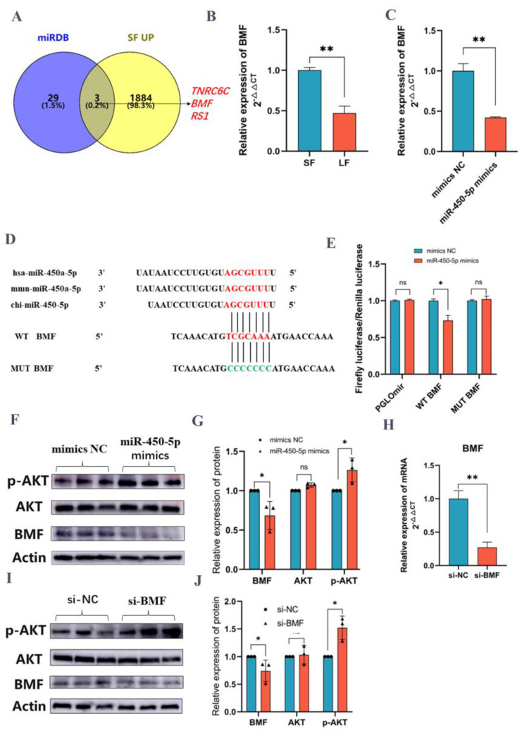
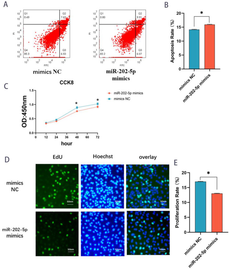
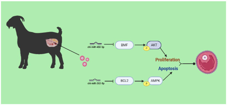
Follicle maturation is a complex biological process governed by numerous factors, and researchers have observed follicle development by studying the proliferation and apoptosis of follicular granulosa cells (GCs). However, the regulatory mechanisms of GCs proliferation and death during follicle development are largely unknown. To investigate the regulatory mechanisms of lncRNAs, mRNAs, and microRNAs, RNA sequencing (RNA-seq) and small RNA-seq were performed on large (>10 mm) and small follicles (<3 mm) of Leizhou black goat during estrus. We discovered two microRNAs, miR-450-5p and miR-202-5p, which can target GCs in goats and may be involved in follicle maturation, and the effects of miR-450-5p and miR-202-5p on ovarian granulosa cell lines were investigated (KGN). Using cell counting kit-8 (CCK-8) assays, 5-Ethynyl-2’-deoxyuridine (EdU) assay and flow cytometry, miR-202-5p overexpression could suppress the proliferation and induce apoptosis of GCs, whereas miR-450-5p overexpression induced the opposite effects. The dual-luciferase reporter assay confirmed that miR-450-5p could directly target the BMF gene (a BCL2 modifying factor), and miR-202-5p targeted the BCL2 gene. A considerable rise in phosphorylated Akt (p-AKT) protein was observed following the downregulation of BMF by miR-450-5p mimics. After BMF gene RNAi therapy, a notable elevation in p-AKT was detected. Mimics of miR-202-5p inhibited BCL2 protein expression, significantly decreasing p-AMPK protein expression. These results imply that during the follicular development in black goats, the miR-450-5p-BMF axis favored GC proliferation on a wide scale, while the miR-202-5p-BCL2 axis triggered GC apoptosis.

Keywords: MiR-202-5p; MiR-450-5p; follicle development; goat; granulosa cells.


Conflict of interest statement

The authors declare no conflict of interest.

Figures

Figure 1
Figure 1
Volcano map and cluster analysis of DEmRNAs, DElncRNAs, and DEmiRNAs. Volcano plots of DEmRNAs (A), DElncRNAs (C) and DEmiRNAs (E); Cluster analysis plots of DEmRNAs (B), DElncRNAs (D) and DEmiRNAs (F).
Figure 2
Figure 2
GO and KEGG enrichment analysis of DEmRNAs and target genes of DElncRNAs and DEmiRNAs. GO analysis of DEmRNAs (A) and predicted target genes of DElncRNAs (C) and DEmiRNAs (E). The top 20 KEGG pathways of predicted target genes for DEmRNAs (B) and DElncRNAs (D), and DEmiRNAs (F). “Count” means the number of predicted target genes enriched in this pathway. The color represents the degree of enrichment, with red representing significant enrichment. Red represents the biological process (BP), blue represents the molecular function (MF), and green represents the cell component (CC).
Figure 3
Figure 3
Differentially expressed mRNAs, lncRNAs and miRNAs were verified by qRT-PCR. (A,B): The expression levels of the 10 mRNAs were verified by qRT-PCR and compared with the results of RNA-seq. (C,D): The expression levels of the five lncRNAs were verified by qRT-PCR and compared with the results of RNA-seq. (E,F): The expression levels of the 2 miRNAs were verified by qRT-PCR and compared with the results of RNA-seq. The results were shown to be consistent with RNA-seq data by RT-qPCR. Data are shown as mean ±SD values (n = 3, * p < 0.05; ** p < 0.01 indicates significant difference).
Figure 4
Figure 4
miR-450-5p stimulates the proliferation of GCs. (A) Expression of miRNA in follicular granulosa cells versus follicular theca cells. Results are shown as Mean ± SD, n = 4, ** p < 0.01 indicates a highly significant difference. (B) CCK8 line graph of miR-450-5p mimics and mimics NC. OD values at 450 nm were determined at 12 h, 24 h, 48 h, and 72 h. (Mean ± SD, ** p < 0.01). (C,D) Results of flow apoptosis measurements of miR-450-5p mimics versus mimic NC with apoptotic cells as a percentage of all cells. (Mean results ± SD, * p < 0.05 indicates significant difference). (E,F) EdU cell proliferation assay. Comparison of miR-450-5p mimics as well as mimics NC in EdU fuel (proliferating cells) as a percentage (%) of hoechst fuel (overall cells). Three replicates per group, mean ± SD,* p < 0.05 indicates a significant difference.
Figure 5
Figure 5
miR-450-5p targets BMF to activate PI3K/Akt signaling pathway (A): Comparison of genes upregulated in small follicles with target genes of miR-450-5p predicted by miRDB database. (B) BMF gene expression on small and large follicles in goats. (C) Expression of BMF gene after transfection with miR-450-5p. (D) Sequence comparison of miR-450-5p and comparison of WT-BMF and MUT-BMF target sequences. (E) Dual fluorescence changes of the original plasmid pGLOmir, wild plasmid WT, and mutant plasmid MUT with miR-450-5p (F,G) Western blot results of p-AKT, AKT, and BMF after transfection of miR-450-5p. (H) Expression of BMF gene after silencing BMF with siRNA. (I,J) Western blot results of p-AKT, AKT, and BMF silenced with siRNA. Reported experiments. Data are shown as mean ± SD values (n = 3). * p < 0.05; ** p < 0.01was considered statistically significant. ns: not significant for statistical results.
Figure 6
Figure 6
miR-202-5p induces the apoptosis of follicular GCs. (A,B) Flow cytometric results of miR-202-5p mimics vs. mimics NC. Flow cytometric results histogram of miR-202-5p mimics vs. mimics NC. (Mean results ± SD, * p < 0.05 indicates significant difference) (C) CCK8 line graph of miR-202-5p mimics and mimics NC. OD values at 450 nm were determined at 12 h, 24 h, 48 h, and 72 h. (Mean ± SD, * p < 0.05). (D,E) EdU cell proliferation assay. Comparison of miR-202-5p mimics as well as mimics NC in EdU fuel (proliferating cells) as a percentage (%) of hoechst fuel (overall cells). Three replicates per group, mean ± SD, * p < 0.05 indicates a significant difference.
Figure 7
Figure 7
miR-202-5p targets the BCL2 gene. (A) miRDB with Target scan 7.2 databases to predict target genes. (B) qPCR results of BCL2 gene after overexpression of miR-202-5p. (C) Sequence comparison of miR-202-5p with dual luciferase results. (D) Fluorescence changes of miR-202-5p and mimics NC after co-staining with PGLOmir, WT-BCL2 and MUT-BCL2. Data are shown as mean ± SD values (n = 6). * p < 0.05 was considered statistically significant. Ns: data results were not significant. (E,F) Western blotting results of p-AMPK, AMPK and BCL2 after overexpression of miR-202-5p.Data are shown as mean ± SD values (n = 3). * p < 0.05 was considered statistically significant. ns: not significant for statistical results.
Figure 8
Figure 8
Diagram of miR-450-5p and miR-202-5p regulatory follicle development pathway.

References

    1. Li L., Shi X., Shi Y., Wang Z. The Signaling Pathways Involved in Ovarian Follicle Development. Front. Physiol. 2021;12:1686–1701. doi: 10.3389/fphys.2021.730196. - DOI - PMC - PubMed
    1. Sun Z., Zhang H., Wang X., Wang Q.-C., Zhang C., Wang J.-Q., Wang Y.-H., An C.-Q., Yang K.-Y., Wang Y. TMCO1 is essential for ovarian follicle development by regulating ER Ca2+ store of granulosa cells. Cell Death Differ. 2018;25:1686–1701. doi: 10.1038/s41418-018-0067-x. - DOI - PMC - PubMed
    1. Lv X., He C., Huang C., Wang H., Hua G., Wang Z., Zhou J., Chen X., Ma B., Timm B.K. Timely expression and activation of YAP1 in granulosa cells is essential for ovarian follicle development. FASEB J. 2019;33:10049–10064. doi: 10.1096/fj.201900179RR. - DOI - PMC - PubMed
    1. Yeung C., Wang G., Yao Y., Liang J., Tenny Chung C., Chuai M. BRE modulates granulosa cell death to affect ovarian follicle development and atresia in the mouse. Cell Death Dis. 2017;8:e2697. doi: 10.1038/cddis.2017.91. - DOI - PMC - PubMed
    1. Zhang Y., Yan Z., Qin Q., Nisenblat V., Chang H.-M., Yu Y., Wang T., Lu C., Yang M., Yang S., et al. Transcriptome Landscape of Human Folliculogenesis Reveals Oocyte and Granulosa Cell Interactions. Mol. Cell. 2018;72:1021–1034.e4. doi: 10.1016/j.molcel.2018.10.029. - DOI - PubMed

Substances

LinkOut - more resources