

A Highly Sensitive Biomarker of Type II Collagen C-Terminal Pro-Peptide Associated with Cartilage Formation
- PMID: 36613894
- PMCID: PMC9820484
- DOI: 10.3390/ijms24010454
A Highly Sensitive Biomarker of Type II Collagen C-Terminal Pro-Peptide Associated with Cartilage Formation
Abstract
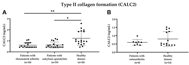
The type II collagen C-terminal pro-peptide is one of the most abundant polypeptides in cartilage. The purpose of this study was to develop a competitive chemiluminescence enzyme-linked immunosorbent assay, CALC2, targeting this pro-peptide as a marker of cartilage formation. Technical assay parameters were evaluated. CALC2 level was measured after in vitro cleavage of recombinant type II collagen with bone morphogenetic protein-1 (BMP-1) and treatment of ex vivo human osteoarthritis (OA) cartilage explant model (HEX) with insulin-like growth factor-1 (IGF-1). Serum CALC2 levels were assessed in 18 patients with rheumatoid arthritis (RA), 19 patients with ankylosing spondylitis (AS), and 18 age- and sex-matched controls in cohort 1 and 8 patients with OA and 14 age- and sex-matched controls in cohort 2. Type II collagen cleavage with BMP-1 increased the CALC2 level. IGF-1 treatment increased the CALC2 levels in HEX compared with the untreated explants (p < 0.05). Results were confirmed using Western blot analysis. CALC2 levels were decreased in the patients with RA and AS compared with the healthy controls (p = 0.01 and p = 0.02, respectively). These findings indicate that CALC2 may be a novel biomarker of type II collagen formation. However, further preclinical and clinical studies are required to validate these findings.
Keywords: C-terminal pro-peptide; ankylosing spondylitis; biomarker; cartilage; osteoarthritis; rheumatoid arthritis; type II collagen formation.
Conflict of interest statement
A.-C.B.-J., Y.H., M.A.K., C.F.T. and S.H.N. are employeesand own stocks in Nordic Bioscience. The funders had no role in the design of the study; collection, analyses, or interpretation of data; in the writing of the manuscript; or in the decision to publish the results. All other authors have no conflict of interest to declare.
Figures


References
-
- Gudmann N.S., Wang J., Hoielt S., Chen P., Siebuhr A.S., He Y., Christiansen T.G., Karsdal M.A., Bay-Jensen A.C. Cartilage turnover reflected by metabolic processing of type II collagen: A novel marker of anabolic function in chondrocytes. Int. J. Mol. Sci. 2014;15:18789–18803. doi: 10.3390/ijms151018789. - DOI - PMC - PubMed
-
- Lian W., Liu H., Sun L.Y., Liu Y.Q., Cui S.L., Wang Y., Song Q.Q., Deng Q., Wang S.P., Cao Y.H., et al. Serum levels of PIICP, PIIANP, and PIIBNP are decreased in patients with an endemic osteochondropathy, Kashin-Beck disease. J. Orthop. Surg. Res. 2018;13:1–7. doi: 10.1186/s13018-018-0840-z. - DOI - PMC - PubMed
-
- Broder C., Arnold P., Goff S.V.L., Konerding M.A., Bahr K., Müller S., Overall C.M., Bond J.S., Koudelka T., Tholey A., et al. Metalloproteases meprin α and meprin β are C- and N-procollagen proteinases important for collagen assembly and tensile strength. Proc. Natl. Acad. Sci. USA. 2013;110:14219–14224. doi: 10.1073/pnas.1305464110. - DOI - PMC - PubMed
MeSH terms
Substances
Grants and funding
LinkOut - more resources
Full Text Sources
Medical
Research Materials
Miscellaneous
