. 2022 Dec 28;24(1):519.
doi: 10.3390/ijms24010519.

Sedative Properties of Dexmedetomidine Are Mediated Independently from Native Thalamic Hyperpolarization-Activated Cyclic Nucleotide-Gated Channel Function at Clinically Relevant Concentrations

Affiliations

Sedative Properties of Dexmedetomidine Are Mediated Independently from Native Thalamic Hyperpolarization-Activated Cyclic Nucleotide-Gated Channel Function at Clinically Relevant Concentrations

Stefan Schwerin et al. Int J Mol Sci. .

Abstract

Dexmedetomidine is a selective α2-adrenoceptor agonist and appears to disinhibit endogenous sleep-promoting pathways, as well as to attenuate noradrenergic excitation. Recent evidence suggests that dexmedetomidine might also directly inhibit hyperpolarization-activated cyclic-nucleotide gated (HCN) channels. We analyzed the effects of dexmedetomidine on native HCN channel function in thalamocortical relay neurons of the ventrobasal complex of the thalamus from mice, performing whole-cell patch-clamp recordings. Over a clinically relevant range of concentrations (1-10 µM), the effects of dexmedetomidine were modest. At a concentration of 10 µM, dexmedetomidine significantly reduced maximal Ih amplitude (relative reduction: 0.86 [0.78-0.91], n = 10, and p = 0.021), yet changes to the half-maximal activation potential V1/2 occurred exclusively in the presence of the very high concentration of 100 µM (-4,7 [-7.5--4.0] mV, n = 10, and p = 0.009). Coincidentally, only the very high concentration of 100 µM induced a significant deceleration of the fast component of the HCN activation time course (τfast: +135.1 [+64.7-+151.3] ms, n = 10, and p = 0.002). With the exception of significantly increasing the membrane input resistance (starting at 10 µM), dexmedetomidine did not affect biophysical membrane properties and HCN channel-mediated parameters of neuronal excitability. Hence, the sedative qualities of dexmedetomidine and its effect on the thalamocortical network are not decisively shaped by direct inhibition of HCN channel function.

Keywords: anesthesia; dexmedetomidine; hcn channel; patch-clamp; thalamocortical relay neuron; thalamus.


Conflict of interest statement

The authors declare no conflict of interest.

Figures

Figure 1
Figure 1
(A) DEX did not affect the resting membrane potential (RMP, control: −58.2 [−59.7–−56.9] mV) over the concentration range from 1 µM to 100 µM after 45 min of exposition, as seen in the left panel. In contrast, the HCN channel blocker ZD7288 (40 µM) hyperpolarized every cell exposed (n = 5) and this effect persisted after coapplication with 10 µM DEX, as seen in the middle panel. For analysis of active and passive biophysical membrane properties, currents from −90 pA to +360 pA (increments of 10 pA) were injected into TC relay neurons, with the corresponding current protocol shown above. On the right, representative voltage traces under the baseline conditions are recorded in the current-clamp mode (for a better overview, only every third voltage trace is included). The RMP was derived from the 0 pA current injection step. Δ: absolute change compared to control and ns: not significant. (B) DEX significantly increased neuronal input resistance at a hyperpolarizing current injection of −90 pA, beginning at a concentration of 10 µM and indicating a decrease in cell leakiness during membrane hyperpolarization. Control: 343.4 (252.3–365.6) MΩ, in the left panel. In the middle panel, the effects of ZD7288 on native recordings and after coapplication with 10 µM DEX are shown. ZD7288 induced a strong increase in neuronal input resistance in all cells. Representative voltage traces under baseline conditions and in the presence of 10 µM DEX (blue), as well as 40 µM ZD7288 (orange), are depicted on the right. The input resistance was derived from the voltage changes following a current injection of −90 pA. The accompanying current protocol is shown above and * p < 0.05. (C) Current-voltage relationships were created by injecting currents from −90 pA to +360 pA (10 pA increments) into TC neurons and the change (Δ V) in membrane potential was recorded. The resting membrane potential was derived from Δ V = 0 mV. Significant increases of Δ V were only reliably observable in presence of 100 µM DEX over the complete current range, whereas the effects of 10 µM DEX were restricted to strongly hyperpolarizing current injections. (D) A total of 100 µM DEX shifted the voltage threshold for action potential (AP) generation toward depolarization, while the threshold remained unchanged at lower concentrations. The AP threshold under control conditions was −39.2 (−40.5–−37.6) mV (left panel). In contrast, ZD7288 shifted the AP threshold in all recorded cells toward hyperpolarization, as shown in the middle panel. Representative voltage traces under control conditions and in the presence of 10 µM DEX are depicted on the right. The threshold is indicated by a dotted line. (E) Similarly, only the concentration of 100 µM DEX significantly decreased the frequency (f) of tonic action potential (AP) firing, induced by depolarizing current injections. Corresponding voltage traces under control conditions and after the application of 10 µM DEX are shown on the right.
Figure 2
Figure 2
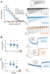
(A) Relative changes of HCN-mediated Ih current amplitudes at respective membrane potentials after DEX application (10 µM and 100 µM) compared to the control and the effect of the selective HCN channel blocker ZD7288 (40 µM). Beginning at −83 mV, 10 µM DEX induced a significant reduction in Ih. On the right, representative current traces from a TC relay neuron under control conditions and after the application of 10 µM DEX for 45 min. In comparison, 40 µM of ZD7288 resulted in a near complete attenuation of Ih currents over the complete voltage range. The voltage-clamp protocol is shown above and * p < 0.05. (B) Relative reduction in the maximal Ih current amplitude (Ih max) was recorded when the cell was hyperpolarized to −133 mV. The observed changes were significant beginning at a concentration of 10 µM DEX. On the right, representative current traces of Ih max under control conditions and in the presence of 10 µM DEX. The reduction observed after adding 40 µM of ZD7288 (orange) was almost complete, as depicted below. (C) At physiological membrane potentials (−73 mV), the reduction in relative Ih amplitude was only significant after the application of 100 µM DEX. HCN channel blocker ZD7288 reduced Ih−73 mV almost completely in all cells (n = 5). On the right, representative current traces of Ih at −73 mV under control conditions and in the presence of 10 µM DEX. Ns: not significant.
Figure 3
Figure 3
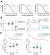
(A) In order to analyze voltage-dependent channel gating and steady-state activation curves of HCN channels under control conditions and after the application of increasing DEX concentrations, normalized tail current amplitudes (Itail) were fitted to a curve using the Boltzmann function. The tail step was set at −103 mV for each voltage step before returning to −43 mV. V1/2: half-maximal activation voltage. (B) Only the high concentration of 100 µM DEX led to a significant left shift (toward hyperpolarization) in V1/2 compared to the control (−86.6 [−85.2–−88.0] mV). Respective current traces under control conditions and after the application of 10 µM DEX are depicted on the right. Tail currents (Itail) were measured subsequent to the hyperpolarizing voltage step at the fixed “tail” step of −103 mV before returning to −43 mV. Δ: absolute change compared to control. Ns: not significant. * p < 0.05. (C) Representative current traces of Ih at the hyperpolarizing voltage step of −133 mV under control conditions and after 45 min of DEX application (10 µM) are depicted above. By fitting a biexponential function to Ih (as indicated by the dotted line), fast (τfast), and slow (τslow) time constants of time-dependent activation were determined. Only the high concentration of 100 µM DEX resulted in a significant deceleration of the fast activation constant compared to the control (231 [214–273] ms), presented as absolute changes. The slow time constant was not affected (control: 1143 [986–1405] ms).
Figure 4
Figure 4
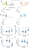
(A) TC replay neurons display an inwardly directed rectification of the membrane potential upon hyperpolarizing current injection (−350 pA, 500 ms, and the current protocol is shown on top), termed voltage sag. The voltage sag is unmasked in the presence of Ba2+ (green, 150 µM), on the left panel. Low-threshold calcium spike rebound burst-firing (rebound burst) is followed by an HCN channel-mediated afterdepolarization. In comparison, there is no quantifiable voltage sag in the presence of HCN channel blocker ZD7288 (orange, 40 µM), corresponding to a complete suppression. (B) Exemplar current-clamp recordings in the absence and after 45 min of 10 µM DEX (blue) administration with no significant changes. (C) DEX had no significant effect on voltage sag amplitude compared to the control (86.4 [70.7–92.9] mV). Δ: absolute change compared to the control. Ns: not significant. (D) Compared to control measurements (29.0 [27.0–30.8] ms), the delay of rebound burst-firing was only prolongated in the presence of 100 µM DEX. * p < 0.05. (E) The median number of action potentials (AP) during rebound burst spiking (control: 9.5 [7.3–12.5]) was only significantly reduced after the application of 100 µM DEX. (F) Accordingly, only the high concentration of 100 µM DEX significantly abbreviated the median rebound burst duration (DUR) compared to the control (225.5 [126.5–399.0] ms).

References

    1. Gerresheim G., Schwemmer U. Dexmedetomidin. Anaesthesist. 2013;62:661–674. doi: 10.1007/s00101-013-2206-6. - DOI - PubMed
    1. Keating G.M. Dexmedetomidine: A Review of Its Use for Sedation in the Intensive Care Setting. Drugs. 2015;75:1119–1130. doi: 10.1007/s40265-015-0419-5. - DOI - PubMed
    1. Huupponen E., Maksimow A., Lapinlampi P., Särkelä M., Saastamoinen A., Snapir A., Scheinin H., Scheinin M., Meriläinen P., Himanen S.-L., et al. Electroencephalogram spindle activity during dexmedetomidine sedation and physiological sleep. Acta Anaesthesiol. Scand. 2008;52:289–294. doi: 10.1111/j.1399-6576.2007.01537.x. - DOI - PubMed
    1. Akeju O., Pavone K.J., Westover M.B., Vazquez R., Prerau M.J., Harrell P.G., Hartnack K.E., Rhee J., Sampson A.L., Habeeb K., et al. A comparison of propofol- and dexmedetomidine-induced electroencephalogram dynamics using spectral and coherence analysis. Anesthesiology. 2014;121:978–989. doi: 10.1097/ALN.0000000000000419. - DOI - PMC - PubMed
    1. Purdon P.L., Sampson A., Pavone K.J., Brown E.N. Clinical Electroencephalography for Anesthesiologists: Part I: Background and Basic Signatures. Anesthesiology. 2015;123:937–960. doi: 10.1097/ALN.0000000000000841. - DOI - PMC - PubMed

MeSH terms

Substances

LinkOut - more resources