. 2022 Dec 30;24(1):632.
doi: 10.3390/ijms24010632.

The Efficacy of Molecular Analysis in the Diagnosis of Bone and Soft Tissue Sarcoma: A 15-Year Mono-Institutional Study

Affiliations

The Efficacy of Molecular Analysis in the Diagnosis of Bone and Soft Tissue Sarcoma: A 15-Year Mono-Institutional Study

Stefania Benini et al. Int J Mol Sci. .

Abstract

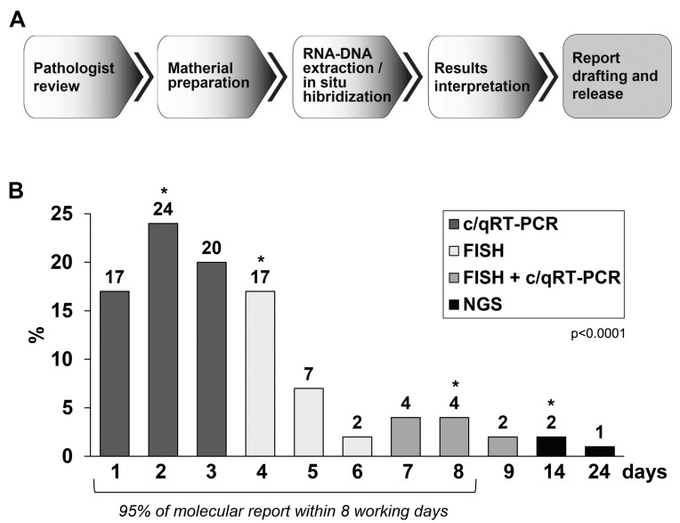
The histological diagnosis of sarcoma can be difficult as it sometimes requires the combination of morphological and immunophenotypic analyses with molecular tests. A total of 2705 tissue samples of sarcoma consecutively collected from 2006 until 2020 that had undergone molecular analysis were assessed to evaluate their diagnostic utility compared with histological assessments. A total of 3051 molecular analyses were performed, including 1484 gene fusions tested by c/qRT-PCR, 992 gene rearrangements analysed by FISH, 433 analyses of the gene status of MDM2, 126 mutational analyses and 16 NGS analysis. Of the samples analysed, 68% were from formalin-fixed, paraffin-embedded tissue and 32% were from frozen tissue. C/qRT-PCR and FISH analyses were conclusive on formalin-fixed, paraffin-embedded tissue in 74% and 76% of samples, respectively, but the combination of the two methods gave us conclusive results in 96% and 89% of frozen and formalin-fixed, paraffin-embedded tissues, respectively. We demonstrate the utility of c/qRT-PCR and FISH for sarcoma diagnosis and that each has advantages in specific contexts. We conclude that it is possible to accurately predict the sarcoma subtype using a panel of different subtype-specific FISH probes and c/qRT-PCR assays, thereby greatly facilitating the differential diagnosis of these tumours.

Keywords: FISH; RT-PCR; formalin-fixed, paraffin-embedded tissue; frozen tissue; fusion transcript; molecular diagnostics; next-generation sequencing; qRT-PCR; sarcomas.


Conflict of interest statement

The authors declare no conflict of interest. The funders had no role in the design of the study; in the collection, analyses, or interpretation of data; in the writing of the manuscript; or in the decision to publish the results.

Figures

Figure 1
Figure 1
Samples and molecular analyses carried out in our institution in 15 years of activity. (A) The diagram shows the distribution of the type of material analysed in this study. (B) Schematic representation of all molecular analyses carried out in our institution in fifteen years of activity. (C) The diagram shows the percentage of molecular investigations performed, based on type of analysis to evaluate the presence of fusion transcript, gene rearrangements, gene amplification and gene variation. (* for NGS the number corresponds to two years of activity).
Figure 2
Figure 2
Evaluation of the analytical quality of the samples under examination comparing frozen tissue vs. FFPE tissue, based on the type of analysis. (A) Analytical quality of c/qRT-PCR tests on frozen tissue samples vs. FFPE tissue. (B) Analytical quality of FISH tests on frozen tissue samples vs. FFPE tissue. (C) Analytical quality of combination of the two methods c/qRT-PCR + FISH tests on frozen tissue samples vs. FFPE tissue. (D) Analytical quality of gene variant tests on frozen tissue samples vs. FFPE tissue. White bars represent frozen tissue samples, and grey bars represent FFPE tissue samples.
Figure 3
Figure 3
Evaluation of the analytical quality of samples analysed for the state of the MDM2 gene by FISH on tissue stored and transported in a traditional way involving the use of formalin solution (light grey bars) vs. sealing tissue specimen under vacuum in plastic bags (dark grey bars). p value refers to the differences in the analytical quality (evaluable, not evaluable and unsuitable material) between the two groups.
Figure 4
Figure 4
Identification of the EWSR1-PBX3 fusion transcript in case 5 (malignant myoepithelioma). (A) Analysis of anchored multiplex PCR result of the Archer FusionPlex Sarcoma Panel showed EWSR1-PBX3 fusion in the biopsy. (B) FISH of interphase nuclei using dual-colour break-apart probe for EWSR1 (22q12) gene reveals 1 non rearranged orange/green fusion signal, 1 separate orange signal and 1 separate green signal, indicating the translocation of EWSR1 in tumour cells. Original magnification: 140. (C) RT-PCR/Sanger sequencing revealed the sequence of the novel EWSR1-PBX3 fusion transcript. Partial sequence chromatogram showing the junction (arrow) of the PBX3 and EWSR1 genes.
Figure 5
Figure 5
Workflow and turnaround time for clinical molecular samples (the year 2019–2020). (A) Workflow labelled with definitions of turnaround time metrics. (B) Histogram of the turnaround time from the start of tissue sample preparation for molecular investigation to report sign out (laboratory time) measured in calendar days. The grey bars represent the samples analysed with c/qRT-PCR, the white bars represent the samples analysed with FISH, the hatched bars represent the samples that required both c/qRT-PCR and FISH investigations, and the black bars represent the NGS analyses. Hatched and filled bars together add up to 100%. An Athena laboratory information system (Dedalus, Firenze, Italy) was used for specimen tracking. Cases were scanned to track their status as they progressed through the steps of the laboratory workflow. Time tracking data were extracted via a query). p value refers to the median differences in time (days) between the 4 groups. * median value of each group.

References

    1. Antonescu C.R., Bridge J.A., Cunha I.W. WHO Classification of Tumours Editorial Board: Soft Tissue and Bone Tumours. 5th ed. International Agency for Research on Cancer; Lyon, France: 2020. Soft tissue tumours. (WHO Classification of Tumours Series).
    1. Bovée J.V.M.G., Flanagan A.M., Lazar A.J., Nielsen G.P., Yoshida A. WHO Classification of Tumours Editorial Board: Soft Tissue and Bone Tumours. 5th ed. International Agency for Research on Cancer; Lyon, France: 2020. Bone tumours. (WHO Classification of Tumours Series).
    1. Sbaraglia M., Bellan E., Tos A.P.D. The 2020 WHO Classification of Soft Tissue Tumours: News and perspectives. Pathologica. 2021;113:70–84. doi: 10.32074/1591-951X-213. - DOI - PMC - PubMed
    1. Antonescu C.R., Dal Cin P. Promiscuous genes involved in recurrent chromosomal translocations in soft tissue tumours. Pathology. 2014;46:105–112. doi: 10.1097/PAT.0000000000000049. - DOI - PubMed
    1. Puls F., Niblett A.J., Mangham D.C. Molecular pathology of bone tumours: Diagnostic implications. Histopathology. 2014;64:461–476. doi: 10.1111/his.12275. - DOI - PubMed