. 2022 Dec 30;24(1):656.
doi: 10.3390/ijms24010656.

Trypanosoma brucei rhodesiense Inhibitor of Cysteine Peptidase (ICP) Is Required for Virulence in Mice and to Attenuate the Inflammatory Response

Affiliations

Trypanosoma brucei rhodesiense Inhibitor of Cysteine Peptidase (ICP) Is Required for Virulence in Mice and to Attenuate the Inflammatory Response

Tatiana F R Costa et al. Int J Mol Sci. .

Abstract

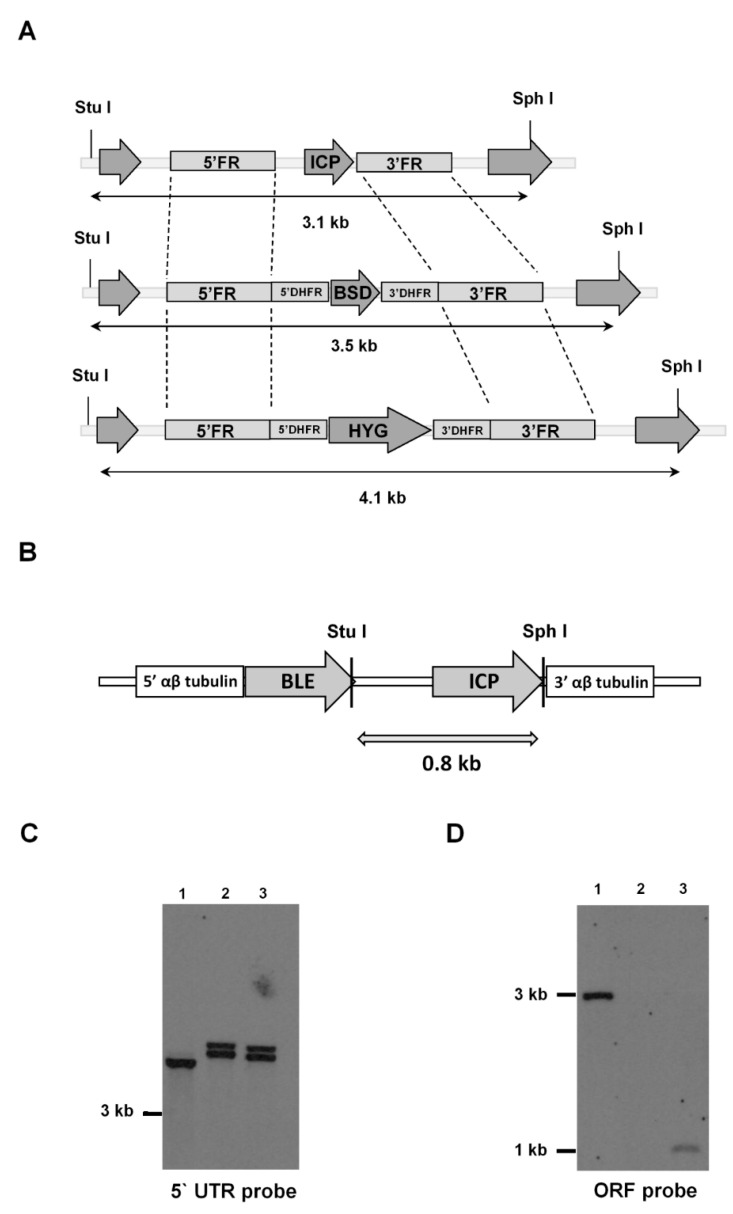
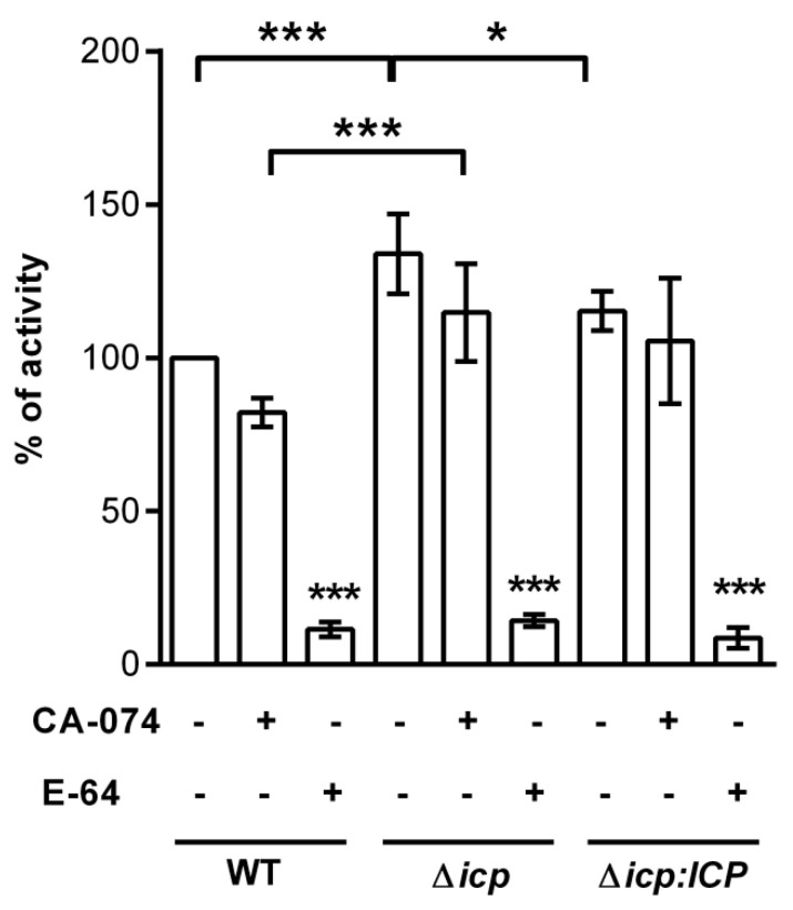
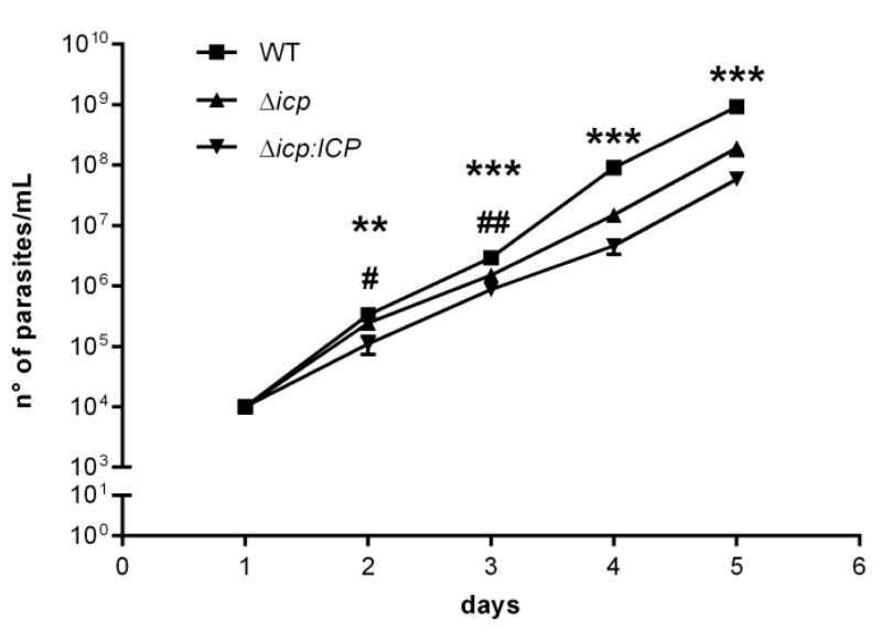
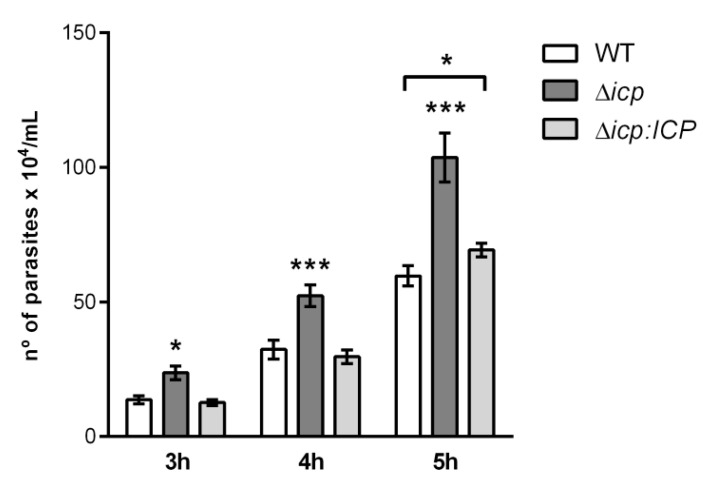
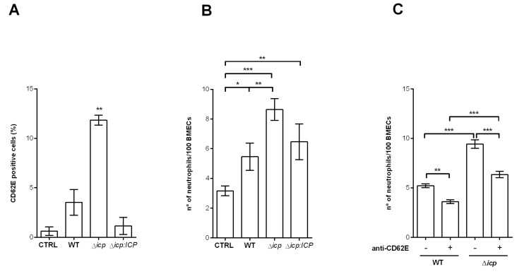
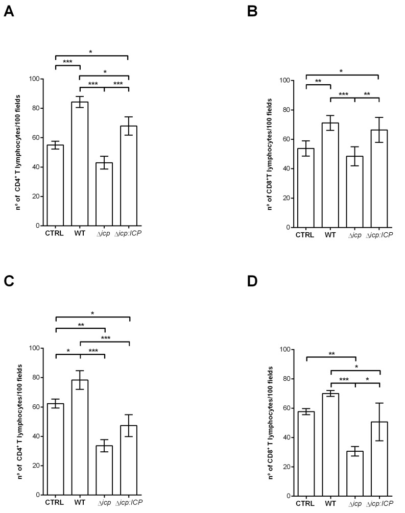
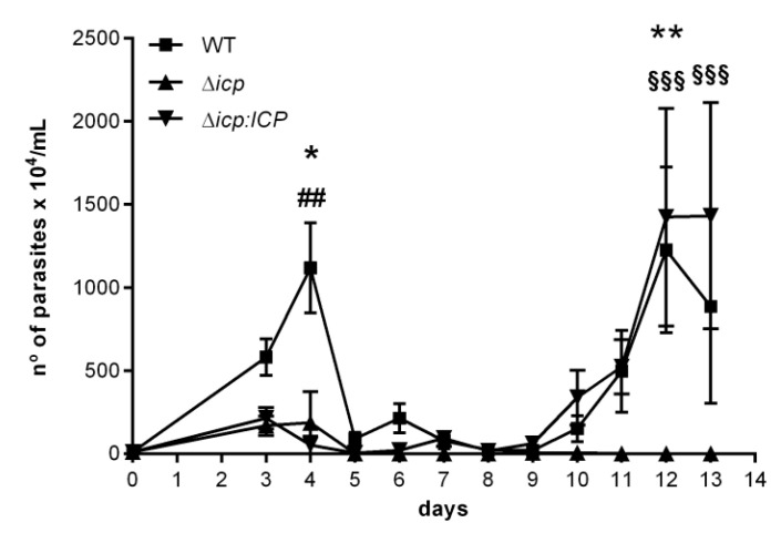
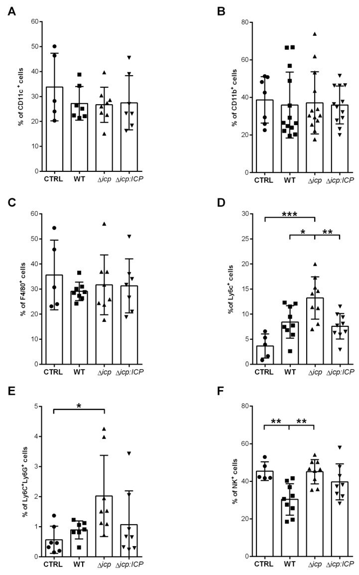
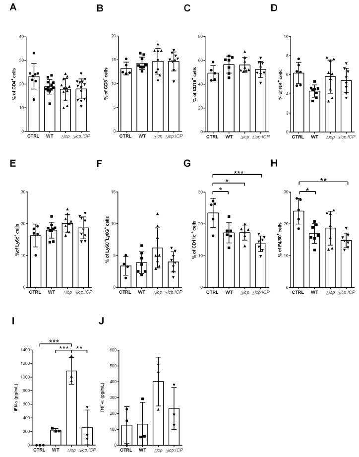
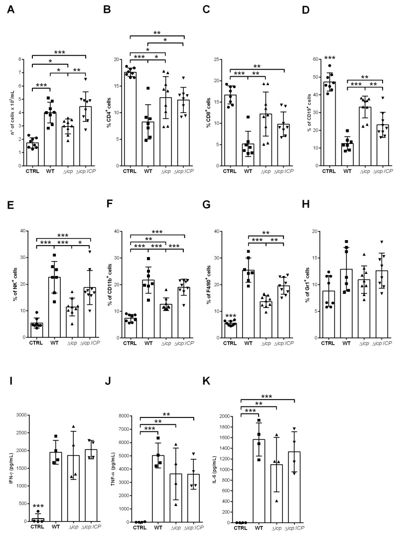
The protozoan Trypanosoma brucei rhodesiense causes Human African Trypanosomiasis, also known as sleeping sickness, and penetrates the central nervous system, leading to meningoencephalitis. The Cathepsin L-like cysteine peptidase of T. b. rhodesiense has been implicated in parasite penetration of the blood-brain barrier and its activity is modulated by the chagasin-family endogenous inhibitor of cysteine peptidases (ICP). To investigate the role of ICP in T. b. rhodesiense bloodstream form, ICP-null (Δicp) mutants were generated, and lines re-expressing ICPicp:ICP). Lysates of Δicp displayed increased E-64-sensitive cysteine peptidase activity and the mutant parasites traversed human brain microvascular endothelial cell (HBMEC) monolayers in vitro more efficiently. Δicp induced E-selectin in HBMECs, leading to the adherence of higher numbers of human neutrophils. In C57BL/6 mice, no Δicp parasites could be detected in the blood after 6 days, while mice infected with wild-type (WT) or Δicp:ICP displayed high parasitemia, peaking at day 12. In mice infected with Δicp, there was increased recruitment of monocytes to the site of inoculation and higher levels of IFN-γ in the spleen. At day 14, mice infected with Δicp exhibited higher preservation of the CD4+, CD8+, and CD19+ populations in the spleen, accompanied by sustained high IFN-γ, while NK1.1+ populations receded nearly to the levels of uninfected controls. We propose that ICP helps to downregulate inflammatory responses that contribute to the control of infection.

Keywords: Trypanosoma; chagasin; inflammation; inhibitor; protease; virulence.


Conflict of interest statement

The authors declare no conflict of interest.

Figures

Figure 1
Figure 1
Generation of T. b. rhodesiense ICP mutants. (A) Schematic representation of the ICP locus in wild-type (WT) T. b. rhodesiense IL1852 and the constructs containing the same 5′ and 3′ flanking regions (FR), and either a blasticidin (BSD) or hygromycin (HYG) resistance cassette, for the homologous recombination to generate an ICP knock-out line (Δicp). The oligonucleotides used to generate the FR for construction of recombination cassettes are listed in Table 1 (methods). Open reading frames (ORFs) of ICP and of flanking genes are shown as arrows. DHFR, dihydrofolate reductase. The predicted DNA fragment sizes after digestion with StuI and SphI are shown. (B) Schematic representation of the construct used, containing the phleomycin (BLE) resistance cassette, for the re-integration of ICP in the tubulin locus of the Δicp line to generate an ICP re-expressing line (Δicp:ICP). The predicted DNA fragment size after digestion with StuI and SphI is shown. (C,D) Genomic DNA isolated from the T. b. rhodesiense WT line, a Δicp clone, and a Δicp:ICP clone was digested with StuI and SphI, separated on a 0.8% agarose gel, transferred to a nylon membrane and probed with alkaline phosphatase labeled 5′ FR of ICP (C) or ICP ORF (D). Lane 1, WT; Lane 2, Δicp; Lane 3, Δicp:ICP.
Figure 2
Figure 2
T. b. rhodesiense lacking ICP have increased papain-like cysteine peptidase activity. Lysates from 106 bloodstream form (BSF) parasites of T. b. rhodesiense WT, Δicp, and Δicp:ICP were tested for peptidase activity in a continuous assay using Z-Phe-Arg-MCA as a substrate. Where indicated, 10 μM of CA-074 or 10 μM of E-64 were added to the samples diluted in assay medium for 10 min prior to the addition of the substrate. Experiments were performed in triplicate, two independent times, and are shown as the mean ± SD. Statistical significance between the controls (lysates only) of Δicp and Δicp:ICP at p < 0.05 (*) and Δicp and WT at p < 0.001 (***), between the E-64 treatments of Δicp and WT at p < 0.001 (***), and for each E-64 treatment in relation to the respective (lysate only) control, as assessed by one-way ANOVA with Bonferroni’s post hoc test.
Figure 3
Figure 3
T. b. rhodesiense Δicp have altered growth in vitro. Growth curves of BSF T. b. rhodesiense WT, Δicp, and Δicp:ICP determined by cell counts on a hemocytometer after cells were seeded in culture medium at 1 × 10⁴ cells/mL. Cultures were diluted to 1 × 10⁴ cells/mL after 3 days and 5 days and the cumulative cell number was determined using the dilution factor. Data are shown as the mean ±SD. Experiment was performed in triplicate and the graph is representative of three independent experiments. Statistical analysis was performed using two-way ANOVA with Tukey’s post hoc test. * indicates significant differences of WT compared to Δicp and Δicp:ICP; and # indicates significant differences between Δicp and Δicp:ICP; where p < 0.05 (#), p < 0.01 (** and ##), and p < 0.001 (***).
Figure 4
Figure 4
T. b. rhodesiense Δicp transmigrate an in vitro blood–brain barrier model more efficiently than wild type. Human brain microvascular endothelial cells (HBMECs) were seeded (5 × 10³) on top of Transwell inserts with 3 μm pores and cultured for 5 days, after which BSF parasites (106) of T. b. rhodesiense WT, Δicp, and Δicp:ICP were added on top of the HBMEC-containing inserts. The number of parasites present in the bottom chamber was determined by counting aliquots on a Neubauer chamber after 3, 4, and 5 h of incubation. The experiments were performed in triplicate, three independent times, and data are shown as the mean ± SD of a representative experiment. Asterisks indicate statistical significance at p < 0.05 (*) and p < 0.001 (***) as assessed by two-way ANOVA with Tukey’s post hoc test.
Figure 5
Figure 5
T. b. rhodesiense Δicp make HBMECs more susceptible to neutrophil adhesion. (A) Human BMECs were cultivated for 3 days, washed, and then incubated with BSF parasites of T. b. rhodesiense WT, Δicp, and Δicp:ICP for 18 h. Monolayers were washed, cells were detached, and cell surface E-selectin (CD62E) was detected by flow cytometry. (B) HBMECs (5 × 10⁴) were seeded on glass coverslips and cultured for 3 days, after which BSF parasites of T. b. rhodesiense WT, Δicp, and Δicp:ICP were added at a 2:1 parasite:cell ratio for 18 h. Cells were then washed and 106 human neutrophils were co-cultivated for 1 h. CTRL refers to control HBMECs not exposed to parasites, and co-cultivated with the neutrophils only. (C) Following cultivation on coverslips, HBMECs were exposed to parasites, washed, incubated with anti-E-selectin antibody for 1 h and washed, prior to the addition of neutrophils, as above. After incubation with the neutrophils, cells were washed, fixed with 70% methanol and then Giemsa stained. The number of adhered neutrophils was determined by the counting of 100 HBMECs per coverslip under a light microscope. The experiments were performed in triplicate, (B) five independent times or (C) two independent times, and data are shown as the mean ± SD of a representative experiment. In (A), asterisks indicate significance to all other points. Asterisks indicate statistical significance at p < 0.05 (*), p < 0.01 (**), and p < 0.001 (***) as assessed by one-way ANOVA with Tukey’s post hoc test.
Figure 6
Figure 6
T. b. rhodesiense Δicp make HBMECs less susceptible to adhesion by T lymphocytes. Human BMECs (5 × 10⁴) were seeded on glass coverslips and cultured for 3 days, after which BSF parasites of T. b. rhodesiense WT, Δicp, and Δicp:ICP were added at a 2:1 parasite:cell ratio for 18 h. CD4+ and CD8+ T cells (4 × 10⁵) were bead-purified from human blood, and were either (A,B) non-activated or (C,D) activated in vitro with 5 µg/mL phytohemagglutinin prior to addition to HBMECs. The cells were then co-cultivated for 2 h. CTRL refers to HBMECs cultivated with either activated or non-activated T cells only. Following this, cells were washed, fixed with 70% methanol and then Giemsa stained. The number of adhered T lymphocytes were determined by the counting of 100 HBMECs per coverslip under a light microscope. The experiments were performed in triplicate, three independent times, and data are shown as the mean ± SD of a representative experiment. Asterisks indicate statistical significance at p < 0.05 (*), p < 0.01 (**), and p < 0.001 (***) as assessed by one-way ANOVA with Tukey’s post hoc test.
Figure 7
Figure 7
T. b. rhodesiense Δicp have reduced virulence in C57BL/6 mice. C57BL/6 mice were infected i.p. with BSF parasites of T. b. rhodesiense WT, Δicp, and Δicp:ICP (1 × 10⁵ parasites per animal) and blood parasitemia was determined from days 3 to 13 of infection (starting with n = 5 per group). The graph shows the mean ±SEM of one experiment and is representative of two independent experiments. No mice died over the experimental period. One-way ANOVA with Tukey’s post hoc test was used to determine significant difference between all three groups; where * indicates a significant difference between WT and Δicp, # between WT and Δicp:ICP, and § between Δicp and Δicp:ICP at p < 0.05 (*), p < 0.01 (** and ##), and p < 0.001 (§§§).
Figure 8
Figure 8
T. b. rhodesiense Δicp induces increased recruitment of monocytes to the inoculation site during early infection of C57BL/6 mice. C57BL/6 mice were infected with BSF parasites of T. b. rhodesiense WT, Δicp, and Δicp:ICP (1 × 10⁵ parasites per animal). On the second day post-infection, mice were euthanized, and the peritoneal cavity was washed with RPMI in order to collect the peritoneal cells. The peritoneal cells were submitted to flow cytometry to assess expression of (A) CD11c, (B) CD11b, and (C) F4/80 within the total cell population, and (D) Ly6C+Ly6G cells and (E) Ly6C+Ly6G+ within the CD11b+ population. (F) Expression of NK1.1 within the total cell population. CTRL represents the non-infected control that was injected with RPMI medium. Graphs show individual points representing each mouse sample and the mean ± SD of two or three combined experiments (n ≥ 3 per group per experiment). Asterisks indicate statistical significance at p < 0.05 (*), p < 0.01 (**), and p < 0.001 (***) as assessed by one-way ANOVA with Tukey’s post hoc test.
Figure 9
Figure 9
T. b. rhodesiense Δicp induces higher inflammatory response in the spleen during early infection of C57BL/6 mice. C57BL/6 mice were infected with BSF parasites of T. b. rhodesiense WT, Δicp, and Δicp:ICP (1 × 10⁵ parasites per animal). On the second day post-infection, mice were euthanized, spleens were removed and macerated, then the splenocytes were submitted to flow cytometry or were cultured for 24 h, after which the supernatant was collected and cytokine levels determined by ELISA. The splenocytes were submitted to flow cytometry to assess expression of (A) CD4, (B) CD8, (C) CD19, and (D) NK1.1 within the total cell population. The proportion of the (E) Ly6C+Ly6G and (F,L) Ly6C+Ly6G+ populations within the CD11b+ was determined, while the expression of (G) CD11c and (H) F4/80 within the total cell population was assessed. Graphs show individual points representing each mouse sample and the mean ± SD of two independent experiments combined (n ≥ 3 per group per experiment). The splenocyte culture supernatants were tested for (I) IFN-γ and (J) TNF-α. CTRL represents the non-infected control that was injected with RPMI medium. Graphs show individual points representing each mouse sample and the mean ± SD of one experiment (n = 3 or 4 per group per experiment) and are representative of the profile of two independent experiments. Asterisks indicate statistical significance at p < 0.05 (*), p < 0.01 (**), and p < 0.001 (***) as assessed by one-way ANOVA with Tukey’s post hoc test.
Figure 10
Figure 10
C57BL/6 infected with T. b. rhodesiense lacking ICP dampen the immune response in the spleen by day 14. C57BL/6 mice were infected with BSF parasites of T. b. rhodesiense WT, Δicp, and Δicp:ICP (1 × 10⁵ parasites per animal). After 14 days, mice were euthanized and the spleens were collected, then macerated in order for (A) the cellularity to be determined. The splenocytes were submitted to flow cytometry (BH) or were cultured for 24 h, after which the supernatant was collected. The splenocytes were submitted to flow cytometry to assess expression of (B) CD4, (C) CD8, (D) CD19, (E) NK1.1, (F) CD11b, (G) F4/80, and (H) Gr1 within the total cell population. Graphs show individual points representing each mouse sample and the mean ± SD of two independent experiments combined (n ≥ 4 per group per experiment). The splenocyte culture supernatants were tested by ELISA to determine the levels of (I) IFN-γ, (J) TNF-α, and (K) IL-6. CTRL represents the non-infected control. Graphs show individual points representing each mouse sample and the mean ± SD of one experiment (n = 4 per group) and are representative of the profile of two independent experiments. Statistically significant differences were determined by one-way ANOVA with Tukey’s post hoc test, where p < 0.05 (*), p < 0.01 (**), and p < 0.001 (***).

References

    1. Kennedy P.G.E., Rodgers J. Clinical and neuropathogenetic aspects of human African trypanosomiasis. Front. Immunol. 2019;10:39. doi: 10.3389/fimmu.2019.00039. - DOI - PMC - PubMed
    1. Trindade S., Rijo-Ferreira F., Carvalho T., Pinto-Neves D., Guegen F., Aresta-Branco F., Bento F., Young S.A., Pinto A., Abbelee J.V.D., et al. Trypanosoma brucei parasites occupy and functionally adapt to the adipose tissue in mice. Cell Host Microbe. 2016;19:837–848. doi: 10.1016/j.chom.2016.05.002. - DOI - PMC - PubMed
    1. Capewell P., Cren-Travaillé C., Marchesi F., Johnston P., Clucas C., Benson R.A., Gorman T.A., Calvo-Alvarez E., Crouzols A., Jouvion G., et al. The skin is a significant but overlooked anatomical reservoir for vector-borne African trypanosomes. Elife. 2016;5:e17716. doi: 10.7554/eLife.17716. - DOI - PMC - PubMed
    1. Onyilagha C., Uzonna J.E. Host immune responses and immune evasion strategies in African Trypanosomiasis. Front. Immunol. 2019;10:2738. doi: 10.3389/fimmu.2019.02738. - DOI - PMC - PubMed
    1. Broadhead R., Dawe H.R., Farr H., Griffiths S., Hart S.R., Portman N., Shaw M.K., Ginger M.L., Gaskell S.J., McKean P.G., et al. Flagellar motility is required for the viability of the bloodstream trypanosome. Nature. 2006;440:224–227. doi: 10.1038/nature04541. - DOI - PubMed

Substances