. 2022 Dec 31;24(1):701.
doi: 10.3390/ijms24010701.

Therapeutic Potency of Induced Pluripotent Stem-Cell-Derived Corneal Endothelial-like Cells for Corneal Endothelial Dysfunction

Affiliations

Therapeutic Potency of Induced Pluripotent Stem-Cell-Derived Corneal Endothelial-like Cells for Corneal Endothelial Dysfunction

Seongjun So et al. Int J Mol Sci. .

Abstract

Corneal endothelial cells (CECs) do not proliferate or recover after illness or injury, resulting in decreased cell density and loss of pump/barrier function. Considering the shortage of donor cornea, it is vital to establish robust methods to generate CECs from induced pluripotent stem cells (iPSCs). We investigated the efficacy and safety of transplantation of iPSC-derived CECs into a corneal endothelial dysfunction (CED) rabbit model. iPSCs were generated from human fibroblasts. We characterized iPSCs by demonstrating the gene expression of the PSC markers OCT4, SOX2, TRA-1-60, and NANOG, teratoma formation, and differentiation into three germ layers. Differentiation of iPSCs into CECs was induced via neural crest cell (NCC) induction. CEC markers were detected using immunofluorescence and gene expression was analyzed using quantitative real-time PCR (qRT-PCR). After culturing iPSC-derived NCCs, we found the expression of zona occludens-1 (ZO-1) and Na+/K+ ATPase and a hexagonal morphology. ATP1A1, COL8A1, and AQP1 mRNA expression was higher in iPSC-derived CECs than in iPSCs and NCCs. We performed an injection of iPSC-derived CECs into the anterior chamber of a CED rabbit model and found improved levels of corneal transparency. We also found increased numbers of ZO-1- and ATP1A1-positive cells in rabbit corneas in the iPSC-derived CEC transplantation group. Usage of the coating material vitronectin (VTN) and fasudil resulted in good levels of CEC marker expression, demonstrated with Western blotting and immunocytochemistry. Combination of the VTN coating material and fasudil, instead of FNC mixture and Y27632, afforded the best results in terms of CEC differentiation's in vitro and in vivo efficacy. Successful transplantation of CEC-like cells into a CED animal model confirms the therapeutic efficacy of these cells, demonstrated by the restoration of corneal clarity. Our results suggest that iPSC-derived CECs can be a promising cellular resource for the treatment of CED.

Keywords: cell injection; cell therapy; corneal endothelial cells; corneal endothelial dysfunction; induced pluripotent stem cell.


Conflict of interest statement

The authors declare no conflict of interest. The funders had no role in the design of the study; in the collection, analysis, or interpretation of data; in the writing of the manuscript; or in the decision to publish the results.

Figures

Figure 1
Figure 1
Generation of corneal endothelial cells (CECs) from induced pluripotent stem cells (iPSCs). (A) Protocol for differentiation of iPSCs to CECs. iPSCs were differentiated into CECs through neural crest cells for 16–18 days. (B) Phase-contrast microscopy at time points during CEC differentiation illustrates likely CECs at day 6, and CECs exhibiting characteristic hexagonal/polygonal morphology at day 10. Scale bars = 500 µm. (C) Immunocytochemistry of pump function protein Na+/K+ ATPase α1 (ATP1A1) and zona occludens-1 (ZO-1; a tight-junction protein) exhibiting hexagonal/polygonal morphology of iPSC-derived CECs. Human primary CEC was used as a control. Cell nuclei were counterstained with DAPI. Scale bars = 100 μm.
Figure 2
Figure 2
Quantitative real-time RT-PCR (qRT-PCR). qRT-PCR analysis of the expression of representative pluripotent stem cell (PSC), neural crest cell (NCC), and corneal endothelial cell (CEC) genes. NCC-related gene expression decreased as differentiation progressed, while CEC-related gene expression increased until 6 days after CEC differentiation, decreasing thereafter (* p < 0.05).
Figure 3
Figure 3
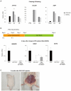
Stability of iPSC-derived CECs. (A) qRT-PCR analysis measuring ATP1A1, COL8A1, and AQP1 expression relative to that of GAPDH, following freezing and thawing of iPSC-derived CECs. Expression of all three genes was maintained after freezing and thawing (* p < 0.05). (B) Experimental scheme for replacing CEC culture medium with iPSC culture medium; qRT-PCR analysis of pluripotency-related gene expression in iPSC-derived CECs 6 days after changing to iPSC culture medium. The medium was changed to iPSC culture medium 2 days after passaging of the iPSC-derived CECs, and then cultured for 6 days to analyze pluripotent gene expression. Pluripotent gene expression did not increase despite change to an iPSC culture medium, and there was no significant difference with iPSC-derived CECs (D6) except for SOX2 (* p < 0.05). (C) In vivo tumorigenicity test of iPSC-derived CECs. Tumor formation was not found even after 2 months after injection of iPSC-derived CECs into the femoral region of immunodeficient mice. Scale bars = 1 cm.
Figure 4
Figure 4
Transplantation of iPSC-derived CECs into a corneal endothelial dysfunction rabbit model. (A) Ocular observation and numerical data at 1, 2, and 3 weeks after transplantation of iPSC-derived CECs. Compared with the non-transplanted control, cloudy corneas were clear in the transplanted eyes after 1 week and the effect increased over time (* p < 0.05; n = 6). Corneal opacity was evaluated on a scale of 0–4 (0 = none; 1 = mild turbidity, iris texture evident; 2 = moderate turbidity, iris texture unclear; 3 = severe turbidity, pupil faint; and 4 = severe turbidity, pupil not visible). (B) The corneal thickness of the cell transplant group was observed histologically using H & E staining. The cornea of the transplanted group (right) is thinner compared with the non-transplanted control group (middle), which closely resembled the healthy cornea (left). Scale bars = 200 μm. (C) Immunohistochemistry for the detection of human CEC markers (ATP1A1 and ZO-1) in the corneas of transplanted and non-transplanted groups. ATP1A1 and ZO-1 were broadly expressed in the cornea of the transplant group, but not in the non-transplant control group. Scale bars = 200 or 100 μm.
Figure 5
Figure 5
Efficient CEC differentiation by replacement of FNC coating reagent into vitronectin (VTN). (A) Pluripotency-related gene expression analysis of iPSC-derived CECs using VTN or FNC as coating reagents. Pluripotent gene expression of iPSC-derived CECs, differentiated using VTN, was lower compared with that of FNCs (* p < 0.05; n = 3). Except for ATP1A1 expression, CEC-related gene expression was significantly higher when VTN was used (* p < 0.05; n = 3). (B) Scheme of the protocol using fasudil or Y27632 as ROCK inhibitor and VTN or FNC as coating material during CEC differentiation. (C) Morphology of differentiated CECs in four different groups of culture conditions: VTN-Y (Y27632), VTN-F (fasudil), FNC-Y, and FNC-F. Scale bars = 100 μm.
Figure 6
Figure 6
Comparison of different ROCK inhibitors and coating materials on CEC differentiation. (A) Immunocytochemistry using antibodies against ZO-1, SLC4A11, N-Cadherin, and ATP1A1. Scale bar = 50 μm for all the images. (B) Western blot using a group of CEC markers: ZO-1, NCAM, SLC4A11, N-Cadherin, and ATP1A1. (C) CCK-8 assay. Fasudil-treated groups had increased levels of cell viability and proliferation compared to Y27632-treated groups. (* p < 0.05; n = 3).
Figure 7
Figure 7
Transplantation of iPSC-induced CECs or iPSC (ZsGreen)-derived CECs produced via a modified protocol (VTN with fasudil) into a corneal endothelial dysfunction rabbit model. (A) Ocular observation. (B) Observation of transplanted iPSC (ZsGreen)-derived CECs expressing green fluorescent protein and DAPI in the iPSC-derived CEC-transplanted cornea using a fluorescence microscope. Scale bars = 400 μm. (C,D) Immunohistochemistry for the detection of human CEC markers (ATP1A1 and ZO-1) in the iPSC-derived CEC-transplanted cornea. ATP1A1 and ZO-1 were broadly expressed in the cornea of the transplant group. (C,D) show results in different corneal regions of rabbits after cell transplantation. Higher density with smaller size of transplanted CECs (C) and lower density with larger sized cells (D) were found in the cornea. The bottom rows show higher magnifications of images of the white rectangles in the top row. Scale bars = 1 mm or 100 μm.

References

    1. Bonanno J.A. Identity and regulation of ion transport mechanisms in the corneal endothelium. Prog. Retin. Eye Res. 2003;22:69–94. doi: 10.1016/S1350-9462(02)00059-9. - DOI - PubMed
    1. Bonanno J.A. Molecular mechanisms underlying the corneal endothelial pump. Exp. Eye Res. 2012;95:2–7. doi: 10.1016/j.exer.2011.06.004. - DOI - PMC - PubMed
    1. Hatou S., Higa K., Inagaki E., Yoshida S., Kimura E., Hayashi R., Tsujikawa M., Tsubota K., Nishida K., Shimmura S. Validation of Na,K-ATPase pump function of corneal endothelial cells for corneal regenerative medicine. Tissue Eng. Part C Methods. 2013;19:901–910. doi: 10.1089/ten.tec.2013.0030. - DOI - PubMed
    1. Srinivas S.P. Cell signaling in regulation of the barrier integrity of the corneal endothelium. Exp. Eye Res. 2012;95:8–15. doi: 10.1016/j.exer.2011.09.009. - DOI - PMC - PubMed
    1. Bourne W.M., Nelson L.R., Hodge D.O. Central corneal endothelial cell changes over a ten-year period. Investig. Ophthalmol. Vis. Sci. 1997;38:779–782. - PubMed