Review
. 2023 Jan 3;24(1):878.
doi: 10.3390/ijms24010878.

Choroid Plexus Aquaporins in CSF Homeostasis and the Glymphatic System: Their Relevance for Alzheimer's Disease

Affiliations
Review

Choroid Plexus Aquaporins in CSF Homeostasis and the Glymphatic System: Their Relevance for Alzheimer's Disease

Cristina Municio et al. Int J Mol Sci. .

Abstract

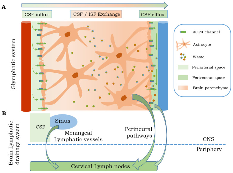
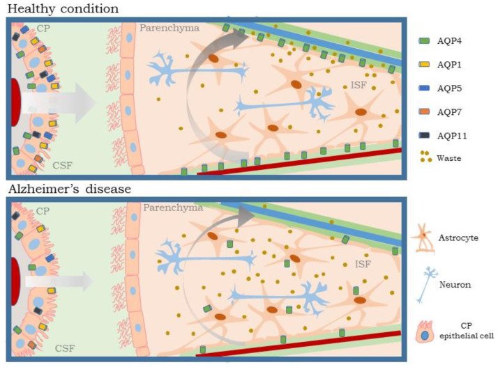
The glymphatic system, a fluid-clearance pathway involved in brain waste clearance, is known to be impaired in neurological disorders, including Alzheimer's disease (AD). For this reason, it is important to understand the specific mechanisms and factors controlling glymphatic function. This pathway enables the flow of cerebrospinal fluid (CSF) into the brain and subsequently the brain interstitium, supported by aquaporins (AQPs). Continuous CSF transport through the brain parenchyma is critical for the effective transport and drainage of waste solutes, such as toxic proteins, through the glymphatic system. However, a balance between CSF production and secretion from the choroid plexus, through AQP regulation, is also needed. Thus, any condition that affects CSF homeostasis will also interfere with effective waste removal through the clearance glymphatic pathway and the subsequent processes of neurodegeneration. In this review, we highlight the role of AQPs in the choroid plexus in the modulation of CSF homeostasis and, consequently, the glymphatic clearance pathway, with a special focus on AD.

Keywords: Alzheimer’s disease; aquaporins; astrocytes; cerebrospinal fluid; choroid plexus; clearance; glymphatic system; homeostasis.


Conflict of interest statement

The authors declare no conflict of interest.

Figures

Figure 1
Figure 1
Illustration of choroid plexus epithelial cells (left) and tight junctions at the luminal membrane (right). CP: choroid plexus; CSF: cerebrospinal fluid; JAMs: junctional adhesion molecules; ZO: zonula occludens.
Figure 2
Figure 2
Brain lymphatic drainage system. (A) Glymphatic system clears solutes and waste of the brain parenchyma due to the influx of CSF from the periarterial space. Once the CSF is mixed with the ISF, they drain into the perivenous space. Part (B) shows a summary of the other brain drainage systems that transport the CSF through the meningeal lymphatic vessels and/or perineural pathways, such as olfactory nerve, to the cervical lymph nodes AQP: aquaporin; CNS: central nervous system; CSF: cerebrospinal fluid; ISF: interstitial fluid.
Figure 3
Figure 3
Relationship between choroid plexus and glymphatic system. In healthy conditions (above), the choroid plexus forms the BCSFB and produces CSF. CSF flows from the periarterial space to the brain parenchyma via AQP4, located in the end-feet of astrocytes. This movement is favored positive pressure of CSF production from the choroid plexus and the arterial pulse. The CSF/ISF mixture and waste products are cleared by passing into the perivenous space to be taken to the lymphatic tissues. In AD (below) the expression of AQPs decreases in choroid plexus epithelial cells causing a reduction in CSF production. The decrease in pressure exerted by the CSF together with the diminution and depolarization of AQP4 in the astrocyte end-feet, reduces the glymphatic function, preventing a correct clearance of the ISF and waste products. AQP: aquaporin; BCSFB: brain CSF barrier; CSF: cerebrospinal fluid; CP: choroid plexus; ISF: interstitial fluid.

References

    1. Jucker M., Walker L.C. Pathogenic protein seeding in Alzheimer disease and other neurodegenerative disorders. Ann. Neurol. 2011;70:532–540. doi: 10.1002/ana.22615. - DOI - PMC - PubMed
    1. Proulx S.T. Cerebrospinal fluid outflow: A review of the historical and contemporary evidence for arachnoid villi, perineural routes, and dural lymphatics. Cell Mol. Life Sci. 2021;78:2429–2457. doi: 10.1007/s00018-020-03706-5. - DOI - PMC - PubMed
    1. Kaur J., Fahmy L.M., Davoodi-Bojd E., Zhang L., Ding G., Hu J., Zhang Z., Chopp M., Jiang Q. Waste Clearance in the Brain. Front. Neuroanat. 2021;15:665803. doi: 10.3389/fnana.2021.665803. - DOI - PMC - PubMed
    1. Iliff J.J., Wang M., Liao Y., Plogg B.A., Peng W., Gundersen G.A., Benveniste H., Vates G.E., Deane R., Goldman S.A., et al. A paravascular pathway facilitates CSF flow through the brain parenchyma and the clearance of interstitial solutes, including amyloid β. Sci. Transl. Med. 2012;4:147ra111. doi: 10.1126/scitranslmed.3003748. - DOI - PMC - PubMed
    1. Nedergaard M. Neuroscience. Garbage truck of the brain. Science. 2013;340:1529–1530. doi: 10.1126/science.1240514. - DOI - PMC - PubMed