Review
. 2022 Dec 22;12(1):91.
doi: 10.3390/jcm12010091.

Contribution of Ultrasound in Current Practice for Managing Juvenile Idiopathic Arthritis

Affiliations
Review

Contribution of Ultrasound in Current Practice for Managing Juvenile Idiopathic Arthritis

Charlotte Borocco et al. J Clin Med. .

Abstract

The interest and application of musculoskeletal ultrasound (MSUS) in juvenile idiopathic arthritis (JIA) are increasing. Numerous studies have shown that MSUS is more sensitive than clinical examination for detecting subclinical synovitis. MSUS is a well-accepted tool, easily accessible and non-irradiating. Therefore, it is a useful technique throughout JIA management. In the diagnostic work-up, MSUS allows for better characterizing the inflammatory involvement. It helps to define the disease extension, improving the classification of patients into JIA subtypes. Moreover, it is an essential tool for guiding intra-articular and peritendinous procedures. Finally, during the follow-up, in detecting subclinical disease activity, MSUS can be helpful in therapeutic decision-making. Because of several peculiarities related to the growing skeleton, the MSUS standards defined for adults do not apply to children. During the last decade, many teams have made large efforts to define normal and pathological US features in children in different age groups, which should be considered during the US examination. This review describes the specificities of MSUS in children, its applications in clinical practice, and its integration into the new JIA treat-to-target therapeutic approach.

Keywords: application; enthesitis; follow-up; gestures; juvenile idiopathic arthritis; peculiarities; reliability; sensitivity; treat-to-target; ultrasonography.


Conflict of interest statement

Linda Rossi-Semerano has received consulting fees from Pfizer, Abbvie, and Novartis, and congress fees from Novartis, SOBI Biovitrum, Abbvie, Pfizer, Nordic, and Amgen. Charlotte Borocco: nothing to declare. Federica Anselmi: nothing to declare.

Figures

Figure 1
Figure 1
Pediatric ultrasound peculiarities. Increased ratio of cartilage to bone (a) and physiological cartilage vascularization (b) in a 6-year-old girl. (c) Physiological irregularity of calcaneus bone surface owing to the presence of ossification centers in a 10-year-old boy. C: cartilage; Tt: tibial tuberosity; Ca: calcaneus; T: tendon; F: fat.
Figure 2
Figure 2
Synovitis of the knee joint in a 12-year-old girl with polyarticular juvenile idiopathic arthritis. (a) Longitudinal suprapatellar scan showing synovitis in the suprapatellar recess on B-mode (grade 2). (b) Physiological aspect of the contralateral joint on B-mode. (c) Transverse lateral parapatellar scan showing pathological vascularization (power Doppler grade 1). (d) Longitudinal suprapatellar scan showing vascularization outside the synovial hypertrophy (power Doppler grade 0). Note that the transverse scan is more sensitive for detecting pathological vascularisation inside synovitis than the longitudinal scan [18]. P: patella; T: tendon; F: femur; *: Synovitis; white arrow: small physiological amount of intra-articular fluid; Red arrows: physiological vascularization.
Figure 3
Figure 3
Longitudinal anterior scan of the tibiotalar joint in a 7-year-old with oligoarticular JIA; (a,b) Severe synovitis (B-mode grade 3) with minimal Doppler activity (grade 1). (c) Physiological aspect of the contralateral joint. C: cartilage; T: tibia; TA: talus; F: fat; * Synovitis.
Figure 4
Figure 4
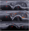
Longitudinal dorsal scan of one metatarsophalangeal joint in a 13-year-old boy with enthesitis-related arthritis. (a,b) Severe synovitis (B-mode grade 3) with moderate power Doppler activity (<30% of the synovial hypertrophy surface, grade 2). (c) Physiological aspect of the contra-lateral joint. C: cartilage; M: metatarsal; P: proximal phalanx; T: tendon; * Synovitis.
Figure 5
Figure 5
(a,b) Enthesitis of the distal patellar ligament in a 14-year-old boy with juvenile enthesitis-related arthritis. Longitudinal scan of the distal patellar ligament showing bilateral enthesitis. PT: patellar tendon; TT: tibial tuberosity; white arrow: secondary ossification center, incompletely ossified tibia.
Figure 6
Figure 6
(a,b). Tenosynovitis of the extensor digitorum and extensor carpi radialis tendons in a 12-year-old girl with JIA. (a) Transverse scan showing tenosynovitis on B-mode; (b) longitudinal scan showing abnormal vascularization on power Doppler mode. Performing the longitudinal scan (B-mode/Doppler mode) is mandatory to confirm the diagnosis of tenosynovitis. ED: extensor digitorum tendon; ER: extensor carpi radialis tendon; (c,d). Tenosynovitis of the tibialis poster tendon in an 8-year-old girl with JIA. Transverse scan of the medial compartment showing tenosynovitis.

References

    1. Thierry S., Fautrel B., Lemelle I., Guillemin F. Prevalence and Incidence of Juvenile Idiopathic Arthritis: A Systematic Review. Jt. Bone Spine. 2014;81:112–117. doi: 10.1016/j.jbspin.2013.09.003. - DOI - PubMed
    1. Petty R.E., Southwood T.R., Manners P., Baum J., Glass D.N., Goldenberg J., He X., Maldonado-Cocco J., Orozco-Alcala J., Prieur A.-M., et al. International League of Associations for Rheumatology Classification of Juvenile Idiopathic Arthritis: Second Revision, Edmonton, 2001. J. Rheumatol. 2004;31:390–392. - PubMed
    1. Aoust L., Rossi-Semerano L., Koné-Paut I., Dusser P. Time to Diagnosis in Juvenile Idiopathic Arthritis: A French Perspective. Orphanet J. Rare Dis. 2017;12:43. doi: 10.1186/s13023-017-0586-4. - DOI - PMC - PubMed
    1. Glerup M., Herlin T., Twilt M. Clinical Outcome and Long-Term Remission in JIA. Curr. Rheumatol. Rep. 2017;19:75. doi: 10.1007/s11926-017-0702-4. - DOI - PubMed
    1. Ohrndorf S., Backhaus M. Pro Musculoskeletal Ultrasonography in Rheumatoid Arthritis. Clin. Exp. Rheumatol. 2015;33:S50–S53. - PubMed

LinkOut - more resources