. 2022 Dec 29;12(1):278.
doi: 10.3390/jcm12010278.

Clinical, Dermoscopic, Ultrasonographic, and Histopathologic Correlations in Kaposi's Sarcoma Lesions and Their Differential Diagnoses: A Single-Center Prospective Study

Affiliations

Clinical, Dermoscopic, Ultrasonographic, and Histopathologic Correlations in Kaposi's Sarcoma Lesions and Their Differential Diagnoses: A Single-Center Prospective Study

Athanasia Tourlaki et al. J Clin Med. .

Abstract

(1) Background: Kaposi's sarcoma (KS) is an angioproliferative neoplasm typically appearing as angiomatous patches, plaques, and/or nodules on the skin. Dermoscopy and ultrasonography have been suggested as an aid in the diagnosis of KS, but there is little evidence in the literature, especially regarding its possible differential diagnoses. Our aim is to describe and compare the clinical, dermoscopic, and ultrasonographic features of KS and KS-like lesions. (2) Methods: we conducted a prospective study on 25 consecutive patients who were first referred to our tertiary care center from January to May 2021 for a possible KS. (3) Results: 41 cutaneous lesions were examined by means of dermoscopy, Doppler ultrasonography, and pathology, 32 of which were KS-related, while the remaining 9 were lesions with clinical resemblance to KS. On dermoscopy, a purplish-red pigmentation, scaly surface, and the collarette sign were the most common features among KS lesions (81.3%, 46.9%, and 28.1%, respectively). On US, all 9 KS plaques and 21 KS nodules presented a hypoechoic image. Dermoscopic and Doppler ultrasonographic findings of KS-like lesions, such as cherry angioma, venous lake, glomus tumor, pyogenic granuloma, and angiosarcoma were also analyzed. (4) Conclusions: dermoscopy and Doppler ultrasonography can be useful to better assess the features of KS lesions and in diagnosing equivocal KS-like lesions.

Keywords: Kaposi’s sarcoma; clinical manifestations; dermoscopy; differential diagnoses; histopathology; ultrasound imaging.


Conflict of interest statement

The authors declare no conflict of interest.

Figures

Figure 1
Figure 1
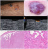
Patch stage Kaposi’s sarcoma. (A,B). Active patches on the feet of a 79-year-old female patient suffering from classic Kaposi’s sarcoma. (C). Dermoscopy of one of the patches revealed a homogeneously structureless pinkish-brown area crossed by fine branching vessels. (D). Ultrasonography showed a thin hypoechoic linear band in the superficial dermis. (E). Histologically, the lesion was characterized by a dermal proliferation of spindle cells forming sinuous vascular spaces and a lymphocytic infiltrate [Hematoxylin-eosin, 40×]. (F). Higher magnification revealing the promontory sign (*) with protrusion of vascular structures into lumens of the few newly formed vascular spaces [H-E, 100×].
Figure 2
Figure 2
Bullous Kaposi’s sarcoma. (A). Angiomatous plaque on the right foot medial surface of a 75-year-old male patient with classic Kaposi’s sarcoma. (B). Magnification of the area showing confluent bullae forming a plaque. (C). Dermoscopy of a bulla showing a target-shaped lesion with whitish, purple and red concentric, structureless areas and short dilated vessels in the periphery. (D). Ultrasonography revealed a homogenous, hypoechoic lesion in the dermis and subcutis. Color-Doppler showed intense intralesional and deep vascular flow. (E). Histological examination showed dilated interstitial spaces delimited by bundles of spindled cells in association with erythrocyte extravasation in the dermis (H-E, 20×). (F). Hemorrhagic-necrotic material in the upper part of the image (H-E, 40×). H. At higher magnification we can appreciate the phenomenon of autolumination (*): the presence of an erythrocyte within a paranuclear vesicle in one spindle cell (H-E, 200×).
Figure 3
Figure 3
Nodular Kaposi’s sarcoma. (A). Hyperkeratotic nodules on the left forearm of an 81-year-old patient suffering from classic Kaposi’s sarcoma. (B). Dermoscopy showed a purplish nodule with whitish-yellow collarette. The rainbow pattern can be clearly seen. (C,D). A hypoechoic nodular lesion, characterized by vascular signal detected with Color-Doppler at the lower pole. (E). Histology showed fascicles of spindle cells forming sinuous vascular spaces in the dermis, especially towards the periphery of the lesion (H-E, 20×). (F). Higher magnification revealed eosinophilic hyaline globules as a result of the autolumination process (H-E, 100×).
Figure 4
Figure 4
Exophytic nodular Kaposi’s sarcoma. (A). Exophytic nodules on the left ankle of an 84-year-old male patient diagnosed with classic Kaposi’s sarcoma. (B). Magnification of a translucent purplish-red nodule characterized by telangiectasias. (C). Dermoscopy showed dilated, serpentine vessels, homogeneously distributed across the nodule. (D). B-mode ultrasonography showed multiple nodules divided by septa and characterized by inhomogeneous hypoechogenicity. (E,F). Histology revealed a well-circumscribed dermal mass formed by monomorphic spindled cells organized in bundles and delimited by dilated confluent vascular spaces (H-E, 20× and 80×).
Figure 5
Figure 5
Deep nodular Kaposi’s sarcoma. (A). Deep nodule on the right heel of a 61-year-old male patient suffering from classic Kaposi’s sarcoma. (B). Dermoscopy without applying pressure showed serpentine vessels with a reticulated distribution. (C) Dermoscopy with downward pressure caused the emptying of the vessels. (D). B-mode ultrasonography revealed a hypoechoic nodule with posterior enhancement. (E). Histology showed some grouped nodules deeply localized in the dermis, and separated by fibrous septa (H-E, 20×). (F). Spindled cells arranged in haphazard fascicles. Intra- and extracellular hyaline globules were detectable (H-E, 100×).
Figure 6
Figure 6
Glomus tumor. (A). A painful nodule located on the left forearm of an 85-year-old woman suffering from classic Kaposi’s sarcoma. (B). Dermoscopy showed a purple-pinkish nodular lesion characterized by arborescent vessels. (C,D). B-mode ultrasonography revealed a horizontally oriented, hypoechoic mass with clear borders. On the left, we can see the afferent artery with the so-called stalk-sign. (E). Histopathology revealed a mixed eosinophilic and basophilic well-defined mass (H-E, 20×). (F). At higher magnification, the specimen was characterized by round glomus cells with pale eosinophilic cytoplasm. Tumor stroma appeared myxoid and edematous (H-E, 80×).
Figure 7
Figure 7
Pyogenic granuloma. (A). A pedunculated, angiomatous, rapidly growing nodular lesion on the right forearm of a 44-year-old male patient suffering from HIV-associated Kaposi’s sarcoma. (B). Dermoscopy of the nodule was characterized by homogeneous white-reddish areas with numerous linear-irregular vessels. Blanching of the blood vessels due to the pressure applied on the skin surface is shown in the left part of the nodule. (C). Ultrasonography showed an inhomogeneous, hypoechoic, and capsulated oval mass. (D). Color-Doppler analysis revealed a highly vascularized peduncle (speed 28.7 cm/s). (E). Histology showed discontinuous parakeratinized stratified squamous epithelium and vascular spaces in the underlying stroma; (F). The nodule did not present fibrous septa, but a fibrinopurulent membrane on the periphery (H-E, 20× and 80×).

References

    1. Etemad S.A., Dewan A.K. Kaposi sarcoma updates. Dermatol. Clin. 2019;37:505–517. doi: 10.1016/j.det.2019.05.008. - DOI - PubMed
    1. Brambilla L., Genovese G., Berti E., Peris K., Rongioletti F., Micali G., Ayala F., Della Bella S., Mancuso R., Calzavara Pinton P., et al. Diagnosis and treatment of classic and iatrogenic Kaposi’s sarcoma: Italian recommendations. Ital. J. Dermatol. Venerol. 2021;156:356–365. doi: 10.23736/S2784-8671.20.06703-6. - DOI - PubMed
    1. Brambilla L., Boneschi V., Taglioni M., Ferrucci S. Staging of classic Kaposi’s sarcoma:A useful tool for therapeutic choices. Eur. J. Dermatol. 2003;13:83–86. - PubMed
    1. Brambilla L., Tourlaki A., Ferrucci S., Brambati M., Boneschi V. Treatment of classic Kaposi’s sarcoma-associated lymphedema with elastic stockings. J. Dermatol. 2006;33:451–456. doi: 10.1111/j.1346-8138.2006.00108.x. - DOI - PubMed
    1. Tourlaki A., Bellinvia M., Brambilla L. Recommended surgery of Kaposi’s sarcoma nodules. J. Dermatol. Treat. 2015;26:354–356. doi: 10.3109/09546634.2014.971698. - DOI - PubMed