. 2023 Jan 3;28(1):440.
doi: 10.3390/molecules28010440.

Effects of Histamine and the α-Tocopherol Metabolite α-13'-COOH in an Atopic Dermatitis Full-Thickness Skin Model

Affiliations

Effects of Histamine and the α-Tocopherol Metabolite α-13'-COOH in an Atopic Dermatitis Full-Thickness Skin Model

Rebecca Riedl et al. Molecules. .

Abstract

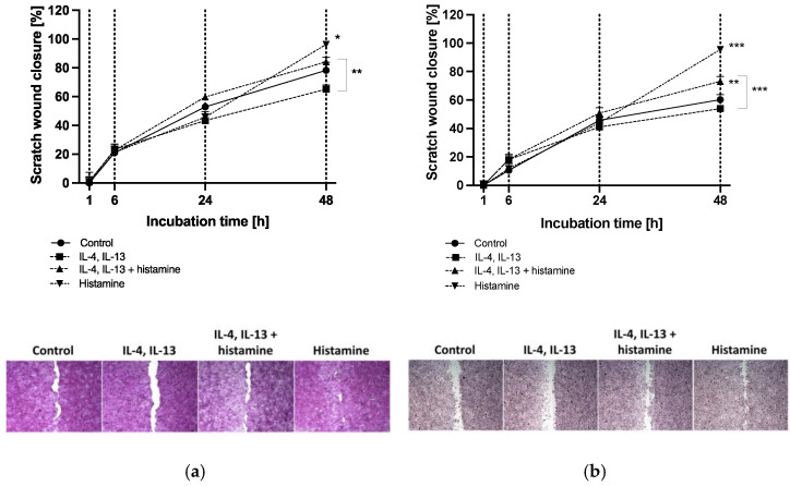
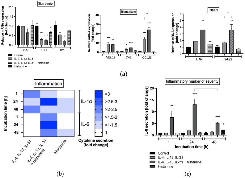
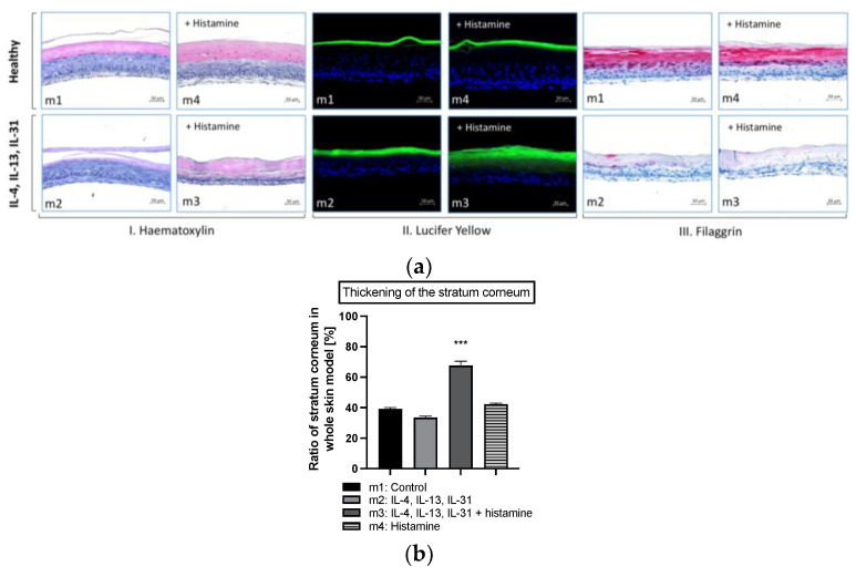
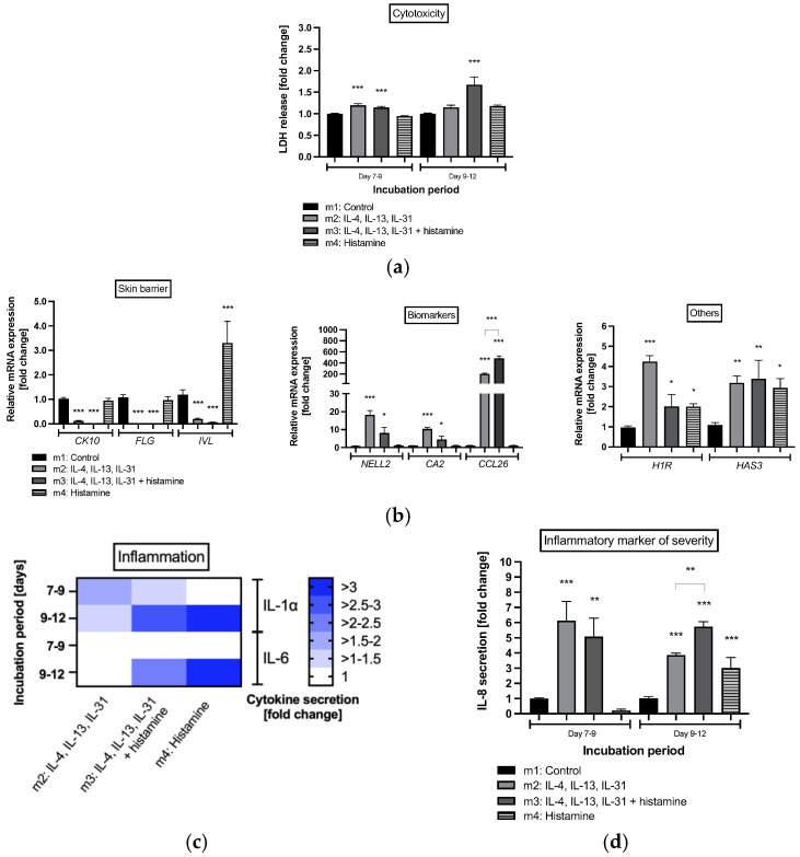
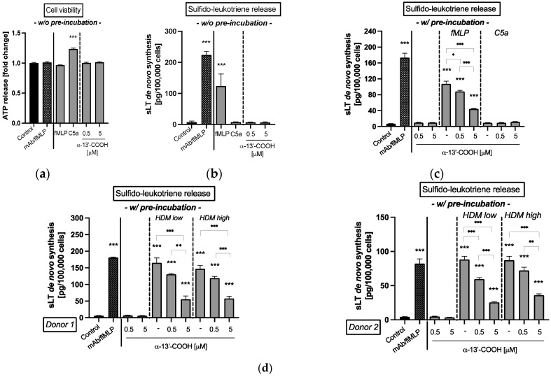
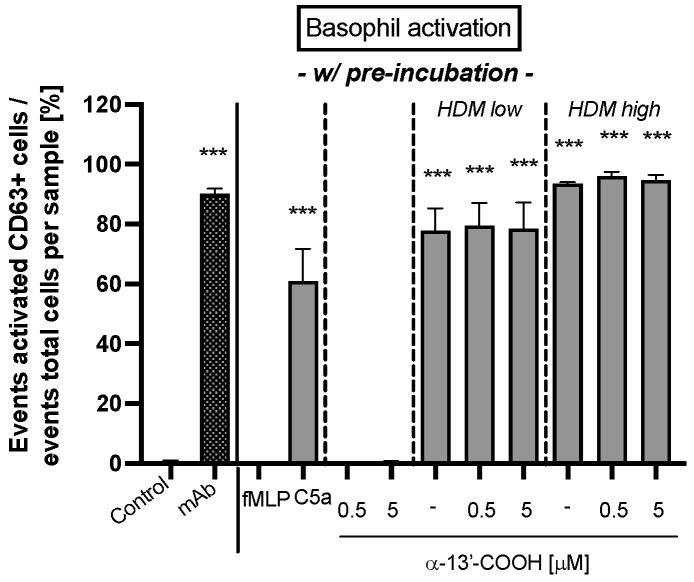
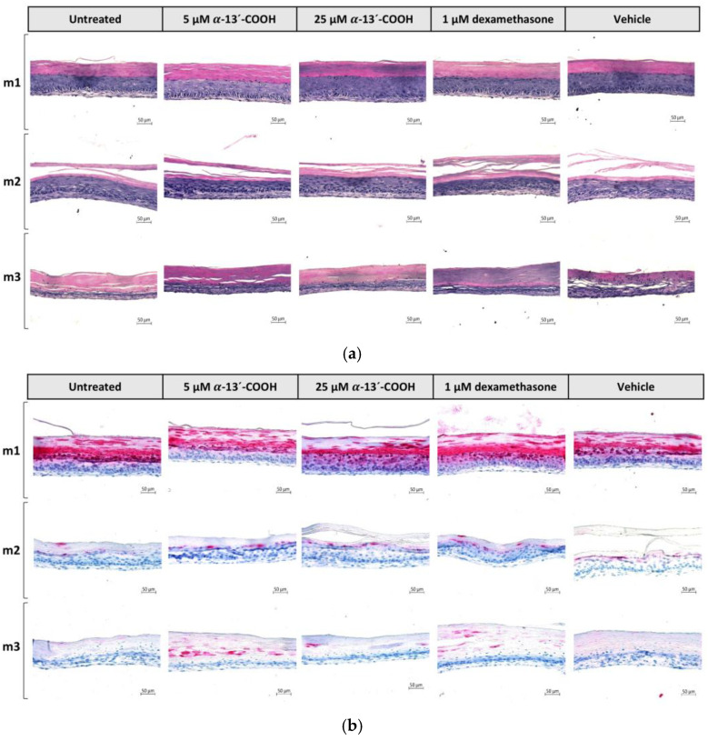
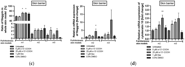
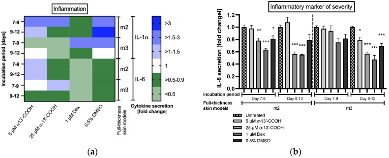
Atopic dermatitis is a T-cell mediated inflammatory skin disease with detected elevated levels of histamine in skin or plasma. In this study, the effects of histamine in a TH2 cytokine environment on human keratinocytes and three-dimensional skin models were investigated. These models were used to explore the anti-inflammatory properties of the α-tocopherol-derived long-chain metabolite α-13'-carboxychromanol (α-13'-COOH). Histamine and TH2 cytokine-induced proliferation of keratinocytes was studied using a scratch assay. The inflammatory marker interleukin-8 was significantly increased in healthy and TH2 cytokine-stimulated keratinocytes and skin models after histamine treatment. The incubation of full-thickness skin models with TH2 cytokines and histamine resulted in morphological changes in the epidermal layer, interpreted as hyperkeratosis. α-13'-COOH significantly decreased interleukin-8 in these disease-associated skin models. Histological staining of filaggrin showed skin-strengthening effects following α-13'-COOH treatment, without changes in mRNA expression. Cytokeratin 10 mRNA expression tended to be increased in response to α-13'-COOH. Anti-allergic properties of α-13'-COOH were studied by pre-incubation of human leukocytes with α-13'-COOH. This resulted in reduced sulfido-leukotriene synthesis. The hyperproliferation effect of histamine in atopic dermatitis skin models may be of further interest to the study of disease-associated morphological changes. Moreover, α-13'-COOH is a promising natural compound for the treatment of inflammatory skin diseases.

Keywords: anti-allergic; anti-inflammatory; atopic dermatitis full-thickness skin model; histamine; hyperkeratosis; α-13’-carboxychromanol.


Conflict of interest statement

M.W. and S.L. have received a research grant from DSM Nutritional Products GmbH. The funder had no role in the design of the study; in the collection, analysis, or interpretation of data; in the writing of the manuscript; or in the decision to publish the manuscript. The remaining authors declare no conflicts of interest.

Figures

Figure 1
Figure 1
(a) HaCaT keratinocyte and (b) primary keratinocyte scratch wound closure after 1, 6, 24 and 48 h of incubation. Keratinocytes were either stimulated with TH2 cytokines (50 ng/mL IL-4, 50 ng/mL IL-13) with or without 10 µM histamine or cultivated under normal medium conditions with 10 µM histamine. The control group was cultivated without any stimulation. Wound closure is displayed as scratch closure and is presented as mean ± standard error of the mean (SEM) in [%]. Two independent experiments were performed as replicates. Scratch assays were evaluated using six images per sample. Asterisks [*] indicate significant deviations from the control at the respective time point (* p < 0.05, ** p < 0.01 and *** p < 0.001). Cells were stained with haematoxylin-eosin. Images show scratch wound closure of (a) HaCaT keratinocytes and (b) primary keratinocytes after 48 h of incubation.
Figure 2
Figure 2
(a) Gene expression profiles of primary keratinocytes after 48 h of incubation and (b) interleukin (IL) IL-1α, IL-6, (c) IL-8 protein expression at after 1, 24 and 48 h of incubation. Keratinocytes were stimulated with TH2 cytokines (50 ng/mL IL-4, 50 ng/mL IL-13, 25 ng/mL IL-31) either with or without 10 µM histamine or under normal medium conditions with 10 µM histamine in the absence of TH2 cytokines. The control group was cultivated under normal medium conditions without any stimulation. (a) Transcript levels are given as normalized relative mRNA expression compared to the untreated control. (b,c) Cytokine levels are given as fold changes compared to the untreated control at the respective time point. (a,c) All data are presented as mean ± SEM [fold change]. (b) All data in the heatmap are presented as mean [fold change]. Two independent experiments were performed as replicates. Asterisks [*] indicate significant deviations from the untreated control (* p < 0.05, ** p < 0.01 and *** p < 0.001).
Figure 3
Figure 3
(a) Histological evaluation of the skin morphology (I. Hematoxylin), skin permeability effects (II. Lucifer Yellow), skin barrier effects (III. Filaggrin) and (b) area ratio of stratum corneum of healthy and atopic dermatitis full-thickness skin models after 12 days of cultivation to air surface (airlift). Atopic dermatitis skin models were stimulated with TH2 cytokines (50 ng/mL IL-4, 50 ng/mL IL-13 and 25 ng/mL IL-31) at day 0, 2, 5, 7 and 9 (m2, m3). Healthy skin model m4 and atopic dermatitis skin model m3 were stimulated with 10 µM histamine at day 9. Healthy skin models were cultivated under normal medium conditions (m1, m4). (a) Skin models for evaluation of skin morphology were stained with haematoxylin/eosin (I), for evaluation of skin permeability with Lucifer Yellow dye (II) and evaluation of skin barrier effects with filaggrin (III). Scale bar: 50 µm. (b) The ratio of the stratum corneum in whole skin models was calculated as the area of the stratum corneum compared to the area of the whole skin model and is given as mean ± SEM in [%] using the image processing program ImageJ (Scale bar: 100 µm). Histological analyses were conducted on three skin models from two independent experiments with four images per skin model. Asterisks [*] indicate significant deviations from the untreated control (*** p < 0.001).
Figure 4
Figure 4
(a) Cell viability, (b) gene expression profiles from healthy and atopic dermatitis full-thickness skin models after 12 days of cultivation to air surface (airlift), and (c) interleukin (IL)-1α, IL-6 and (d) IL-8 protein expression after days 7 to 9 and days 9 to 12 of cultivation to air surface (airlift). Atopic dermatitis skin models were stimulated with TH2 cytokines (50 ng/mL IL-4, 50 ng/mL IL-13, and 25 ng/mL IL-31) at days 0, 2, 5, 7 and 9 (m2, m3). Healthy skin model m4 and atopic dermatitis skin model m3 were stimulated with 10 µM histamine at day 9. Healthy skin models were cultivated under normal medium conditions (m1, m4). (a,c,d) All data are given as fold changes compared to the untreated control at the respective time points, after day 9 (incubation period days 7 to 9) and day 12 (incubation period days 9 to 12) of incubation. (b) Transcript levels are given as relative mRNA expression referred to the untreated control [fold changes]. (a,b,d) All data are presented as mean ± SEM. (c) All data in the heatmap are presented as mean. Two independent experiments were performed as replicates. Asterisks [*] indicate significant deviations from the control at the respective time point (* p < 0.05, ** p < 0.01 and *** p < 0.001).
Figure 5
Figure 5
(a) Cell viability and (bd) de novo synthesis of sulfido-leukotrienes (sLT) from isolated blood leukocytes. (a,b) Leukocytes were simultaneously incubated without any pre-incubation (w/o pre-incubation) with either fMLP, C5a and 0.5 or 5 µM α-13’-COOH for 50 min. (c,d) Leukocytes were pre-incubated with (w/ pre-incubation) 0.5 or 5 µM α-13’-COOH or buffer (untreated) for 50 min. Leukocytes used as control were incubated with buffer only. Incubation with an anti-IgE Receptor mAb/fMLP solution is shown as positive control (mAb/fMLP). (c) A second unspecific stimulation was conducted either with fMLP (5 µM), C5a (10 nM), or buffer for a further 50 min. (d) A second specific stimulation was conducted with an extract from house dust mite (HDM) for specific IgE-mediated response for a further 50 min. Leukocytes from donor 1 and donor 2 were confirmed with a specific IgE level sensitized against HDM extract. HDM extract was applied at two concentrations (HDM low: 2 ng; HDM high: 20 ng). (a) Cell viability is presented as the amount of ATP release given as fold change of the control. (bd) De novo synthesis of sLT is given as absolute amounts per defined cell number. (ad) All data are presented as mean ± SEM. Two independent experiments were performed as replicates. Asterisks [*] indicate significant deviations from the untreated control (* p < 0.05, ** p < 0.01 and *** p < 0.001).
Figure 6
Figure 6
Basophil activation in whole blood from donors with confirmed specific IgE-sensitized immune cells against house dust mite (HDM) extract. Whole blood cells were pre-incubated with 0.5 or 5 µM α-13’-COOH or buffer (untreated control) for 50 min. Blood cells used as controls were pre-incubated with buffer. A second unspecific basophil activation was tested either with fMLP (5 µM), C5a (10 nM), or buffer. Activation of basophils with HDM extract was investigated as specific IgE-mediated response. The HDM extract was applied in two concentrations (HDM low: 2 ng; HDM high: 20 ng). A specific anti-IgE receptor mAb was used as positive control (mAb). Activated basophils were stained with a mixture of monoclonal antibodies against human cluster of differentiation (CD) 63 labeled with fluorescein isothiocyanate (anti-CD63-FITC) and against human C-C chemokine receptor type 3 (CCR3) labeled with phycoerythrin (anti-CCR3-PE). Basophil activation is given as the event of activated CD63-positive basophils in relation to the absolute events of each sample in percent. All data are presented as mean ± SEM. Two independent experiments were performed as replicates. Asterisks [*] indicate significant deviations from the untreated control (*** p < 0.001).
Figure 7
Figure 7
(a,b) Histological, (c) quantitative evaluation, and (d) gene expression profile of filaggrin in healthy and atopic dermatitis full-thickness skin models after 12 days of cultivation to air surface (airlift). Healthy skin models were cultivated under normal medium conditions (m1). Atopic dermatitis skin models were stimulated with TH2 cytokines (50 ng/mL IL-4, 50 ng/mL IL-13 and 25 ng/mL IL-31) at days 0, 2, 5, 7 and 9 (m2, m3). Atopic dermatitis skin model m3 was additionally stimulated with 10 µM histamine at day 9. Skin models m1, m2 and m3 were pre-incubated with either 5 µM α-13’-COOH, 25 µM α-13’-COOH, 1 µM dexamethasone (Dex; positive control), or 0.5% DMSO (vehicle control) at days 7 and 9, or were left untreated as disease control (untreated). (a) Skin models for evaluation of skin morphology were stained with haematoxylin-eosin and (b) for evaluation of skin barrier effects with filaggrin. Scale bar: 50 µm. (c) Ratio of filaggrin expression in whole skin models is given as the area of filaggrin in whole skin models in relation to the area of the whole skin model in [%] using ImageJ software (Scale bar: 100 µm). (d) Transcript levels are given as relative mRNA expression referred to the untreated disease control [fold change]. (c,d) All data are presented as mean ± SEM. Two independent experiments were performed as replicates. Histological analyses were conducted on three skin models from two independent experiments with four images per skin model. Asterisks [*] indicate significant deviations from the untreated disease control (* p < 0.05 and ** p < 0.01).
Figure 7
Figure 7
(a,b) Histological, (c) quantitative evaluation, and (d) gene expression profile of filaggrin in healthy and atopic dermatitis full-thickness skin models after 12 days of cultivation to air surface (airlift). Healthy skin models were cultivated under normal medium conditions (m1). Atopic dermatitis skin models were stimulated with TH2 cytokines (50 ng/mL IL-4, 50 ng/mL IL-13 and 25 ng/mL IL-31) at days 0, 2, 5, 7 and 9 (m2, m3). Atopic dermatitis skin model m3 was additionally stimulated with 10 µM histamine at day 9. Skin models m1, m2 and m3 were pre-incubated with either 5 µM α-13’-COOH, 25 µM α-13’-COOH, 1 µM dexamethasone (Dex; positive control), or 0.5% DMSO (vehicle control) at days 7 and 9, or were left untreated as disease control (untreated). (a) Skin models for evaluation of skin morphology were stained with haematoxylin-eosin and (b) for evaluation of skin barrier effects with filaggrin. Scale bar: 50 µm. (c) Ratio of filaggrin expression in whole skin models is given as the area of filaggrin in whole skin models in relation to the area of the whole skin model in [%] using ImageJ software (Scale bar: 100 µm). (d) Transcript levels are given as relative mRNA expression referred to the untreated disease control [fold change]. (c,d) All data are presented as mean ± SEM. Two independent experiments were performed as replicates. Histological analyses were conducted on three skin models from two independent experiments with four images per skin model. Asterisks [*] indicate significant deviations from the untreated disease control (* p < 0.05 and ** p < 0.01).
Figure 8
Figure 8
(a) Interleukin (IL)-1α and IL-6 protein expression and (b) IL-8 protein expression in atopic dermatitis full-thickness skin models after days 7 to 9 and days 9 to 12 of cultivation to air surface (airlift). Atopic dermatitis skin models were stimulated with TH2 cytokines (50 ng/mL IL-4, 50 ng/mL IL-13 and 25 ng/mL IL-31) at days 0, 2, 5, 7 and 9 (m2, m3). Atopic dermatitis skin model m3 was additionally stimulated with 10 µM histamine at day 9. Skin models m2 and m3 were pre-incubated with either 5 µM α-13’-COOH, 25 µM α-13’-COOH, 1 µM dexamethasone (Dex; positive control), or 0.5% DMSO (vehicle control) at days 7 and 9, or were left untreated as disease control (untreated). Cytokine levels are given as fold change to untreated disease control at the respective time points, after day 9 (incubation period days 7 to 9) and day 12 (incubation period days 9 to 12) of incubation. (a) All data in the heatmap are presented as means. (b) All data are presented as means ± SEM. Two independent experiments were performed as replicates. Asterisks [*] indicate significant deviations from the untreated disease control at the respective time point. (* p < 0.05, ** p < 0.01 and *** p < 0.001).

References

    1. Asher M.I., Montefort S., Björkstén B., Lai C.K., Strachan D.P., Weiland S.K., Williams H. Worldwide Time Trends in the Prevalence of Symptoms of Asthma, Allergic Rhinoconjunctivitis, and Eczema in Childhood: ISAAC Phases One and Three Repeat Multicountry Cross-Sectional Surveys. Lancet. 2006;368:733–743. doi: 10.1016/S0140-6736(06)69283-0. - DOI - PubMed
    1. Lloyd-Lavery A., Solman L., Grindlay D.J.C., Rogers N.K., Thomas K.S., Harman K.E. What’s New in Atopic Eczema? An Analysis of Systematic Reviews Published in 2016. Part 2: Epidemiology, Aetiology and Risk Factors. Clin. Exp. Dermatol. 2019;44:370–375. doi: 10.1111/ced.13853. - DOI - PubMed
    1. Silverberg J.I. Associations between Atopic Dermatitis and Other Disorders. F1000Research. 2018;7:303. doi: 10.12688/f1000research.12975.1. - DOI - PMC - PubMed
    1. Silverberg N.B., Silverberg J.I. Inside out or Outside in: Does Atopic Dermatitis Disrupt Barrier Function or Does Disruption of Barrier Function Trigger Atopic Dermatitis? Cutis. 2015;96:359–361. - PubMed
    1. Buys L.M. Treatment Options for Atopic Dermatitis. Am. Fam. Phys. 2007;75:523–528. - PubMed