. 2022 Dec 23;15(1):71.
doi: 10.3390/nu15010071.

Circulating Extracellular Vesicles Are Increased in Newly Diagnosed Celiac Disease Patients

Affiliations

Circulating Extracellular Vesicles Are Increased in Newly Diagnosed Celiac Disease Patients

Konstantinos Efthymakis et al. Nutrients. .

Abstract

Extracellular vesicles (EVs) are a class of circulating entities that are involved in intercellular crosstalk mechanisms, participating in homeostasis maintenance, and diseases. Celiac disease is a gluten-triggered immune-mediated disorder, characterized by the inflammatory insult of the enteric mucosa following local lymphocytic infiltration, resulting in villous atrophy. The goal of this research was the assessment and characterization of circulating EVs in celiac disease patients, as well as in patients already on an adequate gluten-free regimen (GFD). For this purpose, a novel and validated technique based on polychromatic flow cytometry that allowed the identification and enumeration of different EV sub-phenotypes was applied. The analysis evidenced that the total, annexin V+, leukocyte (CD45+), and platelet (CD41a+) EV counts were significantly higher in both newly diagnosed celiac disease patients and patients under GFD compared with the healthy controls. Endothelial-derived (CD31+) and epithelial-derived (EpCAM+) EV counts were significantly lower in subjects under gluten exclusion than in celiac disease patients, although EpCAM+ EVs maintained higher counts than healthy subjects. The numbers of EpCAM+ EVs were a statistically significant predictor of intraepithelial leukocytes (IEL). These data demonstrate that EVs could represent novel and potentially powerful disease-specific biomarkers in the context of celiac disease.

Keywords: celiac disease; extracellular vesicles; flow cytometry; gluten-free diet regimen (GFD).


Conflict of interest statement

The authors declare no conflict of interest.

Figures
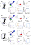
Figure 1
Figure 1
Gating strategy for EV identification. EV identification and sub-typing in (A) healthy subjects, (B) Celiac disease patients and (C) celiac disease patients under a gluten-free diet (GFD patient). (a) Phalloidin-negative events were firstly gated and (b) analyzed for their SSC-H/FSC-H parameters. The scatter area identified on the basis of MegaMix Plus beads (broadly 0.1–1 µm) was selected and named platelet (PLT)-free area. (c) Events were then analyzed on a Phalloidin-H/LCD-H dot-plot and phalloidin– LCD+ vesicles were gated and identified as EVs. EVs were finally sub-typed: (d) platelet-derived EVs (PLT-EVs) were gated on the basis of their combined positivity to CD31 and CD41a. The gate “NOT-PLT-EVs” was obtained and, in such a population, (e) Leukocyte-derived EVs (Leu-EVs) were identified as CD45+ events. By creating a logical gate “Not-Leu-EVs”, (f) endothelial-derived EVs (CD31+, Endo-EVs) and epithelial-EVs (CD326/EpCAM+, Epi-EVs) were also gated. (g) Annexin V positivity was also assessed on an Annexin V-H/CD31-H dot-plot, and the gate was then applied to any identified EV subset. Data are representative of all peripheral blood samples analyzed by flow cytometry.
Figure 2
Figure 2
Total EV and annexin V+ EV counts. Box plots represent total (A) and total Annexin V+ (B) EV concentrations (EVs per µL, log-transformed) in peripheral blood obtained from controls, celiac, and celiac-GFD patients. The horizontal line represents the median value. The asterisk (*) identifies statistically significant differences.
Figure 3
Figure 3
Total CD31+ and CD41a+ EV counts. Box plots represent CD31+ (A) and CD41a+ (B) EV concentrations (EVs per µL, log-transformed) in peripheral blood obtained from controls, celiac, and GFD patients. The horizontal line represents the median value. The asterisk (*) identifies statistically significant differences.
Figure 4
Figure 4
Total CD45+ and EpCAM+ EV counts. Box plots represent CD45+ (A) and EpCAM+ (B) EV concentrations (EVs per µL, log-transformed) in peripheral blood obtained from controls, celiac, and GFD patients. The horizontal line represents the median value. The asterisk (*) identifies statistically significant differences.
Figure 5
Figure 5
Correlation of IEL concentration and R model-predicted EpCAM+ counts (unstandardized predicted value) in CD. In a multiple linear regression model including total, EpCAM+, and CD31+, CD45+ and CD41a+ counts, EpCAM+ EVs represent a statistically significant predictor (IEL: intraepithelial leukocytes).

References

    1. Colombo M., Raposo G., Théry C. Biogenesis, Secretion, and Intercellular Interactions of Exosomes and other Extracellular Vesicles. Annu. Rev. Cell Dev. Biol. 2014;30:255–289. doi: 10.1146/annurev-cellbio-101512-122326. - DOI - PubMed
    1. Romano E., Netti P.A., Torino E. Exosomes in Gliomas: Biogenesis, Isolation, and Preliminary Applications in Nanomedicine. Pharmaceuticals. 2020;13:319. doi: 10.3390/ph13100319. - DOI - PMC - PubMed
    1. Shah R., Patel T., Freedman J.E. Circulating Extracellular Vesicles in Human Disease. N. Engl. J. Med. 2018;379:958–966. doi: 10.1056/NEJMra1704286. - DOI - PubMed
    1. Revenfeld A.L.S., Bæk R., Nielsen M.H., Stensballe A., Varming K., Jørgensen M. Diagnostic and Prognostic Potential of Extracellular Vesicles in Peripheral Blood. Clin. Ther. 2014;36:830–846. doi: 10.1016/j.clinthera.2014.05.008. - DOI - PubMed
    1. Lawson C., Vicencio J.M., Yellon D.M., Davidson S.M. Microvesicles and Exosomes: New Players in Metabolic and Cardiovascular Disease. J. Endocrinol. 2016;228:R57–R71. doi: 10.1530/JOE-15-0201. - DOI - PubMed