. 2022 Dec 24;23(1):181.
doi: 10.3390/s23010181.

Quantitative Evaluation of In Vivo Corneal Biomechanical Properties after SMILE and FLEx Surgery by Acoustic Radiation Force Optical Coherence Elastography

Affiliations

Quantitative Evaluation of In Vivo Corneal Biomechanical Properties after SMILE and FLEx Surgery by Acoustic Radiation Force Optical Coherence Elastography

Yanzhi Zhao et al. Sensors (Basel). .

Abstract

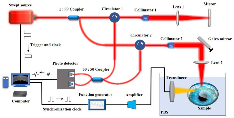
The purpose of this study is to quantitatively evaluate the differences in corneal biomechanics after SMILE and FLEx surgery using an acoustic radiation force optical coherence elastography system (ARF-OCE) and to analyze the effect of the corneal cap on the integrity of corneal biomechanical properties. A custom ring array ultrasound transducer is used to excite corneal tissue to produce Lamb waves. Depth-resolved elastic modulus images of the in vivo cornea after refractive surgery were obtained based on the phase velocity of the Lamb wave. After refractive surgery, the average elastic modulus of the corneal flap decreased (71.7 ± 24.6 kPa), while the elastic modulus of the corneal cap increased (219.5 ± 54.9 kPa). The average elastic modulus of residual stromal bed (RSB) was increased after surgery, and the value after FLEx (305.8 ± 48.5 kPa) was significantly higher than that of SMILE (221.3 ± 43.2 kPa). Compared with FLEx, SMILE preserved most of the anterior stroma with less change in corneal biomechanics, which indicated that SMILE has an advantage in preserving the integrity of the corneal biomechanical properties. Therefore, the biomechanical properties of the cornea obtained by the ARF-OCE system may be one of the essential indicators for evaluating the safety of refractive surgery.

Keywords: FLEx surgery; SMILE surgery; acoustic radiation force; corneal biomechanical properties; optical coherence elastography; optical coherence tomography.


Conflict of interest statement

The authors declare no conflict of interest.

Figures

Figure 1
Figure 1
The ARF-OCE experiment protocol of the SMILE and FLEx surgery, (a) is the SMILE surgery incision structure, the SMILE procedure preserves most of the anterior corneal stroma by creating a small 2–3 mm incision in the anterior corneal stroma and removing the stromal microcrystalline lens; (b) is the FLEx surgery flap structure, the FLEx procedure uses a femtosecond laser to create a corneal flap on the surface of the cornea and then thin the stroma.
Figure 2
Figure 2
The acoustic radiation force based OCE system diagram. The swept laser with a central wavelength of 1310 nm is split by a 1:99 optical coupler and then enters the reference arm and the sample arm, respectively, and the interference signal is detected by an optical balance detector. The laser provides the sampling trigger signal and sampling clock signal. The synchronization control signal of the acoustic radiation force excitation is generated by the computer and synchronized with the OCT sampling clock.
Figure 3
Figure 3
The ARF-OCE experiment results of the normal cornea, (a) is the structure image of the cornea in OCT B-scan, (b) is the vibration mapping in the OCE B-scan, and (c) is the spatial-temporal displacement diagram of the Lamb wave.
Figure 4
Figure 4
The phase velocity calculation of the Lamb wave in normal cornea, (a) is the wave number-frequency map of the Lamb wave, (b) is the phase velocity curve depends on the frequency, and (c) is the depth-resolved elastography result of the normal cornea.
Figure 5
Figure 5
The FLEx surgery corneal images, (a) is the FLEx surgery surface morphology, (b) is the 3D reconstruction structure, (c) is the 2D tomography, (d) is the spatial-temporal displacement diagram of the Lamb wave, (e) is depth-resolved elastography result of the cornea.
Figure 6
Figure 6
The SMILE surgery corneal images, (a) is the SMILE surgery surface morphology, (b) is the 3D reconstruction structure, (c) is the 2D tomography, (d) is the spatial-temporal displacement diagram of the Lamb wave, and (e) is depth-resolved elastography result of the cornea.
Figure 7
Figure 7
Elastic modulus statistical results of the normal cornea, corneal cap/fap and RSB before and after FLEx and SMILE surgery.

References

    1. Nordan L.T., Slade S.G., Baker R.N., Suarez C., Juhasz T., Kurtz R. Femtosecond laser flap creation for laser in situ keratomileusis: Six-month follow-up of initial U.S. clinical series. J. Refract. Surg. 2003;19:8–14. doi: 10.3928/1081-597X-20030101-03. - DOI - PubMed
    1. Sugar A. Ultrafast (femtosecond) laser refractive surgery. Curr. Opin. Ophthalmol. 2002;13:246–249. doi: 10.1097/00055735-200208000-00011. - DOI - PubMed
    1. Sekundo W., Kunert K.S., Blum M. Small incision corneal refractive surgery using the small incision lenticule extraction (SMILE) procedure for the correction of myopia and myopic astigmatism: Results of a 6 month prospective study. Br. J. Ophthalmol. 2011;95:335–339. doi: 10.1136/bjo.2009.174284. - DOI - PubMed
    1. Spiru B., Kling S., Hafezi F., Sekundo W. Biomechanical Differences Between Femtosecond Lenticule Extraction (FLEx) and Small Incision Lenticule Extraction (SmILE) Tested by 2D-Extensometry in Ex Vivo Porcine Eyes. Invest. Ophthalmol. Vis. Sci. 2017;58:2591–2595. doi: 10.1167/iovs.16-20211. - DOI - PubMed
    1. Damgaard I.B., Reffat M., Hjortdal J. Review of Corneal Biomechanical Properties Following LASIK and SMILE for Myopia and Myopic Astigmatism. Open Ophthalmol. J. 2018;12:164–174. doi: 10.2174/1874364101812010164. - DOI - PMC - PubMed

LinkOut - more resources