

LDDNet: A Deep Learning Framework for the Diagnosis of Infectious Lung Diseases
- PMID: 36617076
- PMCID: PMC9824583
- DOI: 10.3390/s23010480
LDDNet: A Deep Learning Framework for the Diagnosis of Infectious Lung Diseases
Abstract
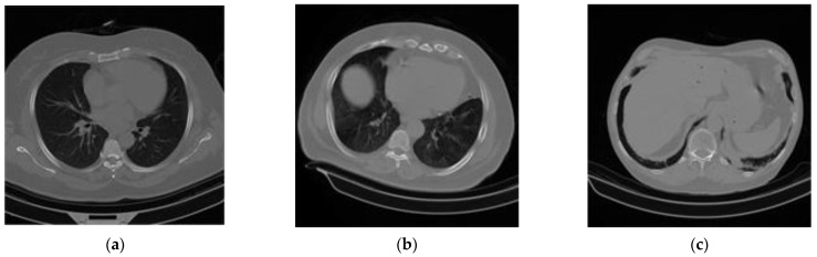
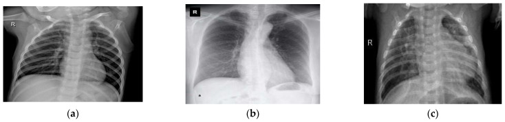
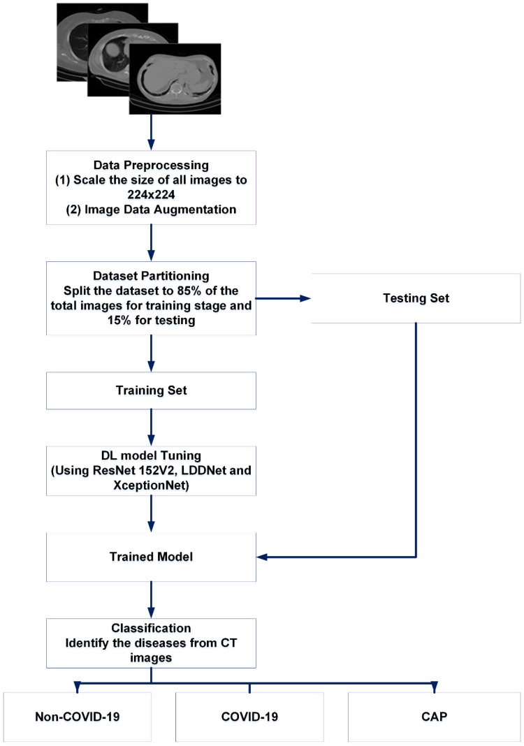
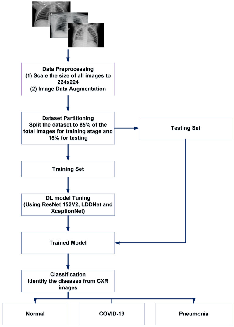
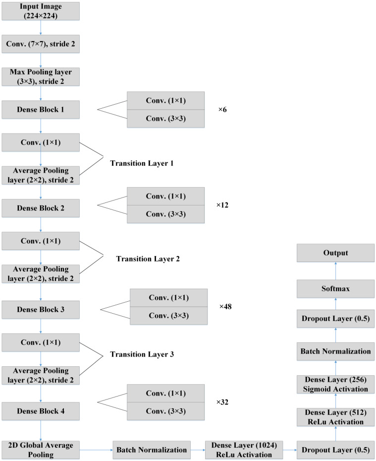
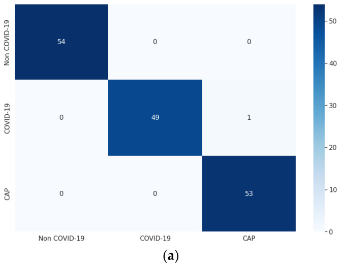
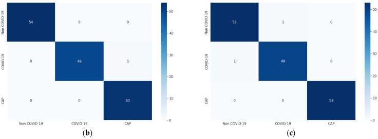
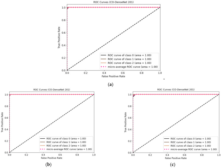
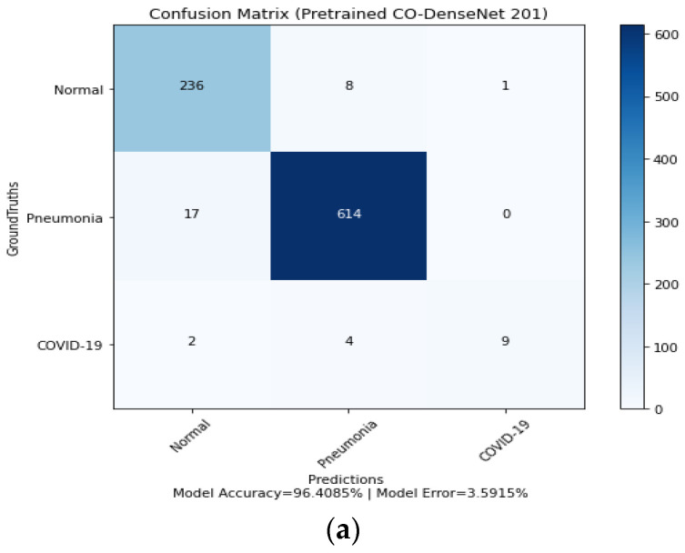
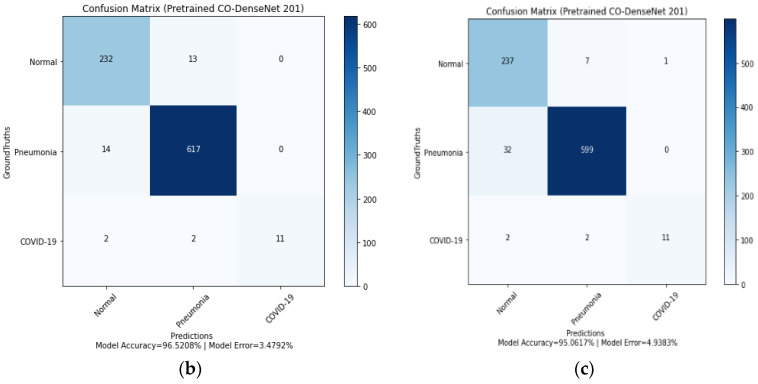
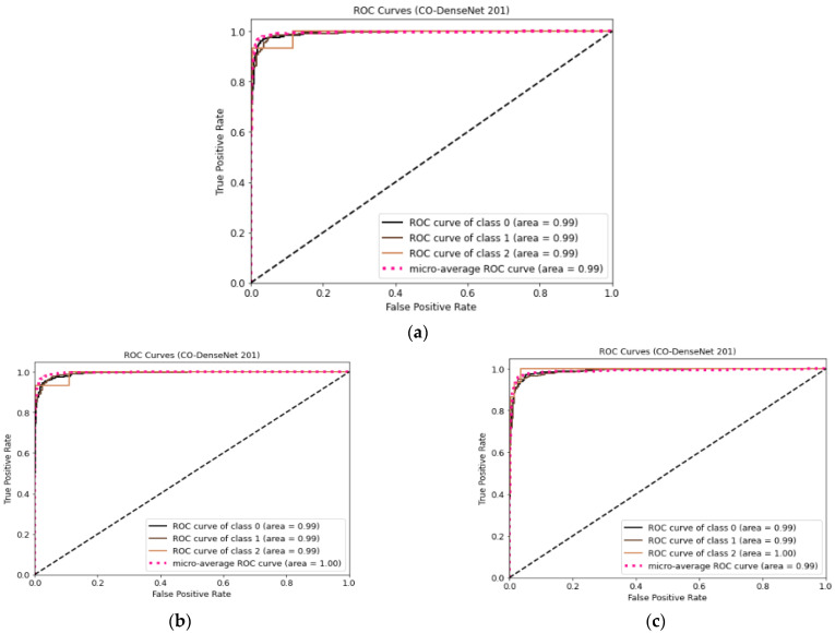
This paper proposes a new deep learning (DL) framework for the analysis of lung diseases, including COVID-19 and pneumonia, from chest CT scans and X-ray (CXR) images. This framework is termed optimized DenseNet201 for lung diseases (LDDNet). The proposed LDDNet was developed using additional layers of 2D global average pooling, dense and dropout layers, and batch normalization to the base DenseNet201 model. There are 1024 Relu-activated dense layers and 256 dense layers using the sigmoid activation method. The hyper-parameters of the model, including the learning rate, batch size, epochs, and dropout rate, were tuned for the model. Next, three datasets of lung diseases were formed from separate open-access sources. One was a CT scan dataset containing 1043 images. Two X-ray datasets comprising images of COVID-19-affected lungs, pneumonia-affected lungs, and healthy lungs exist, with one being an imbalanced dataset with 5935 images and the other being a balanced dataset with 5002 images. The performance of each model was analyzed using the Adam, Nadam, and SGD optimizers. The best results have been obtained for both the CT scan and CXR datasets using the Nadam optimizer. For the CT scan images, LDDNet showed a COVID-19-positive classification accuracy of 99.36%, a 100% precision recall of 98%, and an F1 score of 99%. For the X-ray dataset of 5935 images, LDDNet provides a 99.55% accuracy, 73% recall, 100% precision, and 85% F1 score using the Nadam optimizer in detecting COVID-19-affected patients. For the balanced X-ray dataset, LDDNet provides a 97.07% classification accuracy. For a given set of parameters, the performance results of LDDNet are better than the existing algorithms of ResNet152V2 and XceptionNet.
Keywords: COVID-19; CT scan; DenseNet201; ResNet152V2; X-ray; XceptionNet; infectious disease.
Conflict of interest statement
All the authors in this paper have no conflict of interest.
Figures
Similar articles
-
CO-IRv2: Optimized InceptionResNetV2 for COVID-19 detection from chest CT images.PLoS One. 2021 Oct 28;16(10):e0259179. doi: 10.1371/journal.pone.0259179. eCollection 2021. PLoS One. 2021. PMID: 34710175 Free PMC article.
-
Deep-chest: Multi-classification deep learning model for diagnosing COVID-19, pneumonia, and lung cancer chest diseases.Comput Biol Med. 2021 May;132:104348. doi: 10.1016/j.compbiomed.2021.104348. Epub 2021 Mar 19. Comput Biol Med. 2021. PMID: 33774272 Free PMC article.
-
Deep Learning Algorithm for COVID-19 Classification Using Chest X-Ray Images.Comput Math Methods Med. 2021 Nov 9;2021:9269173. doi: 10.1155/2021/9269173. eCollection 2021. Comput Math Methods Med. 2021. PMID: 34795794 Free PMC article.
-
Development and integration of VGG and dense transfer-learning systems supported with diverse lung images for discovery of the Coronavirus identity.Inform Med Unlocked. 2022;32:101004. doi: 10.1016/j.imu.2022.101004. Epub 2022 Jul 8. Inform Med Unlocked. 2022. PMID: 35822170 Free PMC article. Review.
-
Deep Learning-Driven Automated Detection of COVID-19 from Radiography Images: a Comparative Analysis.Cognit Comput. 2021 Mar 2:1-30. doi: 10.1007/s12559-020-09779-5. Online ahead of print. Cognit Comput. 2021. PMID: 33680209 Free PMC article. Review.
Cited by
-
Contribution to pulmonary diseases diagnostic from X-ray images using innovative deep learning models.Heliyon. 2024 Apr 26;10(9):e30308. doi: 10.1016/j.heliyon.2024.e30308. eCollection 2024 May 15. Heliyon. 2024. PMID: 38707425 Free PMC article.
-
Deep Learning for Pneumonia Detection in Chest X-ray Images: A Comprehensive Survey.J Imaging. 2024 Jul 23;10(8):176. doi: 10.3390/jimaging10080176. J Imaging. 2024. PMID: 39194965 Free PMC article. Review.
-
WPD-Enhanced Deep Graph Contrastive Learning Data Fusion for Fault Diagnosis of Rolling Bearing.Micromachines (Basel). 2023 Jul 21;14(7):1467. doi: 10.3390/mi14071467. Micromachines (Basel). 2023. PMID: 37512779 Free PMC article.
-
Right Information, Right Care, Right Patient, Right Time: Community Preferences to Inform a Self-Management Support Tool for Upper Respiratory Symptoms.Appl Clin Inform. 2025 Jan;16(1):145-155. doi: 10.1055/a-2441-6016. Epub 2024 Oct 29. Appl Clin Inform. 2025. PMID: 39472036
-
Artificial intelligence algorithms for predicting post-operative ileus after laparoscopic surgery.Heliyon. 2024 Feb 22;10(5):e26580. doi: 10.1016/j.heliyon.2024.e26580. eCollection 2024 Mar 15. Heliyon. 2024. PMID: 38439857 Free PMC article.
References
-
- Novel Coronavirus—China. Online. 2020. [(accessed on 11 October 2022)]. Available online: http://www.who.int/csr/don/12-january-2020-novel-coronaviruschina/en/
-
- Srivatsan S., Han P.D., van Raay K., Wolf C.R., McCulloch D.J., Kim A.E., Brandstetter E., Martin B., Gehring J., Chen W., et al. Preliminary support for a “dry swab, extraction free” protocol for SARS-CoV-2 testing via RT-qPCR. BioRxiv. 2020 doi: 10.1101/2020.04.22.056283. - DOI
MeSH terms
LinkOut - more resources
Full Text Sources
Medical
