. 2022 Dec 22:13:1056417.
doi: 10.3389/fimmu.2022.1056417. eCollection 2022.

Progranulin loss results in sex-dependent dysregulation of the peripheral and central immune system

Affiliations

Progranulin loss results in sex-dependent dysregulation of the peripheral and central immune system

Madelyn C Houser et al. Front Immunol. .

Abstract

Introduction: Progranulin (PGRN) is a secreted glycoprotein, the expression of which is linked to several neurodegenerative diseases. Although its specific function is still unclear, several studies have linked it with lysosomal functions and immune system regulation. Here, we have explored the role of PGRN in peripheral and central immune system homeostasis by investigating the consequences of PGRN deficiency on adaptive and innate immune cell populations.

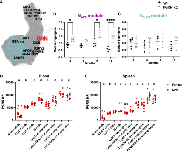
Methods: First, we used gene co-expression network analysis of published data to test the hypothesis that Grn has a critical role in regulating the activation status of immune cell populations in both central and peripheral compartments. To investigate the extent to which PGRN-deficiency resulted in immune dysregulation, we performed deep immunophenotyping by flow cytometry of 19-24-month old male and female Grn-deficient mice (PGRN KO) and littermate Grn-sufficient controls (WT).

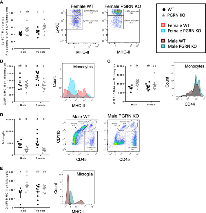
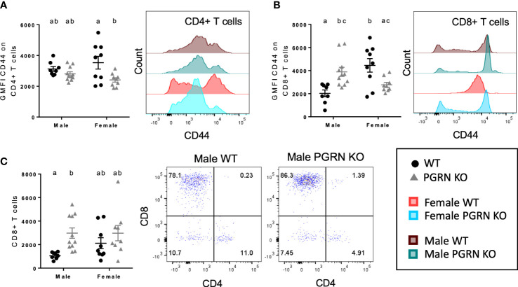
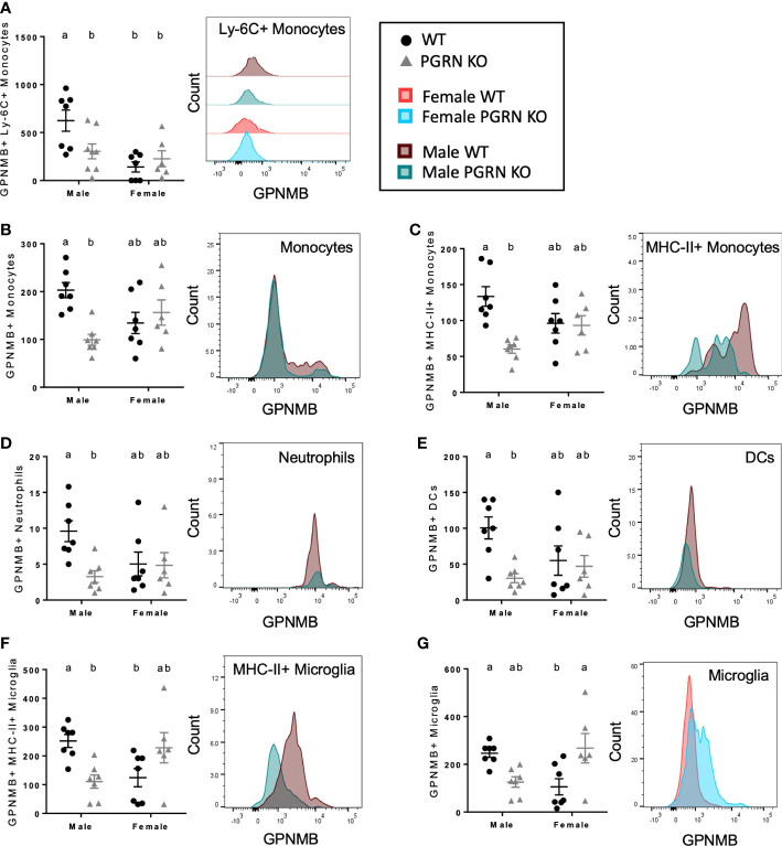
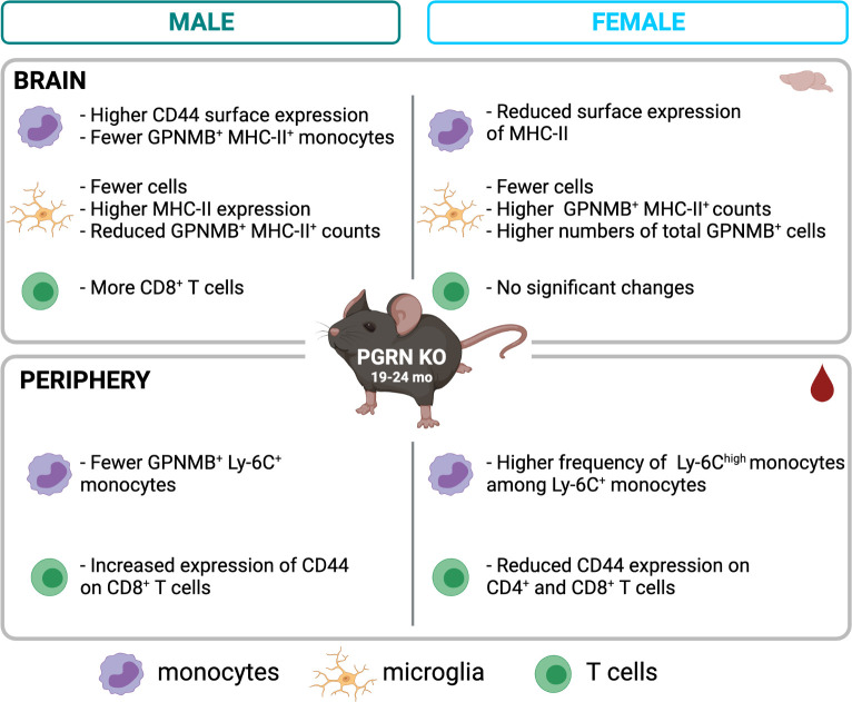
Results: Male PGRN KO mice exhibited a lower abundance of microglial cells with higher MHC-II expression, increased CD44 expression on monocytes in the brain, and more CNS-associated CD8+ T cells compared to WT mice. Furthermore, we observed an increase in CD44 on CD8+ T cells in the peripheral blood. Female PGRN KO mice also had fewer microglia compared to WT mice, and we also observed reduced expression of MHC-II on brain monocytes. Additionally, we found an increase in Ly-6Chigh monocyte frequency and decreased CD44 expression on CD8+ and CD4+ T cells in PGRN KO female blood. Given that Gpnmb, which encodes for the lysosomal protein Glycoprotein non-metastatic melanoma protein B, has been reported to be upregulated in PGRN KO mice, we investigated changes in GPNMB protein expression associated with PGRN deficits and found that GPNMB is modulated in myeloid cells in a sex-specific manner.

Discussion: Our data suggest that PGRN and GPNMB jointly regulate the peripheral and the central immune system in a sex-specific manner; thus, understanding their associated mechanisms could pave the way for developing new neuroprotective strategies to modulate central and peripheral inflammation to lower risk for neurodegenerative diseases and possibly delay or halt progression.

Keywords: GPNMB; T cells; microglia; monocytes; neurodegenaration; peripheral-brain crosstalk; progranulin.


Conflict of interest statement

The authors declare that the research was conducted in the absence of any commercial or financial relationships that could be construed as a potential conflict of interest.

Figures

Figure 1
Figure 1
PGRN regulates inflammatory responses in the periphery and central nervous system. (A) Gene co-expression adjacency network plot showing GRN is in the NSUPP module (turquoise) and highly connected to NACT (pink) module, with genes as nodes and edges between genes proportional to their bi-weighted miscorrelation of their expression in MAPT mutant microglia (see Methods). (B) Module eigengene trajectory of NACT in PGRN knockout, and WT cerebral cortex from Lui et al. (17) (n= 3-6 per condition, two way ANOVA genotype effect p <0.0001 and interaction p = 0.039, Bonferroni post hoc). (C) Module eigengene trajectory of NSUPP in PGRN knockout, and WT cerebral cortex from Lui et al. (17) (n= 3-6 per condition, Mann-Whitney with Holm-Šidák post-hoc). (D) PBMCs and (E) splenocytes from 18–21-month-old female and male B6 mice were isolated, and different immune cell subsets were assessed for PGRN MFI determined by flow cytometry, n= 6 per sex; two-way ANOVA, cell type and sex effect p < 0.05 for all, Bonferroni post hoc. Letter(s) centered above groups reflect results of post hoc tests. Groups that do not share any letters are significantly different from one another. *p<0.05 and **** p<0.0001.
Figure 2
Figure 2
PGRN deficient mice exhibit altered monocytic and microglial immunophenotypes in the peripheral blood and brain. (A) Ly-6Chi frequency of Ly-6C+ monocytes in peripheral blood, geometric mean fluorescence intensity (GMFI) of (B) MHC-II and (C) CD44 on monocytes in the brain, (D) microglia counts, and (E) GMFI of MHC-II on microglia from aged WT and PGRN KO mice (n=8-10) determined by flow cytometry; two-way ANOVA, genotype effect p < 0.01 for all, Tukey’s post hoc. Histograms show distribution of fluorescence intensity per cell. Letter(s) centered above groups reflect results of post hoc tests. Groups that do not share any letters are significantly different from one another.
Figure 3
Figure 3
PGRN deficient mice have altered T cell populations in the peripheral blood and brain. GMFI of CD44 on (A) CD4+ and (B) CD8+ T cells in peripheral blood, and (C) CD8+ T cell counts in the brain from aged WT and PGRN KO mice (n=8-10) determined by flow cytometry; two-way ANOVA, genotype effect p < 0.01 for (A, C), interaction p < 0.0001 for (B), Tukey’s post hoc. Histograms show distribution of fluorescence intensity per cell. Letter(s) centered above groups reflect results of post hoc tests. Groups that do not share any letters are significantly different from one another.
Figure 4
Figure 4
PGRN KO male mice have fewer GPNMB+ immune cells, while female PGRN KO mice increase GPNMB expression on microglia. Counts of (A) GPNMB+ Ly-6C+ monocytes in peripheral blood, and GPNMB+ (B) monocytes, (C) MHC-II+ monocytes, (D) neutrophils, (E) dendritic cells (DCs), (F) MHC-II+ microglia, and (G) microglia in the brain from aged WT and PGRN KO mice (n=6-7) determined by flow cytometry; two-way ANOVA, sex effect p < 0.01 for (A), genotype effect p < 0.05 for (B–E), interaction p < 0.05 for all, Tukey’s post hoc. Histograms show distribution of fluorescence intensity per cell. Letter(s) centered above groups reflect results of post hoc tests. Groups that do not share any letters are significantly different from one another.
Figure 5
Figure 5
Summary of main findings by flow cytometry in aged PGRN KO mice compared to WT. Created with BioRender.com.

References

    1. Chitramuthu BP, Bennett HPJ, Bateman A. Progranulin: a new avenue towards the understanding and treatment of neurodegenerative disease. Brain J Neurol (2017) 140(12):3081–104. doi: 10.1093/brain/awx198 - DOI - PubMed
    1. Du H, Zhou X, Feng T, Hu F. Regulation of lysosomal trafficking of progranulin by sortilin and prosaposin. Brain Commun (2022) 4(1):fcab310. doi: 10.1093/braincomms/fcab310 - DOI - PMC - PubMed
    1. Holler CJ, Taylor G, Deng Q, Kukar T. Intracellular proteolysis of progranulin generates stable, lysosomal granulins that are haploinsufficient in patients with frontotemporal dementia caused by GRN mutations. eNeuro (2017) 4(4):1–22. doi: 10.1523/ENEURO.0100-17.2017 - DOI - PMC - PubMed
    1. Lee CW, Stankowski JN, Chew J, Cook CN, Lam YW, Almeida S, et al. . The lysosomal protein cathepsin l is a progranulin protease. Mol Neurodegener (2017) 12(1):55. doi: 10.1186/s13024-017-0196-6 - DOI - PMC - PubMed
    1. Kao AW, McKay A, Singh PP, Brunet A, Huang EJ. Progranulin, lysosomal regulation and neurodegenerative disease. Nat Rev Neurosci (2017) 18(6):325–33. doi: 10.1038/nrn.2017.36 - DOI - PMC - PubMed

Publication types