Review
. 2022 Dec 23:13:1075260.
doi: 10.3389/fimmu.2022.1075260. eCollection 2022.

No NETs no TIME: Crosstalk between neutrophil extracellular traps and the tumor immune microenvironment

Affiliations
Review

No NETs no TIME: Crosstalk between neutrophil extracellular traps and the tumor immune microenvironment

Qi Fang et al. Front Immunol. .

Abstract

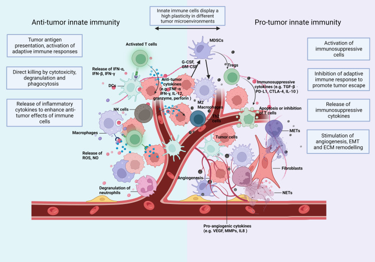
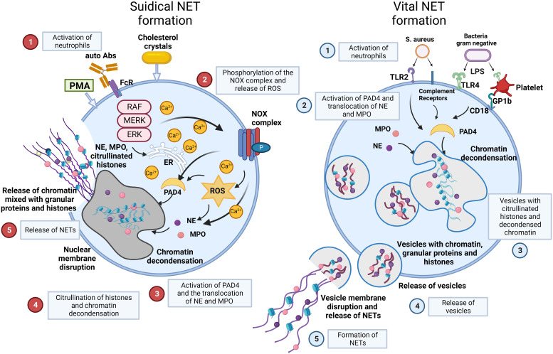
The tumor immune microenvironment (TIME) controls tumorigenesis. Neutrophils are important components of TIME and control tumor progression and therapy resistance. Neutrophil extracellular traps (NETs) ejected by activated neutrophils are net-like structures composed of decondensed extracellular chromatin filaments decorated with a plethora of granules as well as cytoplasmic proteins. Many of these harbour post translational modifications. Cancer cells reportedly trigger NET formation, and conversely, NETs alter the TIME and promote tumor cell proliferation and migration. The specific interactions between NETs and TIME and the respective effects on tumor progression are still elusive. In certain tumors, a CD4+ T helper (Th) 2 cell-associated TIME induces NETs and exerts immunosuppressive functions via programmed death 1 (PD-1)/PD-L1, both associated with poorer prognosis. In other cases, NETs induce the proliferation of Th1 cells, associated with an improved prognosis in cancer. In addition, NETs can drive macrophage polarization and often rely on macrophages to promote cancer cell invasion and metastasis. In turn, macrophages can swiftly clear NETs in an immunologically silent manner. The aim of this review is to summarize the knowledge about the mutual interaction between NETs and TIME and its impact on tumor growth and therapy.

Keywords: adaptive immunity; cancer; immunotherapy; innate immunity; macrophages; neutrophil extracellular traps; neutrophils; tumor microenvironment.


Conflict of interest statement

The authors declare that the research was conducted in the absence of any commercial or financial relationships that could be construed as a potential conflict of interest.

Figures

Figure 1
Figure 1
The TIME: Immune cells and soluble mediators shape the diversity of the TIME. Innate immunity plays multiple roles in cancer and shows a high plasticity depending on changes of the TIME. In Anti-tumorigenic TIMEs, innate immune cells (1) recognize and present tumor cell-derived antigenic peptides to T cells and activate the adaptive immunity (2); eliminate tumor cells directly by phagocytosis and secrete cytotoxic substances, like perforin and granzyme (3); secrete proinflammatory cytokines and, thus, expand and promote a wide variety of Anti-tumor responses. In a Pro-tumorigenic TIME, innate immune cells (1) differentiate towards an immunosuppressive phenotype with the release of immunosuppressive cytokines (2); induce the infiltration of immunosuppressive adaptive immune cells, such as Tregs and Th2 cells (3); increase the formation of extracellular traps (ETs); and (4) promote angiogenesis (5), epithelial-mesenchymal transition (EMT) and (6) extracellular matrix (ECM) remodelling. Consequently, they enhance tumor invasion and migration. The plasticity of innate immunity allows remodelling of the TIME. CTLA-4, cytotoxic T lymphocyte associated antigen 4; DCs, dendritic cells; ECM, extracellular matrix; EMT, epithelial mesenchymal transition; G-CSF, granulocyte colony stimulating factor; GM-CSF, granulocyte-macrophage colony stimulating factor; IFN, interferon; IL, interleukin; MDSC, myeloid-derived suppressor cells; METs, macrophage extracellular traps; MMPs, matrix metalloproteinases; NETs, neutrophil extracellular traps; NK, natural killing; NO, nitric oxide; PD-L1, programmed death-ligand 1; ROS, reactive oxygen species; TGF, transforming growth factor; TNF, tumor necrosis factor; VEGF, vascular endothelial growth factor.
Figure 2
Figure 2
NET formation: suicidal NETosis and vital NET formation establish two main types of chromatin release by neutrophils. Suicidal NETosis is characterized by ROS generation and rupture of neutrophils. Neutrophils are activated by stimuli, such as PMA, cholesterol crystals, certain autoantibodies or immune complexes. These extracellular signals induce the phosphorylation of the NOX complex and the release of ROS. This process depends on a high Ca2+ concentration. Subsequently, PAD4 is activated and causes the translocation of NE and MPO from azurophilic granules to the nucleus. NE and MPO combined with PAD4 result in the citrullination of histones and chromatin decondensation. After rupture of the nuclear membrane, the decondensed chromatin enters the cytoplasm mixed with granular proteins. Finally, the cytoplasma membrane gets leaky, the modified chromatin is released from neutrophils and forms NETs. In contrast, Vital NET formation is executed in a shorter time after activation of neutrophils and can also occur in the absence of the NOX complex and ROS. Vital NET formation is initiated by stimuli, such as S. aureus through TLR2 and complement receptors, or LPS from gram negative bacteria through TLR4 or indirectly through TLR4-activated platelets. PAD4 is activated and NE and MPO translocate to the nucleus to promote chromatin decondensation. The decondensed chromatin decorated with granular proteins and histones is packed in vesicles that bud from nuclei. Subsequently, these vesicles are expelled from intact neutrophils and form NETs in the vicinity of the neutrophils. In consequence, neutrophils stay intact and can exert further functions, such as phagocytosis. Figure adopted with modifications from [36], with permission from Springer Nature, Nature medicine © [2017]. Abs, antibodies; FcR, Fc receptor; GP, glycoprotein; MPO, myeloperoxidase; NE, neutrophil elastase; NOX, NADPH oxidase; P, phosphorylation; PAD4, protein-arginine deiminase 4; PMA, phorbol-12-myristate-13-acetate; ROS, reactive oxygen species; S.aureus, staphylococcus aureus; TLR, toll like receptor.
Figure 3
Figure 3
NETs in colorectal cancer identified by interstitial H3cit are associated with high histopathological tumor grades and lymph node metastasis in human CRC. (A, B) NE, H2B, and H3cit were used to detect NET formation on consecutive sections of CRC by immunofluorescence. Draq5 served as counterstain. Notably, regions with extranuclear H3cit colocalize with NE H2B in consecutive sections. Extracellular DNA detected by anti-DNA antibody is restricted to H3cit positive tissues. Scale bar: 75µm. (C) In human CRC tissues, NETs identified by H3cit positively correlated with high histopathological grading and lymph node metastasis. Results taken from [55], © [2022] Pathological Society of Great Britain and Ireland, first published by John Wiley & Sons Ltd. H2B, histone H2B; H3cit, citrullinated histone 3; NE, neutrophil elastase; NETs, neutrophil extracellular traps. ****p < 0.0001, *p < 0.05.

References

    1. Galon J, Lanzi A. Immunoscore and its introduction in clinical practice. Q J Nucl Med Mol Imaging. (2020) 64(2):152–61. doi: 10.23736/s1824-4785.20.03249-5 - DOI - PubMed
    1. Naschberger E, Liebl A, Schellerer VS, Schütz M, Britzen-Laurent N, Kölbel P, et al. . Matricellular protein SPARCL1 regulates tumor microenvironment–dependent endothelial cell heterogeneity in colorectal carcinoma. J Clin Invest (2016) 126(11):4187–204. doi: 10.1172/jci78260 - DOI - PMC - PubMed
    1. Naschberger E, Croner RS, Merkel S, Dimmler A, Tripal P, Amann KU, et al. . Angiostatic immune reaction in colorectal carcinoma: Impact on survival and perspectives for antiangiogenic therapy. Int J Cancer. (2008) 123(9):2120–9. doi: 10.1002/ijc.23764 - DOI - PubMed
    1. St. Paul M, Ohashi PS. The roles of CD8+ T cell subsets in antitumor immunity. Trends Cell Biol (2020) 30(9):695–704. doi: 10.1016/j.tcb.2020.06.003 - DOI - PubMed
    1. Li C, Jiang P, Wei S, Xu X, Wang J. Regulatory T cells in tumor microenvironment: new mechanisms, potential therapeutic strategies and future prospects. Mol Cancer. (2020) 19(1):116. doi: 10.1186/s12943-020-01234-1 - DOI - PMC - PubMed

Publication types