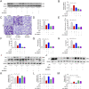
FGF10 ameliorates lipopolysaccharide-induced acute lung injury in mice via the BMP4-autophagy pathway
- PMID: 36618911
- PMCID: PMC9813441
- DOI: 10.3389/fphar.2022.1019755
FGF10 ameliorates lipopolysaccharide-induced acute lung injury in mice via the BMP4-autophagy pathway
Abstract
Introduction: Damage to alveolar epithelial cells caused by uncontrolled inflammation is considered to be the main pathophysiological change in acute lung injury. FGF10 plays an important role as a fibroblast growth factor in lung development and lung diseases, but its protective effect against acute lung injury is unclear. Therefore, this study aimed to investigate protective effect and mechanism of FGF10 on acute lung injury in mice. Methods: ALI was induced by intratracheal injection of LPS into 57BL/6J mice. Six hours later, lung bronchoalveolar lavage fluid (BALF) was acquired to analyse cells, protein and the determination of pro-inflammatory factor levels, and lung issues were collected for histologic examination and wet/dry (W/D) weight ratio analysis and blot analysis of protein expression. Results: We found that FGF10 can prevent the release of IL-6, TNF-α, and IL-1β, increase the expression of BMP4 and autophagy pathway, promote the regeneration of alveolar epithelial type Ⅱ cells, and improve acute lung injury. BMP4 gene knockdown decreased the protective effect of FGF10 on the lung tissue of mice. However, the activation of autophagy was reduced after BMP4 inhibition by Noggin. Additionally, the inhibition of autophagy by 3-MA also lowered the protective effect of FGF10 on alveolar epithelial cells induced by LPS. Conclusions: These data suggest that the protective effect of FGF10 is related to the activation of autophagy and regeneration of alveolar epithelial cells in an LPS-induced ALI model, and that the activation of autophagy may depend on the increase in BMP4 expression.
Keywords: AECs; BMP4; FGF10; acute lung injury; autophagy.
Copyright © 2022 Kong, Lu, Lin, Chong, Wen, Shi, Lin, Zhou, Zhang and Zhang.
Conflict of interest statement
The authors declare that the research was conducted in the absence of any commercial or financial relationships that could be construed as a potential conflict of interest.
Figures


Similar articles
-
RAGE inhibition alleviates lipopolysaccharides-induced lung injury via directly suppressing autophagic apoptosis of type II alveolar epithelial cells.Respir Res. 2023 Jan 24;24(1):24. doi: 10.1186/s12931-023-02332-6. Respir Res. 2023. PMID: 36691012 Free PMC article.
-
FGF10 protects against LPS-induced epithelial barrier injury and inflammation by inhibiting SIRT1-ferroptosis pathway in acute lung injury in mice.Int Immunopharmacol. 2024 Jan 25;127:111426. doi: 10.1016/j.intimp.2023.111426. Epub 2023 Dec 25. Int Immunopharmacol. 2024. PMID: 38147776
-
TIPE2 ameliorates lipopolysaccharide-induced apoptosis and inflammation in acute lung injury.Inflamm Res. 2019 Nov;68(11):981-992. doi: 10.1007/s00011-019-01280-6. Epub 2019 Sep 5. Inflamm Res. 2019. PMID: 31486847 Free PMC article.
-
Cystic fibrosis transmembrane conductance regulator ameliorates lipopolysaccharide-induced acute lung injury by inhibiting autophagy through PI3K/AKT/mTOR pathway in mice.Respir Physiol Neurobiol. 2020 Feb;273:103338. doi: 10.1016/j.resp.2019.103338. Epub 2019 Nov 11. Respir Physiol Neurobiol. 2020. PMID: 31726235
-
FGF10 and Human Lung Disease Across the Life Spectrum.Front Genet. 2018 Oct 31;9:517. doi: 10.3389/fgene.2018.00517. eCollection 2018. Front Genet. 2018. PMID: 30429870 Free PMC article. Review.
Cited by
-
LL37-mtDNA regulates viability, apoptosis, inflammation, and autophagy in lipopolysaccharide-treated RLE-6TN cells by targeting Hsp90aa1.Open Life Sci. 2024 Aug 28;19(1):20220943. doi: 10.1515/biol-2022-0943. eCollection 2024. Open Life Sci. 2024. PMID: 39220589 Free PMC article.
-
FGF21 ameliorates septic liver injury by restraining proinflammatory macrophages activation through the autophagy/HIF-1α axis.J Adv Res. 2025 Mar;69:477-494. doi: 10.1016/j.jare.2024.04.004. Epub 2024 Apr 9. J Adv Res. 2025. PMID: 38599281 Free PMC article.
References
-
- Burnham E., McCord J., Bose S., Brown L., House R., Moss M., et al. (2012). Protandim does not influence alveolar epithelial permeability or intrapulmonary oxidative stress in human subjects with alcohol use disorders. Am. J. physiology Lung Cell. Mol. physiology 302 (7), L688–L699. 10.1152/ajplung.00171.2011 - DOI - PMC - PubMed
LinkOut - more resources
Full Text Sources

