. 2022 Dec;2(4):100147.
doi: 10.1016/j.ynirp.2022.100147. Epub 2022 Nov 15.

Replication of neural responses to monetary incentives and exploration of reward-influenced network connectivity in fibromyalgia

Affiliations

Replication of neural responses to monetary incentives and exploration of reward-influenced network connectivity in fibromyalgia

Su Hyoun Park et al. Neuroimage Rep. 2022 Dec.

Abstract

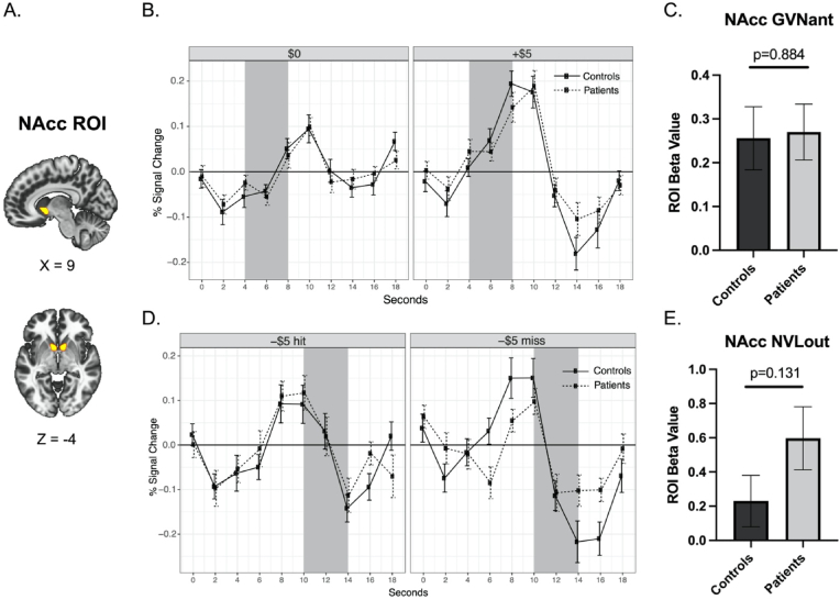
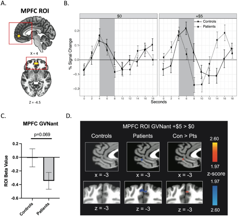
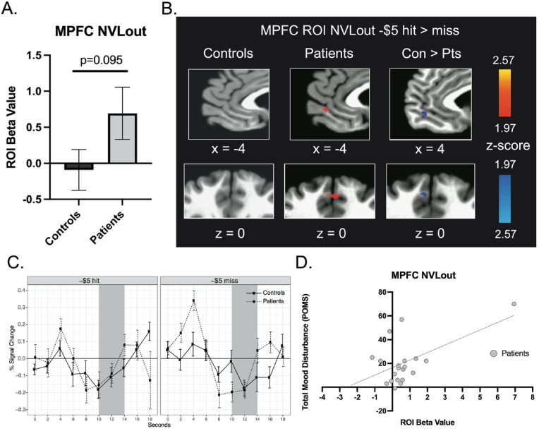
Neuroimaging research has begun to implicate alterations of brain reward systems in chronic pain. Previously, using functional magnetic resonance imaging (fMRI) and a monetary incentive delay (MID) task, Martucci et al. (2018) showed that neural responses to reward anticipation and outcome are altered in fibromyalgia. In the present study, we aimed to test the replicability of these altered neural responses to reward in a separate fibromyalgia cohort. In addition, the present study was conducted at a distinct U.S. location but involved a similar study design. For the present study, 20 patients with fibromyalgia and 20 healthy controls participated in MID task fMRI scan procedures and completed clinical/psychological questionnaires. fMRI analyses comparing patient and control groups revealed a consistent trend of main results which were largely similar to the prior reported results. Specifically, in the replication fibromyalgia cohort, medial prefrontal cortex (MPFC) response was reduced during gain anticipation and was increased during no-loss (non-punishment) outcome compared to controls. Also consistent with previous findings, the nucleus accumbens response to gain anticipation did not differ in patients vs. controls. Further, results from similarly-designed behavioral, correlational, and exploratory analyses were complementary to previous findings. Finally, a novel network-based functional connectivity analysis of the MID task fMRI data across patients vs. controls implied enhanced connectivity within the default mode network in participants with fibromyalgia. Together, based on replicating prior univariate results and new network-based functional connectivity analyses of MID task fMRI data, we provide further evidence of altered brain reward responses, particularly in the MPFC response to reward outcomes, in patients with fibromyalgia.

Keywords: Chronic pain; Medial prefrontal cortex; Monetary incentive delay (MID) task; Nucleus accumbens; Total mood disturbance; Value.


Conflict of interest statement

Declaration of competing interest The authors declare that they have no known competing financial interests or personal relationships that could have appeared to influence the work reported in this paper.

Figures

Fig. 1
Fig. 1
Structure of monetary incentive delay (MID) task trials.
Fig. 2
Fig. 2
Nucleus Accumbens Activity during Reward Anticipation. (A) Bilateral ROI of the NAcc. The same ROI mask that was used in Martucci et al. (2018) was used in the present analyses. (B) Group means and standard error of raw time course plots of NAcc ROI activity to $0 and +$5 anticipation trials. The shaded area represents the estimated anticipation period (presentation of cue and fixation [0–4 s] plus 4 s to account for hemodynamic response function delay). (C) Contrast beta values extracted from the bilateral NAcc ROI during reward anticipation (GVNant, +$5 > $0 trials). (D) Group means and standard error of raw time course plots of NAcc ROI activity to -$5 hit (i.e., no loss) and -$5 miss (i.e., loss) trials. (E) Contrast beta values extracted from the bilateral NAcc ROI during non-punishment outcomes (NVLout; -$5 hit vs. miss). All beta values are shown as 10−3. Abbreviation: NAcc, nucleus accombens; ROI, region of interest.
Fig. 3
Fig. 3
Medial Prefrontal Cortex Activity during Reward Anticipation. (A) Bilateral ROI of MPFC. The same ROI mask from Martucci et al. (2018) was used. Red boxes denote magnification areas of sagittal and axial plane images depicted in Fig. 3D. (B) Group means and standard error for raw time course plots of MPFC ROI activity to $0 and +$5 anticipation trials. The shaded area represents the estimated anticipation period (presentation of cue and fixation [0–4 s] plus 4 s to account for hemodynamic response function delay). (C) Contrast beta values extracted from the MPFC ROI during reward anticipation (GVNant, +$5 > $0 trials). (D) Contrast activation maps of MPFC ROI activity during reward anticipation (GVNant; p < 0.05, uncorrected). All beta values are shown as 10−3. Abbreviation: Con, controls; MPFC, medial prefrontal cortex; Pts, patients; ROI, region of interest. (For interpretation of the references to colour in this figure legend, the reader is referred to the Web version of this article.)
Fig. 4
Fig. 4
Medial Prefrontal Cortex Activity in Response to Non-punishment Outcomes. (A) fMRI beta values extracted from the MPFC ROI during non-punishment outcomes (NVLout contrast, -$5 hit > miss). (B) Contrast activation maps of MPFC ROI activity during non-punishment outcomes (p < 0.05, uncorrected). (C) Group means and standard error of raw time course plots of MPFC ROI activity to outcomes (hits or misses) for -$5 anticipation trials. The shaded area represents the estimated outcome period (presentation of outcome and post-outcome [6–10 s] plus 4 s to account for hemodynamic response function delay). (D) Correlations between MPFC NVLout extracted beta values and total mood disturbance (POMS) in the patient group. One patient with a high MPFC NVLout beta value was kept in the analysis because the fMRI scan volumes were censored for bad movements (greater than a Euclidean norm value of 0.5) during pre-processing, and only 4 vol (among 540 vol) were censored for this individual. Also, this patient did not have as high of a beta value in the NVLout contrast with both -$5 and -$1 data (see results in Supplementary Fig. 4E). Therefore, we believe this to be a true representative signal of MPFC activity for the -$5 NVLout contrast from this individual. Further, the greater total mood disturbance (i.e., worse) in this patient aligns with greater MPFC activation that may be consistent with a greater “relief” experience. Post-hoc analyses excluding this patient resulted in MPFC NVLout beta value for controls vs. patients (p = 0.174) and POMS vs. MPFC NVLout beta values correlation (r = 0.08; p = 0.606). All beta values are shown as 10−3. Abbreviation: Con, controls; MPFC, medial prefrontal cortex; Pts, patients; ROI, region of interest. POMS, Profile of Mood States.
Fig. 5
Fig. 5
Functional Connectivity within the Default Mode Network during MID Task Performance. (A) DMN seeds defined by CONN Toolbox with significant group differences in seed-to-seed connectivity (yellow lines) [Green = MPFC, Blue = Right LP, Peach = Left LP, Magenta = PCC] (B) Significant group differences (fibromyalgia > controls, shown in red with asterisks) in connectivity between DMN seeds (p < 0.05, FDR corrected). Abbreviation: DMN, default mode network; FDR, false discovery rate; LP, lateral parietal cortex; MID, monetary incentive delay; MPFC, medial prefrontal cortex; PCC, posterior cingulate cortex; ROI, region of interest. (For interpretation of the references to colour in this figure legend, the reader is referred to the Web version of this article.)

References

    1. Acikalin M.Y., Gorgolewski K.J., Poldrack R.A. A coordinate-based meta-analysis of overlaps in regional specialization and functional connectivity across subjective value and default mode networks. Front. Neurosci. 2017;11:1. - PMC - PubMed
    1. Albrecht D.S., MacKie P.J., Kareken D.A., Hutchins G.D., Chumin E.J., Christian B.T., Yoder K.K. Differential dopamine function in fibromyalgia. Brain Imaging and Behavior. 2016;10(3):829–839. doi: 10.1007/s11682-015-9459-4. - DOI - PMC - PubMed
    1. Andrews-Hanna J.R. The brain's default network and its adaptive role in internal mentation. Neuroscientist. 2012;18(3):251–270. doi: 10.1177/1073858411403316. - DOI - PMC - PubMed
    1. Baker A.K., Ericksen L.C., Koppelmans V., Mickey B.J., Martucci K.T., Zubieta J.-K., Love T.M. Altered reward processing and sex differences in chronic pain. Front. Neurosci. 2022;16 https://www.frontiersin.org/article/10.3389/fnins.2022.889849 - DOI - PMC - PubMed
    1. Baliki M.N., Petre B., Torbey S., Herrmann K.M., Huang L., Schnitzer T.J., Fields H.L., Apkarian A.V. Corticostriatal functional connectivity predicts transition to chronic back pain. Nat. Neurosci. 2012;15(8):1117–1119. doi: 10.1038/nn.3153. - DOI - PMC - PubMed

LinkOut - more resources